")
Back to Journals » International Medical Case Reports Journal » Volume 17
Bilateral Iris Coloboma in an 11-Year-Old Child with Low Vision and High Intraocular Pressure: A Rare Case Report and Review of Literature
Authors Mushkani TA , Roheen ZUR
Received 12 January 2024
Accepted for publication 5 March 2024
Published 12 March 2024 Volume 2024:17 Pages 157—160
DOI https://doi.org/10.2147/IMCRJ.S453954
Checked for plagiarism Yes
Review by Single anonymous peer review
Peer reviewer comments 2
Editor who approved publication: Dr Scott Fraser
Tawfiq Ahmad Mushkani, Zabih Ur Rahman Roheen
Ophthalmology Department, Kabul University of Medical Science, Kabul, Afghanistan
Correspondence: Tawfiq Ahmad Mushkani, Email [email protected]
Background: Coloboma means curtailed in Greek language. It is mainly used when normal tissue of the eye or another organ is not present since birth. Coloboma is a congenital abnormality mainly caused by incomplete closure of the embryonic fissure of the choroid part of eye.
Purpose: The aim of this case report is to share the clinical findings in a patient with bilateral iris coloboma, low vision, and headache.
Patients and Methods: Case report.
Results: An eleven-year-old boy with low vision and headache visited the University Eye Hospital of Kabul University of Medical Science (UEHKUMS) for consultation. Ophthalmic examination revealed a bilateral iris coloboma without concomitant chorioretinal defect, refractive error, and high intraocular pressure in both eyes. The refractive error of the patient was corrected by advising proper glasses, and the high intraocular pressure was controlled by anti-glaucoma drops. After several follow-up visits, the patient no longer complained of headache and low vision.
Conclusion: Visiting patients with iris coloboma should be considered for intraocular pressure (IOP) check, and screening of other family members is mandatory.
Keywords: iris coloboma, high intraocular pressure, bilateral
Introduction
Eye coloboma is caused by a defect in fetal fissure closure which occurs in the 5th or 6th week of intrauterine life. Sometimes it may extend to the cornea, iris, zonula, ciliary body, choroid, retina, or optic nerve. The overall incidence of this congenital defect is 0.7 per 10,000 live births. Some inherited autosomal and recessive eye disease like retinitis pigmentosa may be associated with the disease. Many problems like cataracts, lens subluxation, strabismus, anisometropia, secondary glaucoma, and amblyopia have been reported with this developmental defect.1 The closure begins at the equator and should continue anteriorly and posteriorly. Closure delays at this time may cause defects of various types and locations. A coloboma may extend from the iris margin to the optic disc and involve one or more defects along fusional lines. Colobomas affecting the posterior segment of the eye can affect one or both eyes. Both eyes are affected in approximately 60% of cases.2
If the coloboma is seen in the retinal pigment epithelium (RPE), neurosensory retina, or choroid, it means that the posterior fetal fissure has failed to close. Patients with the defect may only have sclera covering retinal pigment epithelium (RPE) and absent choroid, but in some cases retina may also be missing.3
A fibrovascular remnant of the anterior hyaloid and pupillary membrane may cause coloboma. The typical form of iris coloboma is located inferonasally, and the pupil of the patient may show the shape of a keyhole, light bulb, or an inverted oil drop. Coloboma may also affect the lens, ciliary body, choroid, and optic nerve. During ophthalmic examination of the patient's family members, we may find small and undiscovered defects in iris and chorioretinal regions. Careful examination of family members is necessary and indicated.4
Case Report
An 11-year-old boy visited the University Eye Hospital of Kabul University of Medical Science for visual blurring and headache for one year. No associated redness was observed in the eyes. According to the patient history, he is suffering from low vision, especially during lessons in class. It was the patient's first visit to an ophthalmic center with no previous ophthalmic consultations.
The distance visual acuity of the right eye of the patient was 20/60 without correction, but with correction of +2 sphere, cylinder of −2.5, and at an angle of 5° patient vision improved to 20/20. The left eye revealed a distance visual acuity of 20/40 without correction and BCVA of 20/20 with a glass of +2.00 (−2.00) 170°.
Slit lamp examination revealed a good conjunctiva, transparent cornea and anterior chamber. The pupil in the right eye was pear-shaped with a notch at the inferonasal location (Figure 1). The pupil in the left eye was pear-shaped with a notch toward the left canthus (Figure 2). The photomotor reflexes were normal. The lens was normal and with no defect. Indirect ophthalmoscopy did not reveal any abnormalities in the choroid and retina. Intraocular pressure was documented at 26 mmHg in the right eye and 24 mmHg in the left eye with both Tonopen and air puff tonometer in three consecutive sessions with the patient fully relaxed. Before starting antiglaucoma drops (Timolol), pachymetry was advised in both eyes and corneal thickness was found within normal range.
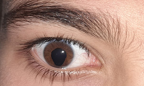 |
Figure 1 Shows inferonasal iris coloboma in the right eye of the patient. |
 |
Figure 2 Shows lateral temporal iris coloboma in the left eye of the patient. |
The younger sibling of the patient was also documented with bilateral iris coloboma without concomitant chorioretinal defect and normal intraocular pressure. The importance of this case report is the unusual location of the left eye iris coloboma, which is located on the lateral-temporal side; the most common side of iris coloboma mentioned in the literature is the inferonasal side. Overall after several follow-ups, the intraocular pressure under Timolol in both eyes is below 20 mmHg and the patient is fully satisfied with medication and glasses advised.
Discussion
Iris colobomas often do not associate with any complication. Association of colobomas with systemic anomalies varies. There are two types of syndromes that coloboma may be associated with: the first one is the CHARGE syndrome (coloboma, heart defect, choanal atresia, retardation of growth and development, genital hypoplasia, and ear abnormalities), and the second is the COACH syndrome (cerebellar vermis hypoplasia, oligophrenia, ataxia, coloboma, and hepatic fibrosis).1 In the present case, no systemic abnormalities were found.
A case report was conducted in a university teaching hospital of Togo regarding a 12-year-old patient with visual impairment of 2 years' duration. During examination the ophthalmologist found bilateral notching of pupil and iris, and the patient was diagnosed with inferonasal iris coloboma.1 The interesting finding compared to the mentioned case is the unusual location of iris coloboma in the left eye of our patient (lateral-temporal).
Chorioretinal colobomas are congenital.5 The patient is exposed to the complications of this anomaly throughout of life.6 Previous studies have found mutations in various genes associated with microphthalmia, anophthalmia, and coloboma (MAC) phenotypes.7 Typical iris and retinochoroidal coloboma is a relatively common finding which results from the failure of choroidal fissure closure when the embryogenesis is complete in the first three months of gestational life.8 However, in our case, except for iris coloboma with high intraocular pressure, no other abnormalities were found in the posterior segment of the eye. Iris colobomas usually do not cause severe low vision and may cause cosmetic blemish. If it is associated with chorioretinal coloboma it may cause severe disturbances of vision, according to some authors.9,10
A similar case of both iris and lens coloboma has been reported in a 28-year-old man by Ould Hamed et al in 2018 in Morocco. In this case the main complaint of the patient was reduced visual acuity.11 Compared to the above-mentioned case report, the main take-away finding which makes our case unique is the high intraocular pressure of our patient.
Conclusion
Iris coloboma in both eyes with high intraocular pressure (IOP) is rare. Visiting patients with iris coloboma should be considered for intraocular pressure check, and screening of other family members is mandatory. Maintaining good visual acuity for the patient is our goal, but sometimes iris coloboma accompanied by posterior segment coloboma or systemic anomalies may significantly affect the prognosis of visual acuity of the patients.
Abbreviations
UEH, University Eye Hospital; KUMS, Kabul University of Medical Science; CHARGE syndrome, coloboma, heart defect, choanal atresia, retardation of growth and development, genital hypoplasia, and ear abnormalities; COACH, cerebellar vermis hypoplasia, oligophrenia, ataxia, coloboma, and hepatic fibrosis syndromes; RPE, retinal pigment epithelium; BCVA, best corrected visual acuity; IOP, intraocular pressure; MAC phenotype, microphthalmia, anophthalmia, and coloboma.
Ethics and Consent Statements
The institutional approval is not required for publication of the case report. Written informed consent for publication of their details was obtained from the father of the patient.
Disclosure
The authors declare no conflicts of interest in this work.
References
1. Messan Amedome K, Assavédo CR, Mensah YA, et al. Bilateral iris coloboma revealed by a decreased vision: about the first case in togo observed in kara university teaching hospital. Open J Ophthalmol. 2021;11(04):249–252. doi:10.4236/ojoph.2021.114020
2. Pavan-Langston D. Manual of Ocular Diagnosis and Therapy. Boston: Little, Brown and Co; 1996:350.
3. Alexander A. Primary Care of the Posterior Segment. Norwalk: Appleton & Lange; 1994:512 p.
4. Hered RW. Pediatric Ophthalmology and Strabismus of Basic and Clinical Science Course. American Academy Of Ophthalmology. 2021;6:266.
5. Gupta V, Gupta A, Dogra MR. Subretinal neovascularization associated with retinochoroidal coloboma. Indian J Ophthalmol. 1997;45:116.
6. Nanda L, Sanjana SM, Srivastava VK. A Case Report of Bilateral iris, lens and chorioretinal Coloboma. J Dent Med Sci. 2015; 14(4):1.
7. Yoo YJ, Han SB, Yang HK, Hwang JM. Ocular coloboma combined with cleft lip and palate: a case report. BMC Ophthalmol. 2020;20:41.
8. Uhumwangho OM, Jalali S. Chorioretinal coloboma in a pediatric population. Eye. 2014;28:728–733. doi:10.1038/eye.2014.61
9. Kumar N, Valliappan A, Bansal R. Unusual superior iris and retinochoroidal coloboma. Indian J Ophthalmol. 2020;68:921. doi:10.4103/ijo.IJO_1876_19
10. Cours du Syndicat national des ophtalmologistes de France (SNOF). Available from: https://www.snof.org/encyclopedie/colobomes.
11. Ould Hamed MA, Soulay AY, Reda K, Oubaaz A. Bilateral Iris, lens and chorioretinal coloboma a case report. J Clin Res Ophthalmol. 2018;5(1):12.
 © 2024 The Author(s). This work is published and licensed by Dove Medical Press Limited. The full terms of this license are available at https://www.dovepress.com/terms.php and incorporate the Creative Commons Attribution - Non Commercial (unported, v3.0) License.
By accessing the work you hereby accept the Terms. Non-commercial uses of the work are permitted without any further permission from Dove Medical Press Limited, provided the work is properly attributed. For permission for commercial use of this work, please see paragraphs 4.2 and 5 of our Terms.
© 2024 The Author(s). This work is published and licensed by Dove Medical Press Limited. The full terms of this license are available at https://www.dovepress.com/terms.php and incorporate the Creative Commons Attribution - Non Commercial (unported, v3.0) License.
By accessing the work you hereby accept the Terms. Non-commercial uses of the work are permitted without any further permission from Dove Medical Press Limited, provided the work is properly attributed. For permission for commercial use of this work, please see paragraphs 4.2 and 5 of our Terms.