")
Back to Journals » Clinical, Cosmetic and Investigational Dermatology » Volume 17
Atypical Large Scrotal Angiokeratomas Treated with Long-Pulse Alexandrite Laser
Authors Molla A
Received 15 January 2024
Accepted for publication 24 March 2024
Published 28 March 2024 Volume 2024:17 Pages 745—750
DOI https://doi.org/10.2147/CCID.S458914
Checked for plagiarism Yes
Review by Single anonymous peer review
Peer reviewer comments 2
Editor who approved publication: Dr Jeffrey Weinberg
Amr Molla
Department of Medicine, College of Medicine, Taibah University, Madinah, Saudi Arabia
Correspondence: Amr Molla, Dermatology, Dermatologic Surgery & Lasers, Universities Road, P.O. Box: 344, Taibah, Madinah, 42353, Saudi Arabia, Tel +966504342992, Email [email protected]
Background: Fordyce angiokeratoma, a benign tiny lesion usually on the scrotum, increases with age and may cause symptoms like itching and bleeding in nearly half of the cases. Although treatment is not always necessary, it is primarily considered for cosmetic reasons in the case of larger or atypical lesions.
Case Report: We present the case of a healthy adult male with multiple large red-blue hyperkeratotic nodules and papules on his scrotal skin, causing bleeding upon minor trauma and personal embarrassment. After confirming the diagnosis of angiokeratomas of the scrotum through histopathology, the patient underwent two sessions of Long-Pulse Alexandrite Laser treatment, resulting in a 90% reduction in lesions, no scrotal bleeding, and a satisfactory cosmetic outcome.
Conclusion: The Long-Pulse Alexandrite Laser is a precise and effective treatment for vascular lesions, like angiokeratomas, offering customizable parameters. However, patient-specific factors and careful evaluation are essential, recognizing the laser’s limitations for optimal results.
Keywords: angiokeratomas, Fordyce angiokeratoma, Alexandrite laser, 755 nm, case report
Introduction
Angiokeratomas are typically characterized as asymptomatic papules measuring 2 to 5 millimeters in size, displaying a blue-to-red coloration and a scaly surface. They are commonly found on the scrotum, shaft of the penis, labia majora, inner thigh, or lower abdomen. Histologically, angiokeratomas of Fordyce consist of dilated thin-walled vessels located in the superficial dermis, accompanied by epidermal hyperplasia in the overlying skin.1
The term “angiokeratoma” serves as an umbrella for a wide range of hyperkeratotic vascular disorders that are typically asymptomatic and share a common histological feature of hyperkeratosis coupled with superficial dermal vascular ectasia.2 Specifically, angiokeratomas can be divided into localized and systemic forms.
Within the category of localized angiokeratomas, there are four major subtypes, each displaying distinct clinical presentations. Firstly, solitary papular angiokeratomas predominantly emerge on the legs. Secondly, Fordyce-type angiokeratomas are typically limited to the scrotum and vulva. Moreover, they are usually tiny red-to-black papules. Thirdly, angiokeratomas circumscriptum naviforme, representing the congenital form, present as multiple hyperkeratotic papules and plaque-like lesions, most commonly found unilaterally on the lower leg, foot, thigh, buttock, and occasionally other areas. Lastly, bilateral angiokeratomas, also known as the Mibelli type, manifest on the dorsa of the fingers and toes.
In contrast, the systemic form, referred to as angiokeratoma corporis diffusum, is often associated with metabolic disorders, with Fabry disease and fucosidosis being the most prevalent. While Fabry disease typically leads to a generalized presentation, a 2010 case report recommends considering Fabry disease as a possibility in all male patients with angiokeratomas, even if the lesions are localized to the scrotum.3 Although the pathogenesis and clinical presentation may vary, the histological features remain consistent across all forms.1,3,4
Without precise epidemiological data, efforts have been made to estimate the morbidity related to angiokeratomas. These estimates mainly address the potential complications like bleeding and discomfort, which can substantially influence the quality of life of affected individuals. Typically, these lesions do not necessitate treatment. However, in rare cases, angiokeratomas may be large and nodular in shape, causing embarrassment and patients concern. If treatment is deemed necessary, locally destructive methods such as electrocoagulation, excision, cryotherapy, or laser therapy may be employed.2,5
Case Report
A 39-year-old healthy man, devoid of any co-morbidities in his medical history, presented himself at the dermatology clinic with a concern about multiple large purple skin lesions on his scrotum, which had persisted for several years. The patient observed that these lesions were progressively increasing in both size and number over time. Furthermore, he was bothered by the lesions as they easily bled with minor trauma and caused him embarrassment due to their appearance. The patient has been in a marital relationship for the past decade with no history of extramarital affairs, and his systemic reviews were unremarkable.
Upon dermatologic examination, the patient has Fitzpatrick skin type 3, with approximately 40 red-blue, non-tender, hyperkeratotic filiform papules and nodules of varying sizes (ranging from 5 mm to 15 mm in diameter) were predominantly located on the left side of the scrotum (refer to Figures 1 and 2). There was no evidence of varicocele, testicular tumors, or inguinal hernias.
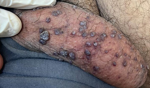 |
Figure 1 Close-Anterior view of the left side of scrotum for 38-year-old man show numerous nodular angiokeratomas presenting as large purple nodules and papules. |
 |
Figure 2 Distance-Lateral view of the left side of scrotum for the same patient. |
Two of the lesions were excised and sent for histopathological examination, revealing cavernous vascular lesions with intravascular thrombosis in the superficial dermis, accompanied by associated epidermal hyperplasia and papillomatosis. The clinical presentation and lesion histopathology correlated, leading to a diagnosis of angiokeratomas of the scrotum.
In order to prevent future episodes of spontaneous bleeding and to enhance the scrotal appearance by addressing the disfigurement, the patient was deemed a suitable candidate for Long-Pulse Alexandrite Laser 755 nm treatment for his scrotal angiokeratomas. Prior to the laser treatment, a topical anesthesia consisting of Lidocaine-prilocaine cream 5% was applied to the entire scrotal skin. Subsequently, the Long-Pulse Alexandrite Laser procedure was conducted, utilizing a hand-piece spot size of 5 mm with a pulse width duration of 10 ms, and a fluence of 175 J/cm² for smaller lesions. For larger lesions, a hand-piece spot size of 10 mm with a pulse width duration of 5 ms and a fluence of 30 J/cm2 were used. During the procedure, the scrotal skin containing lesions was carefully folded from the laser beam by holding it between the fingers to prevent exposure to the testicles. A second session, employing the same technique, was performed after a one-month interval from the first session. The patient experienced no pain during or after the procedures.
A follow-up cutaneous examination was conducted three weeks after the second session, revealing significant improvement with the disappearance of approximately 90% of all treated angiokeratoma lesions on his scrotum, with non-noticeable scarring and few post-inflammatory hypopigmentation (refer to Figure 3). Six months follow-up after the last procedure, the patient reported no further episodes of scrotal bleeding and he expressed high satisfaction with the cosmetic outcome. Upon examination, there were no new lesions and the scrotal appearance remained aesthetically pleasing.
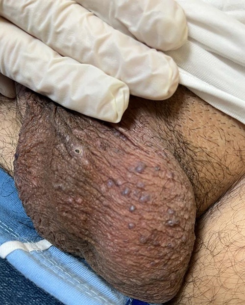 |
Figure 3 View of the left side of scrotum of the same patient after 3 week of the last session. |
Discussion
Angiokeratomas of Fordyce are benign vascular lesions characterized by dilated blood vessels in the superficial dermis and epidermal hyperplasia of the scrotal skin. They often present as raised, tiny red-blue papules measuring 2 to 5 millimeters in size, and rarely be large verrucous nodular lesions, causing cosmetic concerns and, in some cases, bleeding upon minor trauma.1
The principle underlying the Long-Pulse Alexandrite Laser’s operation is selective photothermolysis. This process involves the laser’s energy being predominantly absorbed by the hemoglobin within the blood vessels of angiokeratomas. Subsequently, this absorption induces the heating of the blood, culminating in coagulation and the eventual collapse of the vessel walls. Notably, the surrounding tissues are largely spared from damage, attributable to both the selective nature of absorption and the meticulously controlled pulse duration.6
Moreover, empirical evidence underscores the high efficacy of the Long-Pulse Alexandrite Laser in the treatment of angiokeratomas. Its capacity for precise targeting of vascular lesions manifests in a substantial diminution in both the size and quantity of angiokeratomas, as corroborated by our case study and an additional case report with different type of angiokeratoma.7–10 The employment of topical anesthetics, coupled with a refined technique as delineated in our report, presents the procedure relatively devoid of pain. Furthermore, the rarity of adverse effects, such as scarring and hypopigmentation, together with the high levels of patient satisfaction observed, are testament to the safety and favorability of this laser treatment.
Conclusion
Angiokeratomas are typically small, harmless skin lesions but can occasionally develop into larger nodules, though this is relatively uncommon. In contrast to alternative methods such as electrocautery or cryotherapy, the Long-Pulse Alexandrite Laser presents as a minimally invasive, precise, and cosmetically advantageous treatment. Its versatility is highlighted by adjustable parameters including pulse width, spot size, and fluence, which facilitate custom-tailored treatments. These adjustments are predicated on the unique characteristics of each lesion; thereby support both the efficacy and safety of the procedure.
Although it is highly effective, it remains imperative to consider patient-specific variables such as skin type, lesion dimensions, and anatomical location. The laser’s efficacy may diminish in instances involving particularly large or deeply situated lesions. Thus, meticulous patient selection and comprehensive pre-treatment evaluations are paramount in ensuring optimal treatment outcomes.8–10
In summary, the Long-Pulse Alexandrite Laser signifies a notable progression in the management of vascular lesions, including angiokeratomas. Its targeted approach, combined with a proven efficacy and safety profile, presents it a favored choice amongst both patients and healthcare professionals. Nonetheless, the importance of individualized treatment planning and acknowledgement of the laser’s limitations cannot be overemphasize, as these are critical in realizing the most favorable results.
Data Sharing Statement
The data that support the findings of this case report are available from the corresponding author upon reasonable request. Due to legal and ethical considerations, the data cannot be made publicly available. Requests for data access should be directed to Amr Molla at [email protected].
Statement of Ethics
The case report presented in this manuscript does not require ethical approval in accordance with the prevailing standards of ethics in Saudi Arabia.
Written informed consent was obtained from the patient for the photography and publication of his medical case details, including any accompanying images. A copy of the consent form is available upon request.
Acknowledgment
I would like to extend my heartfelt appreciation to the individuals and institutions that played an instrumental role in the completion of this case report conducted at Saudi German Hospital in Madinah:
- Saudi German Hospital, Madinah: I am deeply grateful to the entire medical and administrative team at Saudi German Hospital, Madinah, for their unwavering support and for providing access to the facilities and resources necessary for this research. The commitment to excellence demonstrated by the hospital staff has been invaluable in the successful completion of this case report.
- Nursing and Support Staff: I wish to acknowledge the dedication and professionalism of the nursing and support staff at Saudi German Hospital, whose tireless efforts contributed to the patient’s well-being throughout the course of their treatment.
- Family of the Patient: I extend my appreciation to the family of the patient for their consent and cooperation, which allowed me to share this case and contribute to medical knowledge.
- This case report would not have been possible without the collaborative efforts and support of the above-mentioned individuals and institutions. Their contributions have greatly enriched the quality and significance of this work.
Funding
The author declares no funding was received for the research.
Disclosure
The author reports no conflicts of interest in this work.
References
1. Wolff K. Fitzpatrick’s Dermatology in General Medicine. McGraw-Hill New York; 2008.
2. Gioglio L, Porta C, Moroni M, et al. Scrotal angiokeratoma (Fordyce): histopathological and ultrastructural findings. Histol Histopathol. 1992;7(1):47–55.
3. Hogarth V, Dhoat S, Mehta AB, et al. Late-onset Fabry disease associated with angiokeratoma of Fordyce and multiple cherry angiomas. Clin Exp Dermatol. 2011;36(5):506–508. doi:10.1111/j.1365-2230.2011.04053.x
4. Jansen T, Bechara FG, Stücker M, et al. Angiokeratoma of the scrotum (Fordyce type) associated with angiokeratoma of the oral cavity. Acta Derm Venereol. 2002;82(3):208–210. doi:10.1080/00015550260132532
5. Agger P, Osmundsen PE. Angiokeratoma of the scrotum (Fordyce). A case report on response to surgical treatment of varicocele. Acta Derm Venereol. 1970;50(3):221–224.
6. Chen C, Ke Y. Applications of long-pulse alexandrite laser in cosmetic dermatology: a review. Clin Cosmet Invest Dermatol. 2023;16:3349–3357. doi:10.2147/CCID.S441169
7. Lorgeou A, Chiaverini C, Le Duff F, et al. Successful treatment of angiokeratoma circumscriptum naeviforme with long pulse alexandrite laser. J Eur Acad Dermatol Venereol. 2017;31(4):e186–e187. doi:10.1111/jdv.13910
8. Gazzani P, Abdullah A. The effective treatment of bleeding angiokeratomas associated with Anderson-Fabry disease using long pulsed alexandrite laser. In: Journal of the American Academy of Dermatology. 360 Park Avenue South, New York, Ny 10010-1710 USA: Mosby-Elsevier; 2015.
9. Baumgartner J, Šimaljaková M, Babál P. Extensive angiokeratoma circumscriptum-successful treatment with 595-nm variable-pulse pulsed dye laser and 755-nm long-pulse pulsed alexandrite laser. J Cosmet Laser Ther. 2016;18(3):134–137. doi:10.3109/14764172.2015.1114643
10. Nguyen J, Chapman LW, Korta DZ, et al. Laser treatment of cutaneous angiokeratomas: a systematic review. Dermatol Ther. 2017;30(6):e12558. doi:10.1111/dth.12558
 © 2024 The Author(s). This work is published and licensed by Dove Medical Press Limited. The full terms of this license are available at https://www.dovepress.com/terms.php and incorporate the Creative Commons Attribution - Non Commercial (unported, v3.0) License.
By accessing the work you hereby accept the Terms. Non-commercial uses of the work are permitted without any further permission from Dove Medical Press Limited, provided the work is properly attributed. For permission for commercial use of this work, please see paragraphs 4.2 and 5 of our Terms.
© 2024 The Author(s). This work is published and licensed by Dove Medical Press Limited. The full terms of this license are available at https://www.dovepress.com/terms.php and incorporate the Creative Commons Attribution - Non Commercial (unported, v3.0) License.
By accessing the work you hereby accept the Terms. Non-commercial uses of the work are permitted without any further permission from Dove Medical Press Limited, provided the work is properly attributed. For permission for commercial use of this work, please see paragraphs 4.2 and 5 of our Terms.