")
Back to Journals » Veterinary Medicine: Research and Reports » Volume 14
Assessment of Humoral Immune Response in Pre- and Post-Vaccinated Cattle Against Lumpy Skin Disease
Authors Moje N , Bari FD, Urge B, Demissie E
Received 30 December 2022
Accepted for publication 24 July 2023
Published 8 August 2023 Volume 2023:14 Pages 133—143
DOI https://doi.org/10.2147/VMRR.S403127
Checked for plagiarism Yes
Review by Single anonymous peer review
Peer reviewer comments 3
Editor who approved publication: Professor Young Lyoo
Nebyou Moje,1 Fufa Dawo Bari,1 Beksisa Urge,2 Ejigayehu Demissie2
1College of Veterinary Medicine and Agriculture, Addis Ababa University, Bishoftu, Ethiopia; 2Holeta and Debre Zeit Agricultural Research Centers, EIAR, Oromia, Ethiopia
Correspondence: Nebyou Moje, Email [email protected]
Introduction: Lumpy skin disease (LSD) is viral disease affecting cattle production and productivity in Ethiopia. As a prevention method, vaccinations have been used for a long period with a questionable output due to the existence of LSD outbreaks in vaccinated herds in different parts of Ethiopia.
Methods: A longitudinal study was performed from October 2019 to April 2020 with the objective of assessing the humoral immune response of cattle with a serum neutralization test (SNT) from different management systems in central Ethiopia. In this study, theserum was collected from 113 cattle (extensive (60/113) and intensive (53/113) management systems) before and after vaccination.
Results and Discussion: From collected sera, a limited number of cattle had seroconversion before vaccination (7.08%). On the other hand, it is obvious the seroconversion rises post vaccination. Accordingly, seroconversion starts to increase after a week (8.85% at 7 dpv) post-vaccination which proceeds to significantly increase at 30 days post vaccination (dpv) (41.65% (25/60)). Furthermore, the risk factor study before and after vaccination showed intensively managed cattle with significantly higher levels of antibody titer at 7 dpv (OR = 1.17; 95% CI = 0.22, 6.2; p = 0.016) and 30 dpv (OR = 3.67; 95% CI = 1.1, 12.29; p = 0.035) compared with that of extensively managed cattle. The other animal-related risk factor that showed a significant difference was breeds and a specific age group ([4½, 7] years) at 15 dpv (OR = 6.69; 95% CI = 2.02, 22.08; p = 0.002) and 30 dpv (OR = 4.24; 95% CI = 1.22, 14.71; p = 0.023); respectively.
Conclusion: This study showed an overall lower antibody detection across the study, posing a question on the current LSD-vaccine efficacy. Therefore, a circulating strain of LSDV should be cross-checked with the vaccine strain and adaptations should be made from it.
Keywords: age, cross-bred, humoral immune response, longitudinal study, LSD, SNT
Introduction
In sub-Sahara African (SSA) countries including Ethiopia, livestock plays a multifunctional activity with an irreplaceable role in the livelihood of rural communities and further into the country’s economy.1 Animal-based agricultural activity is the major livelihood of Ethiopian farmers; hence, the contribution of livestock to the livelihoods of the people is in terms of income generation, food, employment, transport, determining social status; and source of energy from the use of manures. Considering this fact, cattle production and use is at the center of the agricultural sector of the Ethiopian economy.2 There are more than 56 million heads of cattle in Ethiopia, providing over 3.8 billion liters of milk2–4 and roughly one million tons of beef per year.5 These resources of livestock are backed by the fact that the country is leading in cattle population in Africa and fifth in the world.6 This livestock sector has contributed a considerable amount to the country’s economy and promises to bring together the economic development of the country. Concerning this, 85% of the country’s economy depends on farming and animal husbandry.7 Additionally, Ethiopian livestock plays an important role in providing export commodities, such as live animals, hides, and skins to earn foreign exchange to the country.4 However, livestock diseases in the country pose a huge problem in the sector that plays a crucial role to lift the country from poverty. Lumpy skin disease (LSD) is one of the most important viral diseases hampering livestock production and productivity with its high morbidity rates.8,9
The disease is characterized by clinical signs including eruptive, infectious, and occasionally causing death to affected animals. It is caused by the family Poxviridae and genus Capripoxvirus with a strain of Neethling virus, a double-stranded DNA virus.10 It is considered a list A disease by Office International des Epizooties (OIE) because of its huge impact on the socio-economic status of the community. The economic implication of this disease on the cattle industry is usually related to debilitating chronic impacts on the animal that causes production problems. These problems include reduction in milk production, abortion, temporary or permanent sterility, and damaged hides.8 LSD presents itself in different forms including acute, sub-acute, or in apparent diseases in which their severity is dependent on the virus strain and susceptibility of affected cattle breeds. As mentioned by Radostits et al,11 Bos indicus is known to be less susceptible to clinical disease than Bos taurus. Furthermore, Carn and Kitching12 identified that even the lactation status of a cow influences the severity of this disease and showed lactating cows are more at risk. LSD is less contagious with low mortality (less than 10% in most reports) and varying morbidity rates (1–90% mostly and few reports of 100%).13–15 Apart from factors related to animals, environmental factors such as the season can have an impact on LSD occurrence. As such, the LSD caseload is expected to be higher in wet than dry seasons which is related to favorable conditions for biting insects that are thought to transmit the disease.16
The first reported cases of LSD in the world were in Africa specifically Northern Rhodesia (now Zambia) in 1929 and spread to other African countries as well as other parts of the world by different means. For example, the introduction of the disease into the Middle East in 1991 was correlated with live animal importation from affected areas.13,17,18 However, the way LSD was introduced to Ethiopia is unknown, it was first observed in the northwestern part of the country (southwest of Lake Tana) in 1983.19 It has spread to almost all regions and agroecological zones of the country. This is related to the existence of LSD virus (LSDV) host, cattle in almost all agroecological parts of the country that mainly rely on cattle production. Since almost all farmers depend on their cattle population as a means to accomplish their livelihood, including food and agricultural processes, the impact of LSD is one of the most economically important livestock diseases in the country.16
The diagnosis of Capripox virus (CaPVs) diseases can be achieved by identifying the specific clinical signs followed by confirmation with standard virological and/or serological methods. One of the major problems encountered in the CaPVs diagnosis is poor seroconversion, for this reason, the confirmation of the disease is generally based on the detection of Capripox virions or antigens through electron microscopy, virus isolation, and real-time polymerase chain reaction (PCR).20 The poor seroconversion is expected to relate to the fact that the immune response against CaPVs is predominantly cell-mediated. Yet, humoral immunity can also play its role in fighting the antigen. The later types of immunity present serum antibody titration which can reflect the protection level of the individual animal. This mechanism of humoral immunity is used in the blood test for LSDV antibody detection.21
There are different methods to detect LSD virus antibodies from blood samples. These are serum neutralization test (SNT), indirect fluorescent antibody test (IFAT), and ELISA.20,22 From these tests, SNT is considered to be a reference and the unique validated serological test that has been used to evaluate immune status in individual animals or post-vaccinated populations. It was also mentioned in OIE20 that SNT is the only serological test validated by the OIE with high specificity for detecting Capripoxvirus-specific antibodies. This is mostly due to its strong specificity that can reach 100% but less sensitivity between 70% and 96% for CaPVs. However, coupling with the use of standard viral strain and pre-vaccination antibody controls can improve the sensitivity of the test.21
Ethiopia has been striving to control LSD using mass vaccination at a specified season as well as following a report of the case. Most research findings also characterize the LSD virus following an outbreak. Yet few authors23 have checked for immune responses of cattle after vaccination aiming mainly at different Capripox vaccine comparisons. Post-vaccination evaluation of LSD vaccine on crossbred cattle on the station was reported by limited authors.23,24 In addition to this, post-vaccination outbreaks of LSD have been reported in different parts of Ethiopia.7,25,26 Evaluating the effect of breeds, management system, and other risk factors on immunological reaction have to be done to unlock the existing differences in immunological responses of vaccinated and unvaccinated animal states. Therefore, the objectives of the current study were to measure and compare LSD-specific antibody responses of cattle before and after vaccination, and evaluation of induced antibody persistence in vaccinated cattle using a serum neutralization test. Furthermore, the effects of various risk factors were also evaluated across different quantities of antibodies in vaccinated cattle.
Methods
Study Area
The study was conducted in two purposely selected peasant associations (PAs) of Ada’a district (Dire and Bekajo) and at Debre-Zeit Agricultural Research Center (DZARC) dairy farm, Bishoftu, East Shoa zone, Oromia (Figure 1). The selection considers the absence of previous outbreaks of LSD to the sites and accessibility to reach the sites for sampling. The stationed farm was taken to look at and compare the differences between the management systems. Both PAs had never reported LSD outbreaks while the farm reported outbreaks at different times. East Shewa zone is located in the middle of Oromia. Ada’a district has a bimodal rainfall season: the long rainfall season extends from late June to late September and the short rainy season extends from February to April, with a mean annual rainfall ranging from 450 mm to 1000 mm and a temperature range of 17 °C to 30 °C.27
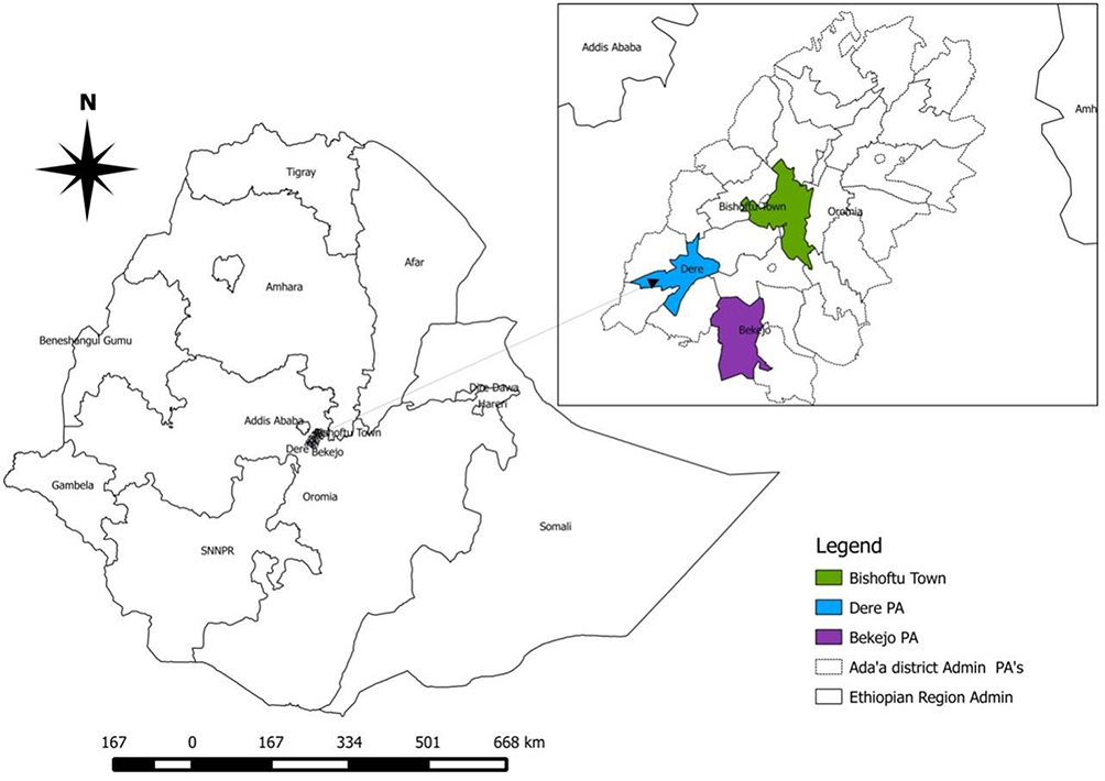 |
Figure 1 Map of the study area (the map was made with the use of QGIS free software tool). |
Study Animals
In this study, both male and female, and local and crossbred cattle from 6 months to 12 years old were included. These cattle were sampled from Dire and Bekajo representing extensive management and those from the DZARC dairy farm of Bishoftu representing intensive management systems. These cattle in extensive management were used mainly for agricultural purposes including plowing while those kept intensively were kept for dairy farming and research. Animal identification, including breed, age, sex, body condition score (BCS), parity, lactation status, and location (kebele or farm) was recorded with a format designed for this purpose. BCS was measured before sampling and scored on a scale from 1–5 categorized as good (for BCS 4 and 5), medium (for BCS 3), and poor (for BCS 1 and 2).28 Additionally, the age of cattle was estimated based on dentition according to the guide given by Johnson.29
Sample Size Determination
A total of 113 cattle (30 from Dire, 30 from Bekajo, and 53 from DZARC) were sampled on different days pre- and post-vaccination. The sampled animals were mostly zebu cattle (all from both Dire and Bekajo and 23 Borena cattle from DZARC) while the remaining (30 cattle) from DZARC were cross-bred with a range of 50% to 75% blood level.
Thirty cattle per the two sites (Bekajo and Dire) from an extensive management system (30 from Bekajo and 30 from Dire) were followed and had blood samples taken four times (once pre-vaccination and three times post-vaccination), equating to 240 serum samples. Additionally, the 53 cattle from the intensive management system were included in this study from the DZARC dairy farm and sampled three times (one pre-vaccination and twice post-vaccination), equating to 159 sera. Accordingly, a total of 399 sera were collected from 113 cattle from both extensive and intensive management systems.
Sampling Methods
Purposive sampling was made to select the study sites taking information from the district about LSD less reported areas and accessibility into consideration. Additionally, cattle selection per household took into consideration the owner’s closeness to the veterinary clinic and their willingness for repeated sampling. After this, cattle registration with their households was made on the first day. Cattle to be sold soon, calves below six months, diseased ones, and pregnant were ruled out of the study. A total of 4 to 15 cattle per owner were selected and tagged to make the cattle traceable for repeated sampling.
Blood Sample Collection
Blood samples were collected from the selected cattle while they were restrained inside the crush following all animal welfare protocols. About 5 to 7 mL of blood was collected from the jugular vein. Each sample was labeled appropriately in a similar fashion to the data collection paper with a permanent marker. Collection of samples was made at two stages; before vaccination (day 0) and after vaccination with Capripox-LSD vaccine on days 7, 15, and 30. Cattle were vaccinated (1 mL per animal subcutaneous) with the Capripox-LSD vaccine, a homologous LSD vaccine produced from attenuated Kenyan sheep and goat pox strain virus (KSGPV), manufactured in Ethiopia. On days 0, 7, and 15 the blood samples for serum separation were collected from all (n = 113 cattle (Bekajo (n = 30), Dire (n = 30), and DZARC (n = 53)) and on day 30, only 60 samples were collected from DZARC (total 399 samples). The collected samples were transported to DZARC under a cold chain and allowed to clot for 16–24 hrs at room temperature and then the sera were separated and the samples were transferred to labeled cryovials and kept at −20 °C until transported to the national veterinary institute (NVI) for a serum neutralization test (SNT test).
Lumpy Skin Disease Vaccine
LSD vaccine is one of the vaccines produced locally in Ethiopia. The produced vaccine is the live attenuated vaccine of Kenyan sheep and goat Poxvirus strain/KSGP that is used in the country for LSD prevention and control. According to Gelaye et al,30 KSGP was identified as a Neethling virus strain rather than the previous claim of sheep and goat pox.
Study Design
Multiple sampling of longitudinal study design was used to determine LSD antibody levels before and after vaccination.
Laboratory Diagnosis
Cell Culture Preparation and SNT
Reviving Vero cell culture: Cryovial containing Vero cells (Vero cells were originally donated from the Pan African Veterinary Center of the African Union/AU-PANVAC and its use was approved by the ethics committee of the institution) was warmed in a water bath heated to 37 °C until the suspension was thawed completely. The laminar airflow as well as the surface of the cryovial was disinfected with 70% ethanol. After disinfection, the vial was opened in a laminar airflow cabinet and the Vero cell was transferred with a sterile pipette to a universal bottle containing 10% Glasgow modified essential medium (GMEM). This was followed by centrifugation of about 200 gm of a cell for 10 min. at 4 °C. The supernatant with preservative was discarded and resuspension of the Vero cell was made at the required concentration in the fresh complete sterile medium (10% GMEM) inside the laminar airflow cabinet. This was followed by cell suspension adjusted to the desired concentration of 20 × 106 cell/mL for diploid cell lines (this was labeled for the type of cell line, date of incubation, and passage) and then incubated at 37 °C.
Subculture Preparation
In the subculture preparation, 0.05% trypsin solution was pre-warmed for sub-culturing and complete media for cell growth at 37 °C. For this purpose, a cell culture flask was taken with a confluent monolayer from the incubator followed by the decanting of old media. Next, the cells were washed with PBS free of Ca++ and Mg++ to remove residual serum and bivalent ions which inactivate the trypsin and versene, respectively. Additionally, a sufficient amount of trypsin was added slowly to the opposite wall of the attached cell monolayer culture flask and the action of the trypsin was observed for two minutes. This added trypsin was decanted, followed by placing the flask inverted until the cell detached from it inside the laminar flow cabinet. Finally, growth medium (10% GMEM) (with sterile newborn calf serum) was added, and vigorously pipetted with a pipette mechanically until the monolayer became dispersed to a single cell. This monolayer was used for the serum neutralization test (SNT).
Serum neutralization test (SNT): SNT, recommended by OIE10 as the gold standard serological technique to detect antibodies against CaPV, was used to assess the Ab level against LSD in cattle. Furthermore, SNT has been used by different researchers as an effective method to determine the level of antibody titer in LSD.23,31 This method of assessing antibody titer has a strong specificity though sensitivity is relatively less. The recorded specificity of SNT is more than 97% while the sensitivity ranges from 70% to 96%.21,22,25 However, the use of pre-vaccination antibodies as a reference can improve the sensitivity of the test.22
The laboratory process started by thawing the frozen (−20 °C) sera samples at room temperature followed by serial five-fold dilutions of 1/5, 1/25, 1/125, 1/625, and 1/3125 in Glasgow’s minimum essential cell culture medium (GMEM). Starting from the fourth day; the monolayer cells were checked for cytopathic effect (CPE) under an inverted microscope. This checking for CPE was continued and the final check was made on the ninth day. This last day examination was recorded from the highest dilution which inhibited CPE in both or either of the duplicate wells and recorded as the reciprocal of the log titration. The final interpretation was made by considering wells without CPE at a dilution of ≥1/25 (log52) as positive. This means the antibody against the LSDV has reacted with the vaccine strains and inhibited the growth of the virus thereby showing the absence of a cytopathic effect on the cell culture.10,16
Data Management and Analysis
The collected data were entered and stored into Microsoft Office Excel 2010 spreadsheet and thoroughly screened before being subjected to statistical analysis. Descriptive analysis was made to determine the proportion of cattle with levels of SNT antibody titers across each sampling day (pre- and post-vaccination sampling). SNT cut-off value by Gari et al16 and Zenebe et al22 referred from OIE10 (≥log52 antibody titers) was used. This value is considered to be an effective antibody concentration that gives protection to LSD. Based on this value, vaccinated animals have been classified as either protected or at-risk groups on each sampling day. Furthermore, the association between the development of LSD-specific antibodies with different risk factors (BCS, age, lactation status, management system, and parity) were made with STATA Corp. version 13.32 Throughout the analysis, 95% of confidence intervals (CI) with 5% of precision that sets p < 0.05 as the significant difference between the risk factors and antibody titer level was used.
Limitations of the Study
Limitation of the present study was related to the absence of sampling for a prolonged period of more than 30 days. Additionally, the cellular immune response was not addressed in this study.
Results
Longitudinal Study
Serum neutralization test/SNT result: A total of 113 cattle representing extensive (60/113) and intensive (53/113) management systems were included in this study. From the sera collected, a limited number of cattle had seroconversion before vaccination while the highest seroconversion of 41.65% (25/60) was observed in 60 sera collected from extensively managed cattle on day 30 suggesting the presence of the LSD-antibody (Table 1).
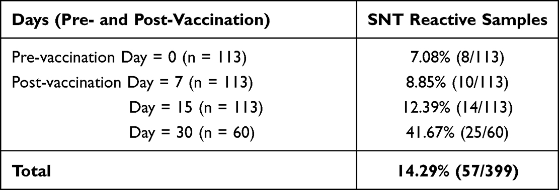 |
Table 1 Serum Neutralization Test/SNT Result Before and After Vaccination of Study Animals (Till 30 Days) (n = 399). |
The last day of sampling on day 30th was done only from 60 cattle. Relatively, the number of cattle with LSD-antibody increased through time from dpv 7 (n = 10) to 30 (n = 25) (Table 1 and Figure 2).
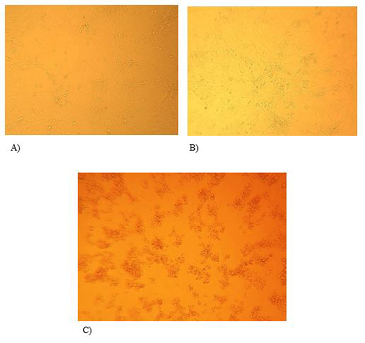 |
Figure 2 Microscopic observation of Vero cell culture monolayer during serum neutralization test (100x) (A) negative control, only Vero cell culture without the virus, no CPE (ie cells appear to be morphologically normal), (B) positive result on day 9, no CPE and (C) negative result on day 9; with CPE (ie cell started to degrade and made clustered appearance). |
The presence and absence of the cytopathic effect (CPE) were assessed after incubation of the test sera on monolayer Vero cells injected with vaccine strains between days 4 and 9. With CPE under close observation via an inverted microscope, the final interpretation was made by considering wells without CPE at a dilution of ≥1/25 (neutralization index of ≥1.5) as positive. Therefore, the absence of CPE in cell cultures indicate the presence of a protective antibody in the test sera that neutralizes the injected vaccine strain virus which in turn leaves the cells intact without damage by the virus (ie absence of CPE) (Figure 2).
Sero-Conversion Relationship with Risk Factors
The proportion of cattle with antibodies against LSD differs across risk factors before and after vaccination. Accordingly, most antibody production was seen in intensively managed, crossbred, and those aged ≤4½ years except at day 30. However, lactating cows and those having ≥1 calf showed higher antibody production both before and after vaccination. The maximum number of cattle that showed LSD-antibodies in either the sampling stage (before or after vaccination sampling) once or more was 25 animals (Table 2).
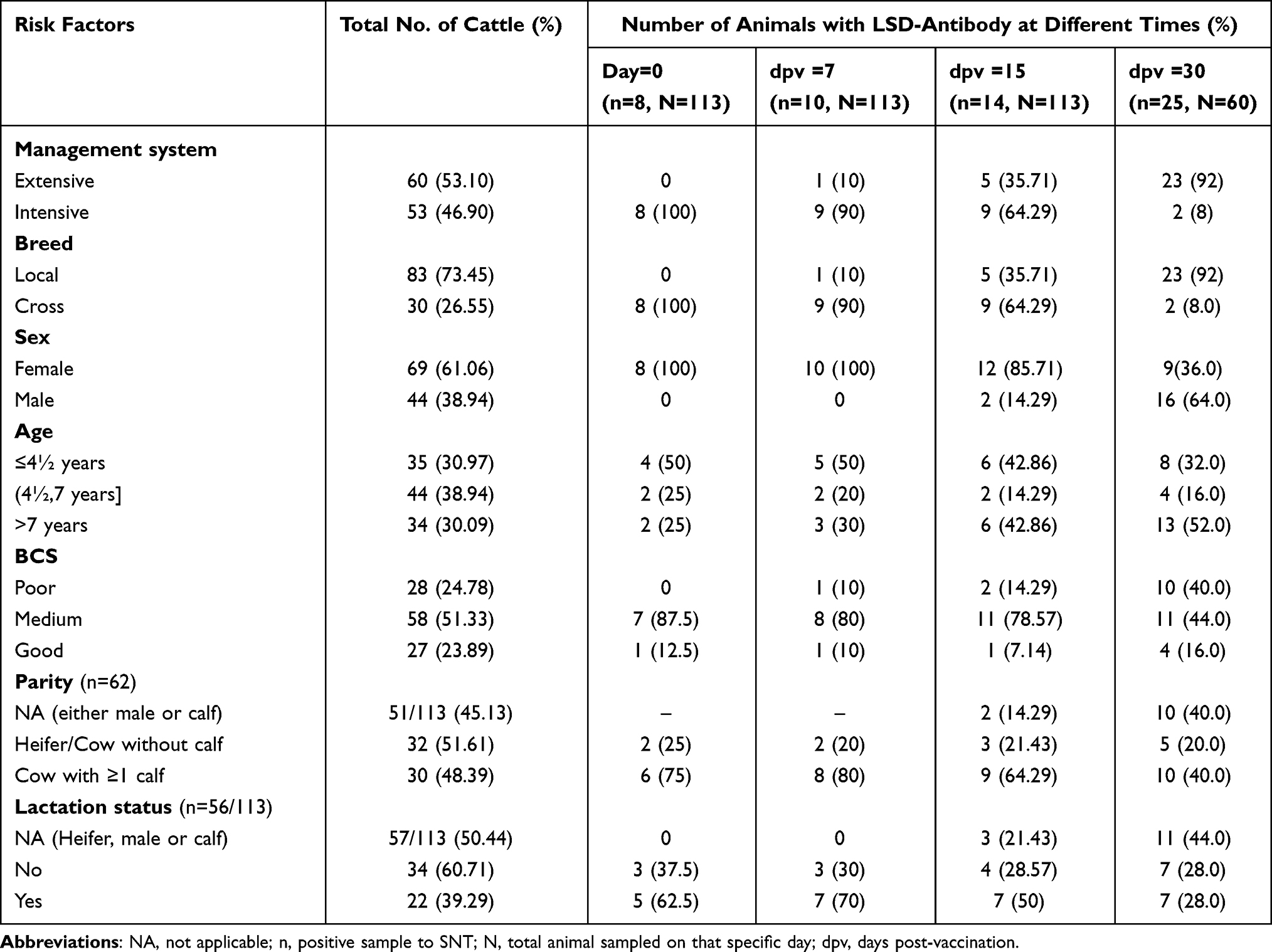 |
Table 2 Risk Factors Associated with Detected Immune Response Against LSD (N =113) |
Regression analysis showed the presence of a significant difference between the SNT test results of the three cattle management systems. Accordingly, p-value showed a significant difference in SNT results between cattle managed by intensive and extensive management systems at day 7 (OR = 15.5; 95% CI = 1.65, 145.5; p = 0.016) and 30 (OR = 3.67; 95% CI = 1.1, 12.29; p = 0.035) of post-vaccination. Additionally, different breeds and age groups ((4½, 7) years) of cattle showed a significant SNT results at day 15 (OR = 6.69; 95% CI = 2.02, 22.08; p = 0.002) and 30 (OR = 4.24; 95% CI = 1.22, 14.71; p = 0.023); respectively. However, the rest of the risk factors (sex, BCS, >7 years of age, parity, management system, breed, and lactation status) did not show any significant difference in SNT results (p > 0.05) (Table 3).
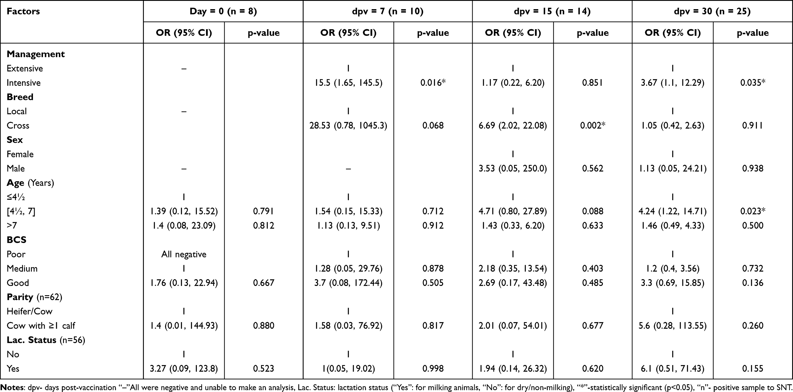 |
Table 3 Regression Analysis of SNT Results at Pre-and Post-Vaccination |
Discussion
Vaccination is the most effective and economical method of disease control both in humans and animals worldwide including this country, Ethiopia. Ethiopia is endemic to many internationally important livestock diseases including LSD and vaccination is an important recommendation to effectively control the disease.10,17 Vaccination against LSD is an ongoing task in some areas of Ethiopia but vaccine failures were suspected due to the occurrence of the LSD outbreak in vaccinated herds. In this study, the KSGP vaccine strain produced locally was used to vaccinate cattle. Furthermore, the immune status and response were assessed with a serum neutralization test (SNT) before and after vaccination in the intensive and extensive cattle management systems. Accordingly, the longitudinal study showed fewer cattle (22.12%) with LSDV-specific antibodies via SNT during pre-vaccination and/or post-vaccination. The specific period of sampling comparison showed lesser samples with LSD-antibody before vaccination (7.08%) than post-vaccination sampling that ranges from 8.85% to 41.67% (for 7 to 30 dpv). This could be related to the antibody level being raised in vaccinated than in unvaccinated animals.33,34 Furthermore, Samojlovic et al35 correlated their finding of fewer cattle presentations with the specific antibody to the duration of detectable humoral immune response to be less than a year as vaccinations were commonly done annually. This statement was backed by OIE20 which put a time range of about 7 months of detectable antibodies after vaccination.
The current SNT results showed the maximum number of cattle with LSD-antibodies in either of the sampling stages (before or after vaccination sampling) once or more was 25 cattle. The highest seroconversion was detected at 30-days post-vaccination. However, antibody detection was observed in fewer samples at the first sampling post-vaccination (7 days post-vaccination (dpv)). Similarly few findings of LSD-specific antibodies at 7 days post-vaccination was reported by Zenebe et al23 though the current finding reported less number of cattle with specific antibodies as the former based their research on stationed cattle.
In this study, a higher number of seroconversion was observed on the 30th dpv. Likewise, Zenebe et al23 and Samojlovic et al35 also reported the highest seroconversion on the 30th day after vaccination. This relative increase in the number of sera with antibody appearance during post-vaccination Milovanović et al36 considered as an indication of a better immune response to vaccination. However, the overall antibody detection appeared to be lower as a maximum of 41.67% on 30 dpv while much less antibody detection was detected at 7 and 15 dpv. Accordingly, a dominant number of collected sera showed the absence of specific LSD-antibodies both pre-and post-vaccination. This was explained by Babiuk et al37 with the probable existence of lower neutralizing humoral immunity following vaccination with live attenuated Capripox vaccines. Additionally, Gari et al31 suggested the high amount of missing seroconversion with poor immunogenicity of the vaccine was due to over-attenuation of Neethling and KSGP vaccine strains in its process of production.
In the current study, the SNT result of antibody-specific detection was compared across different risk factors. Accordingly, intensively managed cattle showed a significantly higher level of antibody titer post-vaccination compared with extensively managed cattle (p < 0.05). This could be related to the good husbandry care in intensively managed cattle that probably opened the way for boosting the build-up of immunity in general. Additionally, certain groups of age and breeds were found to cause a significant effect on the development of antibodies. In agreement with this, Molla et al9 put the importance of host-related factors including age on the immune system reaction. Likewise, different breeds and age groups of cattle showed significant SNT results at different dpv (p < 0.05). In contrast to the current finding, Zenebe et al23 did not find any effect of host risk factors on antibody production. This might relate to Zenebe et al’s23 focus on stationed cattle with a good husbandry status.
Conclusion
The current longitudinal study on protective antibodies showed a variability between pre- and post-vaccinated sera as well as among different days post vaccinated samples. In general, the number of cattle that showed a protective antibody post vaccination indicated the slow buildup or lesser level of protection gained from this available vaccine. As this study still left some questions behind; we recommend an extended period of sampling to look for protective antibodies as well as to exhaust the study regarding cellular immune response against the LSD vaccine.
Abbreviations
CPE, cytopathic effect; DZARC, Debre Zeit/Bishoftu Agricultural Research Center; GMEM, Glasgow modified essential medium; HH, household; KSGP, Kenyan sheep and goat Poxvirus strain; SNT, serum neutralization test; LSD, lumpy skin disease; LSDV, LSD virus; OR, odds ratio; NVI, national veterinary institute.
Data Sharing Statement
All data generated and analyzed during this study are included in this article. Further information could be gathered from the corresponding author.
Ethics Approval and Consent to Participate
Animal blood sampling followed international standards for animal welfare conduct without any welfare deterioration where the proposal was passed through the Ethiopian Institute of Agricultural Research (EIAR) and College of Veterinary Medicine and Agriculture, Addis Ababa University (CVMA-AAU) research ethics committee for the consideration of animal welfare during blood sampling. Accordingly, cattle blood sampling was done according to international guidelines. Antiseptic procedures and standard antiseptics were applied before and after blood sampling to cattle at the jugular vein site. A minor swelling that only remained for a maximum of 30 to 45 minutes was observed that was relieved by itself. Furthermore, verbal and written informed consent was obtained from the owners of cattle and/or managers of the dairy farms.
Acknowledgments
This work was part of a thesis and we want to acknowledge some specific institutions and individuals such as Ethiopian Agricultural Research Centers, the animal health sector for laboratory cost coverage, DZARC for the field trip as well as sampling equipment, and Mr. Takele Beyene for providing sampling materials (tubes and needles). We also extend our appreciation to Dr. Zerihun Assefa for his input on part of the location mapping and discussion. Finally, we would like to appreciate the farmers (in Dire and Bekajo), Ada’a animal health field practitioners, Dr. Esayas, and Dr. Belayneh with their team (NVI) for their all round support for the success of this work.
Funding
This study was supported by Addis Ababa University, College of Veterinary Medicine and Agriculture (researcher per diem and field sample collection materials), Hawassa University (researcher feild per diem), and Ethiopian Agricultural Research Centers (contribution to part of sample collection and entire laboratory processing fees).
Disclosure
The authors declare that they have no competing interests.
References
1. Food and Agriculture Organization of the United Nations. Livestock in Balance. Vialedelleterme di Caracalla: Food and Agriculture Organization of the United Nations; 2009:23.
2. FAO. The Future of Livestock in Ethiopia, Opportunities, and Challenges in the Face of Uncertainty. FAO; 2019.
3. FAO, New Zealand Agricultural Greenhouse Gas Research Centre. Supporting low emissions development in the Ethiopian dairy cattle sector – reducing enteric methane for food security and livelihoods. Rome; 2017:34. Available from: http://www.fao.org/3/ai6821e.pdf.
4. Central Statistical Agency (CSA). Report on Livestock and Livestock characteristics (Private peasant holdings). Agricult Sample Survey. 2018;II:9–22.
5. Shapiro BI, Gebru G, Desta S, et al. Ethiopia Livestock Master Plan. ILRI Project Report. Nairobi, Kenya: International Livestock Research Institute (ILRI); 2015.
6. World Cattle Inventory (WCI). Ranking of countries (FAO) by Rob Cook; 2015. Available from: https://www.drovers.com/article/world-cattle-inventory-ranking-countries-fao.
7. Ayelet G, Haftu R, Jemberie S, et al. Lumpy skin disease in cattle in central Ethiopia: outbreak investigation and isolation and molecular detection of the virus. Rev Sci Tech. 2014;33:877–887. doi:10.20506/rst.33.3.2325
8. Tuppurainen ESM, Oura CAL. Review; Lumpy skin disease: an emerging threat to Europe, Middle East and Asia. Trans Emerg Dis. 2012;59:40–48.
9. Molla W, Frankena K, Gari G, Kidane M, Shegu D, De Jong MCM. Sero-prevalence and risk factors of lumpy skin disease in Ethiopia. Prev Vet Med. 2018;160:99–104. doi:10.1016/j.prevetmed.2018.09.029
10. OIE. Lumpy Skin Disease; Manual of Diagnostic Tests and Vaccines for Terrestrial Animals. Paris: World Organization for Animal Health; 2010:1–13.
11. Radostits OM, Gay CC, Hinchcliff KW, Constable PD. Veterinary Medicine: A Textbook of Diseases of Cattle, Horses, Sheep, Pigs and Goat.
12. Carn VM, Kitching RP. An investigation of possible routes of transmission of LSD (Neethling). Epid Infect. 1994;114:219–226. doi:10.1017/S0950268800052067
13. Davies FG. Lumpy skin disease of cattle: a growing problem in Africa and the near east. World Anim Rev. 1991;68:37–42.
14. Coetzer JAW. Lumpy skin disease. In: Coetzer JAW, Justin C, editors. Infectious Diseases of Livestock.
15. Babiuk S, Bowden TR, Boyle DB, Wallace DB, Kitching RP. Capripoxviruses: an emerging worldwide threat to sheep, goats and cattle. Trans Emerg Dis. 2008a;55(7):263–272. doi:10.1111/j.1865-1682.2008.01043.x
16. Gari G, Waret-Szkuta A, Grosbois V, Jacquiet P, Roger F. Risk factors associated with observed clinical lumpy skin disease in Ethiopia. Epidemiol Infect. 2010;138:1657–1666. doi:10.1017/S0950268810000506
17. Brenner J, Haimovitz M, Oron E, et al. Lumpy skin disease in a large dairy herd in Israel. Isr J Vet Med. 2006;61:73–77.
18. El-Kholy AA, Soliman HMT, Abdelrahman KA. Polymerase chain reaction for rapid diagnosis of a recent lumpy skin disease virus incursion to Egypt. Arab J Biotechnol. 2008;11:293–302.
19. Mebratu G, Kassa B, Fikre Y, Berhanu B. Observations on the outbreak of lumpy skin disease in Ethiopia. Vet Tropicaux. 1984;37:395–399.
20. OIE. Manual of diagnostic tests and vaccines for terrestrial animals. In: Chapter 2.4.13. Paris: OIE; 2017.
21. Bhanuprakash V, Indrani BK, Hosamani M, Singh RK. The current status of sheep pox disease. Comp Immunol Microbiol Infect Dis. 2006;29:27–60. doi:10.1016/j.cimid.2005.12.001
22. Gari G, Biteau-Coroller F, LeGoff C, Caufour P, Roger F. Evaluation of indirect fluorescent antibody test (IFAT) for the diagnosis and screening of lumpy skin disease using Bayesian method. Vet Micr. 2008;129(3–4):269–280. doi:10.1016/j.vetmic.2007.12.005
23. Zenebe T, Bayssa B, Keskis S, Duguma R. Towards effective vaccine production: a controlled field trial on the immunological response of three lumpy skin disease vaccine strains in dairy farms. Acad J Anim Dis. 2014;3(3):17–26. doi:10.5829/idosi.ajad.2014.3.3.85134
24. Tegegne B. Outbreak Investigation of Lumpy Skin Disease; Isolation and Molecular Characterization of the Virus in South Wollozone, Northern Ethiopia [MSc Thesis]. College of Veterinary Medicine and Agriculture, AAU; 2018.
25. Gari G, Grosbois V, Waret-Szkuta A, Babiuk S, Jacquiet P, Roger F. Lumpy skin disease in Ethiopia: seroprevalence study across different agro-climate zones. Acta Trop. 2012;123(2):101–106. doi:10.1016/j.actatropica.2012.04.009
26. Molla W, Mart CM, Gari G, Frankena K. Economic impact of lumpy skin disease and cost effectiveness of vaccination for the control of outbreaks in Ethiopia. Prev Vet Med. 2017;147(1):100–107. doi:10.1016/j.prevetmed.2017.09.003
27. Eastern Shewa agricultural bureau report (Unpublished Office data records). 2014.
28. Mishra S, Kumari K, Dubey A. Body condition scoring of dairy cattle review, research and reviews. J Vet Sci. 2016;2(1):1–8.
29. Johnson RF. The Stockman’s Handbook by Ensminger.
30. Gelaye E, Belay A, Ayelet G, et al. Capripox disease in Ethiopia: genetic differences between field isolates and vaccine strain, and implications for vaccination failure. Antivir Res. 2015;119:28–35. doi:10.1016/j.antiviral.2015.04.008
31. Gari G, Abie G, Gizaw D, et al. Evaluation of the safety, immunogenicity and efficacy of three capripoxvirus vaccine strains against lumpy skin disease virus. Vaccine. 2015;33:3256–3261. doi:10.1016/j.vaccine.2015.01.035
32. StataCorp. Stata Statistical Software: Release 13. College Station: StataCorp LP; 2014.
33. Tuppurainen ESM, Venter EH, Coetzer JAW. The detection of lumpy skin disease virus in samples of experimentally infected cattle using different diagnostic techniques. Onderstepoort J Vet Res. 2005;72:153–164. doi:10.4102/ojvr.v72i2.213
34. Kitching RP, Hamond JM. Poxvirus, infection and immunity. In: Roitt IM, Delves PJ, editors. Encyclopaedia of Immunology. Vol. 3. London: Academic Press; 1991:1261–1264.
35. Samojlovic M, Polacek V, Gurjanov V, et al. Detection of antibodies against lumpy skin disease virus by virus neutralization test and ELISA methods. Acta Vet Beograd. 2019;69(1):47–60. doi:10.2478/acve-2019-0003
36. Milovanović M, Dietze K, Milićević V, et al. Humoral immune response to repeated lumpy skin disease virus vaccination and performance of serological tests. BMC Vet Res. 2019;15:80. doi:10.1186/s12917-019-1831-y
37. Babiuk S, Bowden TR, Parkyn G, et al. Quantification of lumpy skin disease virus following experimental infection in cattle. Trans Emerg Dis. 2008b;55:299–307.
 © 2023 The Author(s). This work is published and licensed by Dove Medical Press Limited. The full terms of this license are available at https://www.dovepress.com/terms.php and incorporate the Creative Commons Attribution - Non Commercial (unported, v3.0) License.
By accessing the work you hereby accept the Terms. Non-commercial uses of the work are permitted without any further permission from Dove Medical Press Limited, provided the work is properly attributed. For permission for commercial use of this work, please see paragraphs 4.2 and 5 of our Terms.
© 2023 The Author(s). This work is published and licensed by Dove Medical Press Limited. The full terms of this license are available at https://www.dovepress.com/terms.php and incorporate the Creative Commons Attribution - Non Commercial (unported, v3.0) License.
By accessing the work you hereby accept the Terms. Non-commercial uses of the work are permitted without any further permission from Dove Medical Press Limited, provided the work is properly attributed. For permission for commercial use of this work, please see paragraphs 4.2 and 5 of our Terms.