")
Back to Journals » Veterinary Medicine: Research and Reports » Volume 15
A Comprehensive Review of the Common Bacterial Infections in Dairy Calves and Advanced Strategies for Health Management
Authors Robi DT , Mossie T, Temteme S
Received 2 December 2023
Accepted for publication 16 January 2024
Published 24 January 2024 Volume 2024:15 Pages 1—14
DOI https://doi.org/10.2147/VMRR.S452925
Checked for plagiarism Yes
Review by Single anonymous peer review
Peer reviewer comments 2
Editor who approved publication: Professor Young Lyoo
Dereje Tulu Robi,1 Tesfa Mossie,2 Shiferaw Temteme1
1Ethiopian Institute of Agricultural Research, Tepi Agricultural Research Center, Tepi, Ethiopia; 2Ethiopian Institute of Agriculture Research, Jimma Agriculture Research Center, Jimma, Ethiopia
Correspondence: Dereje Tulu Robi, Email [email protected]
Abstract: Dairy farming faces a significant challenge of bacterial infections in dairy calves, which can have detrimental effects on their health and productivity. This review offers a comprehensive overview of the most prevalent bacterial infections in dairy calves, including Escherichia coli, Salmonella typhimurium, Salmonella dublin, Salmonella enterica, Clostridium perfringens, Pasteurella multocida, Listeria monocytogenes, Mycoplasma bovis, and Haemophilus somnus. These pathogens can cause various clinical signs and symptoms, leading to diarrhea, respiratory distress, septicemia, and even mortality. Factors such as management practices, environmental conditions, and herd health influence the incidence and severity of the infections. Efficient management and prevention strategies include good colostrum and nutrient feeding, early detection, appropriate treatment, hygiene practices, and supportive care. Regular health monitoring and diagnostic tests facilitate early detection and intervention. The use of antibiotics should be judicious to prevent antimicrobial resistance and supportive care such as fluid therapy and nutritional support promotes recovery. Diagnostic methods, including immunological tests, culture, polymerase chain reaction (PCR), and serology, aid in the identification of specific pathogens. This review also explores recent advancements in the diagnosis, treatment, and prevention of bacterial infections in dairy calves, providing valuable insights for dairy farmers, veterinarians, and researchers. By synthesizing pertinent scientific literature, this review contributes to the development of effective strategies aimed at mitigating the impact of bacterial infections on the health, welfare, and productivity of young calves. Moreover, more research is required to enhance the understanding of the epidemiology and characterization of bacterial infections in dairy calves.
Keywords: bacteria, diarrhea, pneumonia, septicemia, vaccination, dairy calve
Introduction
Dairy farming plays a crucial role in meeting the growing global demand for milk and dairy products.1 However, one of the major challenges faced by dairy farmers is the prevalence of bacterial infections in dairy calves.2 Bacterial infections can have severe effects on the respiratory, digestive, and immune systems of animals that reduce the growth rates, and increase morbidity and mortality of dairy calves. This results in significant economic losses for the dairy industry, attributable to treatment costs and mortality.2 Therefore, understanding the common pathogenic bacterial infections affecting dairy calves, their effects, and effective management strategies is essential for ensuring the well-being and performance of the dairy industry.3
To comprehensively address this issue, it is necessary to explore the most prevalent bacterial infections encountered in dairy calves. Studies have identified several primary bacterial pathogens responsible for infections in calves, including Escherichia coli, Salmonella spp., Clostridium spp., and various species of Pasteurella and Mycoplasma.4,5 These pathogens can cause a range of clinical signs and symptoms, such as diarrhea, respiratory distress, septicemia, and even mortality in severe cases. The incidence and severity of bacterial infections in dairy calves are influenced by various factors, including management practices, environmental conditions, and the general health status of the herd.6
An efficient management and prevention strategy for bacterial infections in dairy calves requires a multifaceted approach. These include good colostrum feeding, proper nutrition, and optimal hygienic practices that can significantly reduce the risk of infections.7 Early detection of infections through regular health monitoring and diagnostic tests also allows for timely intervention and treatment of emerging, reemerging, and novel infectious diseases.8 Antibiotics are commonly employed to treat bacterial infections in dairy calves, but their use should be judicious to prevent the development of antimicrobial resistance and ensure animal welfare.9 Furthermore, supportive care, including fluid therapy and nutritional support, plays a vital role in managing infected calves and promoting their recovery. This review aims to provide a comprehensive overview of the most common bacterial infections observed in dairy calves, their effects on calf health and productivity, and current management practices. To explore recent advancements in the diagnosis, treatment, and prevention of bacterial infections in dairy calves, we conducted a comprehensive review of the latest research findings and emerging technologies. The information discussed here serves as a valuable resource for dairy farmers, veterinarians, and researchers, aiding in the development of effective strategies to mitigate the impact of bacterial infections on calf health and welfare.
Common Causes of Bacterial Infections in Dairy Calves
Escherichia coli (E. coli)
Escherichia coli is a gram-negative, rod-shaped, motile or non-motile, non-spore-forming, facultative anaerobic bacterium that commonly inhabits the gastrointestinal tract of calves (Table 1). E. coli is a type species that belongs to Enterobacteriaceae family and Escherichia genus. It can be pathogenic and non-pathogenic.10 Non-pathogenic strains are part of the normal flora of the gut, aiding the hosts by producing vitamin K2 and preventing the establishment of pathogenic bacteria in the intestine E. coli can be differentiated into 190 serotypes (serogroups) based on somatic (O), capsular (K), and flagellar (H) antigens. About 80 different capsular polysaccharide (K) antigens are present. There are about 174 O antigens and 56 H antigens. It is serotyped depending on the combination of O, H, and K antigens (eg, O157: H7). Serotyping of E. coli with molecular and phage typing is a useful epidemiological tool.6
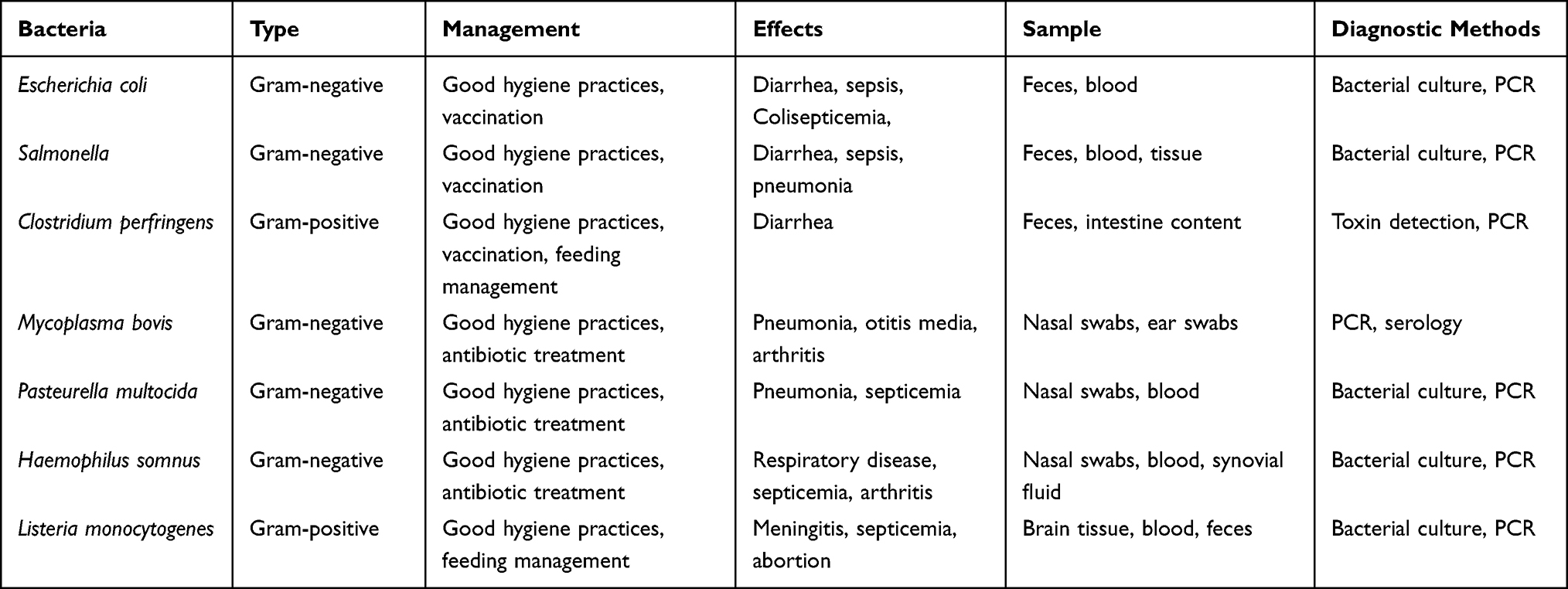 |
Table 1 Common Bacterial Species in Dairy Calves with Their Type, Management Practices, Effects, and Diagnostic Methods |
Escherichia coli can be grouped into six pathotypes depending on virulence factors. These are enterotoxigenic E. coli (ETEC), shigatoxin-producing E. coli (STEC), enteropathogenic E. coli (EPEC), enteroinvasive E. coli (EIEC), enteroaggressive E. coli (EAEC), and enterohaemorrhagic E. coli (EHEC).11 ETEC, EPEC, and EHEC are the diarrheagenic pathotypes occurring in young farm animals. ETEC strains are the most common cause of neonatal diarrhea because they produce the K99 (F5) adhesion antigen and heat-stable enterotoxin.12,13 The virulence factor facilitates sticking of bacteria to the villous epithelial cells in the intestine to prevent elimination by peristalsis and production of heat-stable and heat-labile enterotoxin. During the first four days after birth, newborn calves are especially vulnerable to ETEC infection and have “watery” diarrhea if infected.14
The prevalence of enterotoxigenic E. coli in diarrheic calves exhibits considerable variation geographically and across different herds, depending on the age of the animals. Consequently, enterotoxigenic colibacillosis is a leading cause of diarrhea in calves below 3 days of age, but it does not trigger diarrhea outbreaks in older calves12,15 suggests that the presence of E. coli in dairy calves can be associated with age groups, environmental and management conditions of the farms, and inadequate intake of colostrum by calves. Poor hygiene practices can also lead to the accumulation of pathogenic strains in the surroundings of young animals.15,16 Furthermore, according to17 a significant amount of pathogenic E. coli may surpass the colostrum immunity. In severe cases of E. coli outbreaks, even calves as young as 16 to 24 hours can be affected, with younger calves having a higher risk of dying from severe dehydration that progressively sets in.18
The transmission of the organism in a herd occurs through fecal-oral route from contaminated objects such as bedding, pails, boots, tools, clothing, feed, and water supplies. Poor sanitation increases the likelihood of newborn calves contracting E. coli scours infections from their environment.19,20 E. coli strains that cause diarrhea initially colonize the gut of the calf by attaching to the intestinal wall using pili or fimbriae, which are fine, fuzz-like protrusions. The K99+ antigen designates these pili, and the strains that possess them are called enterotoxigenic E. coli (ETEC).21 Good hygiene practices and vaccination can help prevent the infection caused by E. coli. However, if infected, calves may suffer from diarrhea, and sepsis. Feces and blood are the preferred sample types for diagnosis.22,23
Cultural isolation, serological test, and molecular diagnostic approaches can be used to diagnose E. coli. Serological diagnosis of the bacteria is carried out using latex agglutination, fluorescent antibody technique, and an ELISA test to detect fimbrial antigens (K99) or enterotoxin directly from fecal samples or isolated colonies.24,25 ELISA using monoclonal antibodies is the most sensitive diagnosis method.17 Conventional diagnostic procedures such as cultural isolation, serological tests, and pathotyping methods are time consuming, expensive, and cannot distinguish definitively strains of E. coli infection. Therefore, rapid nucleic-based tests have been developed around the world to detect circulating bacterial strains based on amplification and detection of nucleic acid.26 Sensitive and specific molecular diagnostic techniques like PCR and sequencing play a significant role in the detection and differentiation of strains compared to conventional diagnostic tools. The most commonly used molecular diagnostic technique for the detection of E. coli strains is PCR. The assays are more sensitive, specific, and less labor-intensive compared to conventional diagnostic methods. The development of biosensor devices has the potential to become indispensable in detecting low colony-forming units of pathogenic E. coli in environmental samples (Table 1).27
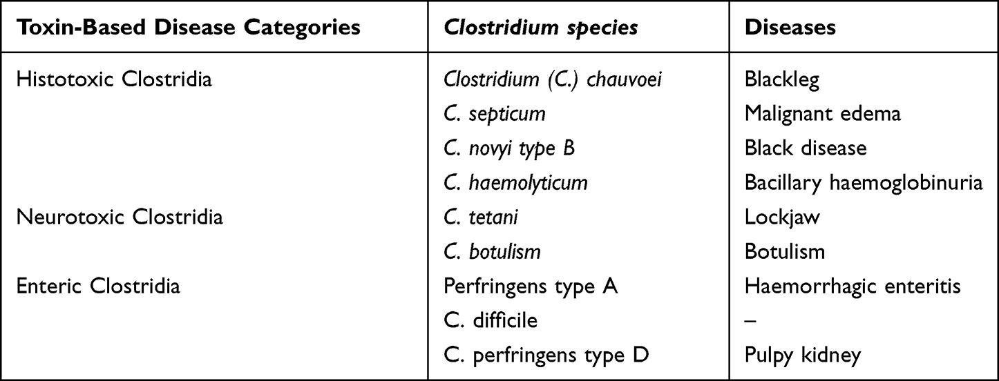 |
Table 2 Pathogenic Clostridium Species and Diseases |
Salmonella
Members of the genus Salmonella are gram-negative, facultative, and rod-shaped short-bacilli bacteria belonging to the family Enterobacteriaceae28 (Table 1). There are three central species of Salmonella: Salmonella enterica, Salmonella bongori, and Salmonella subterranean (S. subterranean) based on differences in their 16S rRNA sequence analysis. The type species of Salmonella enterica is again subdivided into six subdivisions of Salmonella enterica subspecies based on their genomic relatedness and biochemical properties the type species.29 Salmonella enterica subspecies enterica is the dominant subspecies affecting humans and domestic animals.30 Currently, there are more than 2700 Salmonella serovars that are serologically identified by antigenic variation in O (lipopolysaccharide), H (flagella), and VI (capsular) antigens in accordance with the Kauffmann-White scheme.30 A Salmonella infection is a common problem in captive animals like dairy calves that cause enteritis. The issue is particularly severe in calves younger than one month old.31 The infected calves develop fibrin purulent necrotizing enteritis, which is characterized by a severe diffuse infiltrate mainly composed of neutrophils.32 The genus Salmonella is motile except S. enterica ser. Pullorum and S. enterica ser. Gallinarum, which lacks flagella.33
The most common isolated Salmonella serotypes in cattle are Salmonella typhimurium and Salmonella dublin. These serotypes exhibit multiple resistances to commonly used antibiotics.34 Salmonella dublin and S. typhimurium are host-specific and non-host-specific, respectively, that affect calves severely between six and twelve weeks of age. Salmonella infection has a wide range of clinical manifestations, from asymptomatic to clinical salmonellosis. Acute diarrheal disease is most common with S. typhimurium and systematic disease with S. dublin in cattle. The disease is mainly found in dairy cattle rather than beef cattle and is associated with management practices. The infective dose, predisposing factors, and immunity status of the hosts determine the outcome of the infection. Salmonella infection has per acute (diarrhea and septicemia), acute (fever), and chronic (unthrifty scruffy hair) forms.5,35,36
The pathogenicity of Salmonella serotypes differs depending upon the difference in virulence potential of serovars and susceptibility of an infected host. The pathogenesis of Salmonella infection is governed by many virulence factors including type three secretion systems (T3SS), virulent plasmids, flagella, capsule, and other adhesion systems. Parts of the adhesion system produced by most serovars of Salmonella enterica include adhesins, invasins, toxins, fimbriae, and hemagglutinins. These virulence factors enable Salmonella to colonize the host cell, bypassing the defense mechanisms of the host. Genes contained on a wide range of genetic elements, including bacterial chromosomes, plasmids, prophages, and other SPIs, encode these bacterial virulence factors.37
The persistence of Salmonella in the environment plays a significant role in the epidemiology of calf salmonellosis. They are commonly found in farm effluents and human sewage. Salmonellosis is most prevalent in intensive animal husbandry, especially dairy, poultry, and swine production.38 This makes the epidemiology of salmonellosis complex. The fecal wastes of infected animals and humans are potential sources of contamination of the environment and the food chain.39 Sick animals shed organisms through their saliva, nasal secretions, colostrum, and milk, which can lead to oral transmission of the disease in the dairy.40,41 Fecal-oral transmission is the primary mode of direct animal-to-animal transmission.42 Furthermore, indirect transmission of Salmonella can occur via contaminated feed and water supplies, pasture contaminated by slurry or sewage, and wildlife vectors like small mammals and birds.12
Different approaches to Salmonella diagnosis, including cultural isolation, immunology-based assays, nucleic acid-based assays, miniaturized biochemical assays, and biosensors, are present. Conventional cultural isolation protocols are laborious and time-consuming.43 Immunology-based assays that use specific mono- or polyclonal antibodies binding to somatic or flagellar antigens are widely used for the detection of Salmonella species from representative specimens. The assays include ELISA, latex agglutination tests, immunodiffusion, and immunochromatography (dipstick). The assays are a valuable option for detecting non-cultural Salmonella cells (Table 1). The studies showed immunology-based assays are more rapid and specific than cultural methods to isolate Salmonella species in conjunction with immune-magnetic separation techniques. The shortcomings of the assays are cross-reactions with closely related antigens, a longer enrichment time to get a number of cells, and a high cost for automation. The methods handle large samples and are readily automated to reduce time and labor. ELISA test is used to determine the infection status of individual animals and the entire herd. The test can distinguish between recently infected (increased Ab. titer) and convalescent (decreased Ab. titer) calves. Constant titers are recognized in carrier hosts.31,44
The nucleic acid-based assays utilize a specific nucleic acid target sequence to detect the organisms directly from samples or colonies. The assays are more sensitive, specific, and inclusive than other methods.43 Direct hybridization (DNA probe) and amplification of genetic materials (PCR) methods are the two main diagnostic techniques.45,46 The PCR test is a primer mediated enzymatic amplification of specific segments of DNA for the detection of Salmonella pathogens.47 Salmonella invasion gene (invA) is highly conserved among Salmonella species and could serve as a reliable and accurate target gene for molecular detection of the genus Salmonella.48 The amplified products are detected using either gel-based systems or real-time PCR. The assays detect a very low number of organisms in the sample. This capability shortens the enrichment time to reach the Salmonella concentration needed for reliable detection by PCR compared to other methods.
A biosensor is an integrated receptor-transducer device made up of a detector and a biological recognition system (receptor). Without any additional processing stages or reagents in between signal sampling and output, it converts the biochemical (biological) response into a quantifiable output signal.49 When a particular analyte attaches to the biological recognition element, a recognition signal is generated. According to,43 the signal can take the form of changes in mass, oxygen consumption, potential difference, refractive index, pH, and other parameters. Current immunology and nucleic acid-based diagnostics are replaced by the biosensor diagnostic approach. A biosensor device can identify biological components such as enzymes, nucleic acids, entire cells, tissue, and biomimetic materials.50 Salmonella prevention and control can be achieved by adopting the principles of Hazard Analysis Critical Control Point (HACCP). Strict biosecurity measures, appropriate disinfection, avoiding stressful conditions, and frequent immunization play a significant role in Salmonella prevention and control programs.51
Clostridium perfringens
The genus Clostridium contains a diverse group of gram-positive anaerobic rods that form heat-resistant endospores.52 It is widespread in the environment and is normally found in soil and feces. The genus causes different types of Clostridium bacterial diseases through one or more of the several species of Clostridium and their potent toxins.53 The diseases include tetanus, botulism, blackleg, malignant edema, and bacillary hemoglobinuria in humans and animals (Table 2). Clostridium diseases can be neurotoxic, histotoxic, and enteric diseases. The genus Clostridium contains about 200 species of spore-forming anaerobic rods. However, most of the species are non-pathogenic bacteria living in the environment. The clostridial species that have veterinary and medical importance are Clostridium perfringens, Clostridium tetani, Clostridium difficile, Clostridium chauvoei, Clostridium novyi type A, Clostridium haemolyticum, and Clostridium septicum.53 Among these, Clostridium perfringens and Clostridium difficile species are the most important pathogens in dairy calves.
Members of the species Clostridium perfringens (C. perfringens) are gram-positive, rod-shaped, non-motile, spore forming, and anaerobic bacterium belonging to the genus Clostridium and the family Clostridiaceae (Table 1). The species is commonly found in the intestines of calves. The bacteria are the most widespread with a ubiquitous environmental distribution in soil, sewage, water, preserved feeds, contaminated colostrum or milk, and calf housing environments.54,55 Although these bacteria are typically not harmful in small quantities in the intestine, they can multiply and thrive under certain circumstances, leading to enterotoxaemia. This condition occurs when the bacteria produce specific toxins in the small intestine, resulting in both localized and systemic effects.56 The pathogen proliferates during warm weather after heavy rainfall.57 Clostridium infections are common in young animals, particularly in calves under the age of two weeks. However, they have also been observed in calves up to two months old.3
The virulence of C. perfringens is mediated by toxins and degradative enzymes. Five different types of toxinotypes (A, B, C, D, and E) depend on the production of four major toxins, namely alpha (CPA), beta (CPB), epsilon (ETX), and iota (ITX)54 (Table 3). Besides the major toxins, C. perfringens can produce other toxins such as enterotoxin, necrotic enteritis like B toxin (NetB), and beta-2 toxins that play a significant role in the pathogenesis of C. perfringens-associated diseases in animals and humans.58 Among Clostridium species, C. perfringens is the major toxin producer. Type C is frequently encountered and highly virulent in calves under 10 days old, usually under five days old.54 It is responsible for inducing hemorrhagic enteritis and sudden death in newborn calves. The epsilon toxin may spread through contaminated food, water, or aerosol transmission. The organism enters the body through ingestion from the soil or fecal contamination on the surface of the dam’s udder since the organism is ubiquitous. This makes it easy for calves to be exposed to and ingest varying amounts of the pathogen.59 It then attaches to the epithelial cells of the intestinal villus, although toxin production and mucosal damage may occur before attachment.54
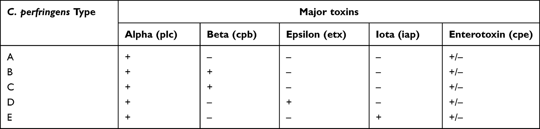 |
Table 3 Types of C. Perfringens and Major Toxins Produced |
The affected calves display sudden symptoms of depression, weakness, abdominal pain, and bloating.55 If diarrhea does develop, it might contain streaks of blood and tissue. Typically, Clostridia bacteria inhabit the gastrointestinal tract of cattle as part of their natural microbiota. However, under conditions such as dietary stress, injury, management changes, parasitism, or other unusual circumstances that create conducive growth environment, they can produce potent toxins that cause problems.23
Immunological and molecular biological tests are the main detection methods for C. perfringens from representative samples. Toxins or antigens secreted by different C. perfringens can be detected using immunological assays.57 The main immunological detection methods are the serum neutralization test, Nagler’s test, and the ELISA test. SDS-PAGE electrophoresis technology was used to identify the type of C. perfringens. The detection of major toxin in C. perfringens can also be done using PCR and multiplex PCR60. PCR detects alpha toxins of the species via amplification of alpha toxin gene.61 Good hygiene practices, feeding management, and vaccination can prevent the infection caused by C. perfringens.4,62 Preventive strategies for pathogens depend on good farm management practices, limiting their susceptibility to infection through vaccination, high hygienic levels of tools and structures for handling of the animal can avoid risk of infections (Table 1). Good management practices in harvesting, storing, and feeding the animals prevent diseases associated with enterotoxic clostridia.63,64
Mycoplasma bovis
The genus Mycoplasma is a gram-negative bacterium that belongs to the Mycoplasmataceae family (Table 1). The genus Mycoplasma is characterized by a small genome size, a lack of a cell wall, and a low G+C content. Currently, the genus Mycoplasma contains at least 130 species, and Mycoplasma bovis (M. bovis) is considered one of the important causes of bovine mycoplasmosis. The classification is based on 16S rRNA gene sequencing for differentiation between closely related species.65
Mycoplasma bovis is a significant worldwide opportunistic pathogen of intensively reared beef and dairy calves. It is a significant cause of pneumonia, otitis media, and arthritis in young dairy calves less than three months of age.66 The disease caused by M. bovis is chronic, debilitating, and unresponsive to antimicrobial therapy. The disease can persist for a very long period in a herd, with the possibility of the infected animals shedding microorganisms for a few weeks to several months.67 The virulence factors of M. bovis are adhesion, host cell invasion, host immune system modulation, production of secondary metabolites, biofilm formation, and synergistic infections with other viral and/or bacterial microorganisms that enable evading the host immune system.68 Actinomyces pyogenes, Haemophilus somnus, and Pasteurella species may synergistically act with M. bovis to cause bovine pneumonic pasteurellosis, bovine enzootic bronchopneumonia, or bovine respiratory disease (BRD). The disease is also significant from an animal welfare point of view, as it often results in calves that are subject to severe, chronic disease for which veterinarians can only provide limited relief. It is a major concern in the dairy industry, as it can lead to economic losses due to decreased milk production and increased morbidity and mortality in calves. Therefore, improved preventive and therapy strategies are needed for the disease.69
Mycoplasma bovis infections in dairy calves can present with a range of clinical signs. Respiratory disease is the most common manifestation, which can range from mild to severe.70 Affected calves may show signs of coughing, nasal discharge, fever, otitis media in young calves, polyarthritis in adult animals, and mastitis in dairy cows, arthritis, lameness, joint swelling, pain, and difficulty breathing. In severe cases, calves may develop pneumonia and require intensive care. In some cases, the infection can spread to the central nervous system, leading to neurological signs such as incoordination and seizures.71 Mycoplasma bovis can be transmitted directly through nose-to-nose contact or aerosols, or indirectly through contaminated utensils and the respiratory secretions of infected animals. A major transmission route for M. bovis from cows to calves is thought to be ingestion of contaminated milk.72 Colonization of the respiratory tract occurs more often in calves fed milk infected with M. bovis compared to calves fed non-infected milk. It can survive for a long period of time in a protected environment, with the greatest survival in humid and cool conditions. Apparently healthy animals can harbor the organism in the upper respiratory tract for long periods of time, acting as a reservoir for infection in the herd.66,73
The diagnosis of M. bovis infection is challenging as the clinical signs are not specific. Cultural isolation, serological tests, and molecular diagnostic approaches can be used to diagnose Mycoplasma bovis infections from nasal swabs, joint fluid, and blood samples. PCR is a molecular technique that can detect the DNA of Mycoplasma bovis in samples. Serology involves testing for antibodies against the bacterium in the blood.70,74
Antibiotics are used for the treatment of Mycoplasma bovis infections in dairy calves, although they are difficult to treat. However, the choice of antibiotics can be challenging since Mycoplasma bovis is resistant to many commonly used antibiotics.75 The bacteria are resistant to several antimicrobial classes, including fluoroquinolones, macrolides, tetracyclines, and β-lactams, because of the uncontrolled usage of antimicrobial agents in the animal industry. The cost of infection is primarily associated with the intensive treatment of affected calves coupled with the culling of animals that are unresponsive to treatments.76 Antimicrobial susceptibility testing is recommended to guide antibiotic selection. Supportive care is also an important component of treatment. Calves with respiratory disease may require oxygen supplementation, nebulization, and bronchodilators. Those with arthritis may require pain management and joint support.77 Preventing and controlling Mycoplasma bovis infections in dairy calves requires a comprehensive approach that includes biosecurity measures, vaccination, and appropriate antibiotic use. Good hygiene practices, such as cleaning and disinfecting equipment and surfaces, can reduce the risk of transmission. Vaccination against Mycoplasma bovis can also be effective in reducing the severity of infections in calves (Table 1). However, there is no single vaccine available that provides complete protection against all strains of the bacterium.66,70
Pasteurella multocida
The genus Pasteurella belongs to the family Pasteurellaceae. Pasteurella species are a short rod/coccobacilli-shaped bacterium that is capsulated, non-spore-forming, gram-negative, non-motile, facultative anaerobic. They are also sugar fermentative, oxidase and catalase positive, and bipolar in gram stain.17 The species also exists as a commensal and opportunistic pathogen found in the upper respiratory and proximal gastrointestinal tracts of animals. They are responsible for major bacterial causative agents of bovine respiratory disease.78 The most important members of the Pasteurellaceae family in the livestock industry include M. haemolytica, P. multocida, and B. trehalosi. These species are the most common cause of pneumonic and hemolytic septicemia in cattle and small ruminants that pose serious hazards to the livestock sector.76 Pasteurella species are widespread and cause a wide range of economically significant endemic and epizootic diseases in ruminants all over the world. Pasteurella species cause respiratory, systemic, and local infections in various animal species, including dairy calves.77 P. multocida and M. haemolytica are two primary bacterial pathogens causing pneumonia and hemorrhagic septicemia in calves that affect the health, welfare, and productivity of calves.79
Hemorrhagic septicemia (HS) is an acute, fatal, and septicemic disease of cattle and buffaloes in tropical regions of the world. Hemorrhagic septicemia is the most economical contagious bacterial disease caused by B2 and E2 serotypes of P. multocida,80 and Serotype E2 is predominantly found in Africa. The disease is characterized by the sudden onset of fever, profuse salivation, severe dyspnea, and death in about 24 hours. Hemorrhagic septicemia is list B disease by OIE since it is a primary pasteurellosis with 100% mortality in infected animals in endemic areas of Africa and Asia. Thus, HS is the most economically important bacterial diseases.81 The morbidity and mortality rates of the disease range between 50% and 100%, respectively. Morbidity depends on the immune status of the animal, either acquired naturally or induced by vaccination. The occurrence of the disease is often associated with wet humidity during the rainy season, transportation, and other stress factors. Pneumonic pasteurellosis is responsible for huge mortality in feedlot cattle. In general, pneumonic pasteurellosis and hemorrhagic septicemia diseases cause significant economic losses in the cattle sector due to high morbidity and mortality rates across the world.82
Virulence genes are significant in the pathogenesis of P. multocida. The virulent genes of P. multocida include fimbriae and adhesions (nanB and nanH), outer membrane proteins (OMP) and toxins (toxA).83,84 These virulent factors promote P. multocida colonization and invasion by impairing host defense mechanisms, destroying host tissues, and inducing a lethal host inflammatory response.85 The virulent determinants also keep organisms in the respiratory tract by preventing phagocytosis and increasing resistance to complement and bactericidal effects of the host defense mechanisms.86
Identifying the organisms’ circulating serotypes is critical for effectively preventing and controlling pasteurellosis in domestic ruminants,87 and also important for the selection of vaccine strains. P. multocida is divided into five serogroups based on capsular structure (A, B, D, E, and F) using indirect hemagglutination test and further subdivided into 16 serotypes based on lipopolysaccharide composition by Heddleston gel diffusion precipitation assay.88,89 B2 and E2 cause hemorrhagic septicemia in addition to the possible pneumonia or septicemia caused by the remainder of the capsular serogroups and somatic serotypes.90
Mannheimia haemolytica is a major pathogen in ruminants of all age groups, and it is the main cause of bovine and ovine pneumonic pasteurellosis.91,92 It has two biotypes (A and T) based on fermentation of arabinose and trehalose, respectively.93 Based on their surface antigen, these biotypes are further subdivided into 17 serotypes. Thirteen of these are biotype A, while the remaining four are biotype T (3, 4, 10, and 15). The most prevalent serotypes of M. haemolytica are A1 and A2, which are found worldwide. Bovine serotype A1 is considered to be the most prevalent cause of bovine pasteurellosis. M. haemolytica serotype A2 typically causes pneumonic pasteurellosis in small ruminants of all age groups.94 M. haemolytica uses capsules, adhesions, lipopolysaccharides (LPS), OMP, and various protease virulent determinant factors to colonize and infect the lungs.95 The pathogenicity of M. haemolytica is associated with various virulence genes. These include leukotoxin (lkt), leukotoxin C (lktC), putative adhesion (ahs), and outer membrane lipoprotein (gs60). The identification of these genes reveals crucial information about M. haemolytica’s pathogenicity.96
The diagnosis of pneumonic pasteurellosis is based on the history of patients, clinical signs, and conventional bacterial isolation. Recently advanced and more confirmatory diagnostic assays are PCR, RT-PCR, RFLP, real-time PCR, gene sequencing, and phylogenetic analysis.97 The phenotypic and genetic characterization of P. multocida serotypes that are currently circulating in the field is critical for disease prevention and control strategy. It is also important to precisely distinguish the phylogeny of fastidious pathogens and virulence genes as a future step in vaccine development.98 Calves infected with P. multocida may develop pneumonia and septicemia. Nasal swabs and blood are the preferred sample types for diagnosis. The transmission of P. multocida can occur through direct contact with infected animals, contaminated equipment, or aerosolization of the bacteria. The risk of infection is higher in calves that are stressed, malnourished, or have weakened immune systems.99
The major clinical signs in the infected dairy calves are fever, cough, nasal discharge, and difficulty breathing. The infection can also cause local infections including abscesses and septicemia, which can lead to significant economic loss in the dairy industry.75 The risk factors that increase infection of dairy calves are stress (environmental and management problem), overcrowding, poor ventilation, and co-infection with other pathogens.100 Good management practices, such as vaccination, proper nutrition, and hygiene, can help prevent Pasteurella infections and reduce the severity of BRD in dairy calves.101 Treatment of Pasteurella infection in dairy calves typically involves the use of antibiotics. Supportive care, including fluid therapy and nutritional support, may also be necessary for severely affected calves.102
Listeria monocytogenes
The genus listeria is an intracellular gram-positive bacterium that belongs to the family Listeriaceae. The genus is characterized by non-capsulated, coccoid to rod shaped cells, facultative anaerobes, flagellated, and non-spore forming bacterium103 (Table 1). The genus contains six species including Listeria monocytogenes, L. ivanovii, L. innocua, L. welshimeri, L. seeligeri, and L. grayi. Listeria monocytogenes and L. ivanovii are two main pathogenic species for animals and humans.104,105 Listeria monocytogenes (L. monocytogenes) is ubiquitous and widely spread in the environment such as soil, mud, plant decaying vegetation, raw and treated sewages, water, food processing facilities.106 The organism also possesses unique physiological characteristics that can grow at refrigerator temperatures. The species L. monocytogenes is the causative agent of listeriosis in humans and animals.107 It is one of the most significant emerging and opportunistic food-borne bacterial diseases in humans. The infection can result in septicemia, meningitis, uterine infection (abortion and still birth), subclinical mastitis, and sometimes death in dairy calves and cow. Listeriosis usually occurs in calves, pregnant cows and immunocompromised cattle.108
The virulence potential of L. monocytogenes is determined by several molecular determinants. Hemolysin (listeriolysin O), two phospholipase, protein (ActA), internalin A (InlA), ternalin B (InlB), and cytotoxins are the main virulence factors of L. monocytogenes.109 The virulence factors are essential for intracellular motility of the pathogen. The hemolysin toxin lyses tissue and blood cells. The cytotonic toxin stimulates cyclic AMP production similar to cholera toxin. All serotypes of L. monocytogenes have the ability to provoke monocytosis. Host and environment-related risk factors that expose animals to infection are poor nutritional status, age, parity, sudden change in weather, late pregnancy, and parturition stress, transport, and overcrowding.108
Listeria monocytogenes is transmitted by ingestion, inhalation, or direct contact with infected animals. Ingestion of contaminated silage, feed, water, or bedding is the primary modes of transmission.110 Calves that develop the septicemic L. monocytogenes disease may acquire infection from contamination of the cow teat from the ingestion of milk containing the organism or from a cow with subclinical bacteremia, through the navel from the environment, and also from congenital infection.23 The clinical signs of listeriosis depend on pathogenic strains and immune status of the host. The clinical manifestation of listeriosis in dairy cows is neonatal septicemia, encephalitis, and abortion in late pregnancy.111 L. monocytogenes can also cause depression, fever, diarrhea in some cases, anorexia, meningitis, and neurological signs in calves. These neurological signs include circling, head pressing, and ataxia.112
Listeria monocytogenes can be diagnosed via bacterial culture, animal inoculation, immunological assays, and molecular tests from brain tissue, blood, and feces specimens.102 The most recommended drugs for the disease listeriosis include penicillin G, aminoglycosides, trimethoprim, sulfamethoxazole, and tetracycline in dairy calves.113 Prevention of infection in dairy calves can be achieved through good management practices, such as maintaining clean and hygienic facilities and equipment, ensuring proper colostrum management, and minimizing stress on the calves (Table 1). Vaccination may also be effective in preventing infection in calves.114
Haemophilus somnus
Haemophilus somnus (H. somnus) is a gram-negative bacterium that causes respiratory and joint infections in calves115 (Table 1). This bacterium is considered an opportunistic pathogen, which means it can cause disease when the immune system of the calf is compromised.116 Good hygiene practices and antibiotic treatment can prevent H. somnus infection.117 Calves infected with H. somnus may develop respiratory disease, septicemia, and arthritis. Nasal swabs, blood, and synovial fluid are the preferred sample types for diagnosis.118 Bacterial culture and PCR are commonly used diagnostic methods for H. somnus infection119.
Respiratory disease caused by H. somnus is characterized by fever, depression, coughing, and nasal discharge. In severe cases, the calf may develop pneumonia, which can be fatal. Furthermore, the bacterium can invade the nervous system of the calf, leading to septicemia, meningitis, and other neurological symptoms.118 Several risk factors have been associated with H. somnus infection in dairy calves, including stress, transportation, overcrowding, and poor ventilation.120 Diagnosis of H. somnus infections in dairy calves can be challenging, as the bacterium can be difficult to isolate and culture from clinical samples.121 Molecular techniques such as PCR and DNA sequencing have been developed to aid in the diagnosis of H. somnus infections, but these methods can be expensive and require specialized equipment and expertise.119
Studies have shown that the use of antimicrobial agents can be effective in treating H. somnus infection in dairy calves. However, the emergence of antimicrobial resistance in this bacterium has raised concerns about the long-term effectiveness of treatments.122 Prevention and control of H. somnus infections in dairy calves involve a multi-faceted approach, including vaccination, biosecurity measures, and appropriate management practices.101 Vaccines against H. somnus are available and can be effective in reducing the incidence and severity of BRD and reproductive diseases.123 Biosecurity measures, such as quarantine and disinfection protocols, can help to prevent the introduction and spread of H. somnus on dairy farms (Table 1). Management practices, such as minimizing stress and optimizing nutrition, can also help to maintain the health and immune function of dairy calves.124
Conclusion
The review highlights the significant challenges of bacterial infections in dairy calves, emphasizing common bacterial infections, clinical manifestations, and factors influencing their occurrence. Effective management strategies, including early detection, proper treatment, and preventive measures such as good colostrum feeding, are crucial. The review also underscores the importance of judicious antibiotic use, supportive care, and advancements in diagnostics. Furthermore, it explores recent advancements in diagnosis and prevention, providing valuable insights for dairy farmers, veterinarians, and researchers. Comprehending the common bacterial pathogens, their methods of transmission, and the factors that impact infection rates is essential for the successful implementation of management strategies. Further study is essential for a deeper understanding of the epidemiology and characterization of these infections in dairy calves.
Acknowledgments
We would like to express our heartfelt gratitude to the Ethiopian Institute of Agricultural Research (EIAR) for its invaluable assistance during this review. Their expertise and support have been instrumental in maintaining the accuracy and quality of our work. Furthermore, we extend our sincere thanks to all individuals and organizations that have contributed to this review. Whether by providing data, perspectives, or comments, their assistance has been essential in helping us achieve the objectives of this review.
Disclosure
The authors have not revealed any possible conflicts of interest.
References
1. Brown KA, Venkateshmurthy NS, Potubariki G, et al. The role of dairy in healthy and sustainable food systems: community voices from India. BMC Public Health. 2022;22(1):806. doi:10.1186/s12889-022-13194-w
2. El-Seedy FR, Abed AH, Yanni HA, Abd El-Rahman SAA. Prevalence of Salmonella and E. coli in neonatal diarrheic calves. Beni-Suef Univ J Basic Appl Sci. 2016;5(1):45–51. doi:10.1016/j.bjbas.2015.11.010
3. Cho YI, Yoon KJ. An overview of calf diarrhea infectious etiology, diagnosis, and intervention. J Vet Sci. 2014;15(1):1–17. doi:10.4142/jvs.2014.15.1.1
4. McGuirk SM. Disease management of dairy calves and heifers. Vet Clin North Am Food Anim. 2008;24(1):139–153. doi:10.1016/j.cvfa.2007.10.003
5. Fentie T, Guta S, Mekonen G, et al. Assessment of major causes of calf mortality in urban and periurban dairy production system of Ethiopia. Vet Med Int. 2020;2020:1–7. doi:10.1155/2020/3075429
6. Liu D. Diarrhoeagenic Escherichia coli, molecular medical microbiology. Elsevier Ltd. 2014. doi:10.1016/B978-0-12-397169-2.00064-0
7. LeBlanc SJ, Lissemore KD, Kelton DF, Duffield TF, Leslie KE. Major advances in disease prevention in dairy cattle. J Dairy Sci. 2006;89(4):1267–1279. doi:10.3168/jds.S0022-0302(06)72195-6
8. Lemon SM, Hamburg MA, Sparling PF, Choffnes ER, Mack A. Global infectious disease surveillance and detection: assessing the challenges. Workshop summary. In: Global Infectious Disease Surveillance and Detection: Assessing the Challenges. Workshop summary: National Academies Press; 2007.
9. Kasimanickam V, Kasimanickam M, Kasimanickam R. Antibiotics use in food animal production: escalation of antimicrobial resistance: where are we now in combating AMR? Med Sci. 2021;9(1):14. doi:10.3390/medsci9010014
10. Aditya A, Tabashsum Z, Martinez ZA, et al. Diarrheagenic Escherichia coli and their antibiotic resistance patterns in dairy farms and their microbial ecosystems. Journal of Food Protection. 2023;86(3):100051. doi:10.1016/j.jfp.2023.100051
11. Reid G, Howard J, Gan BS. Can bacterial interference prevent infection? Trends Microbiol. 2001;9(9):424–428. doi:10.1016/S0966-842X(01)02132-1
12. Radostitis OM, Gay CC, Henchiliff W, Constable D. A Text Book of Disease of Cattle, Horses, Sheep, Pigs and Goats. london: Harcourt Publisher Ltd; 2007:837–846.
13. Gomes V, Barros BP, Castro-Tardon DI, et al. The role of anti-E. Coli antibody from maternal colostrum on the colonization of newborn dairy calves’ gut with Escherichia coli and the development of clinical diarrhea. Ani-Open Spac. 2013;2:100037.
14. Foster DM, Smith GW. Pathophysiology of diarrhea in calves. Vet Clin North Am Food Anim Pract. 2009;25(1):13–36. doi:10.1016/j.cvfa.2008.10.013
15. Gebregiorgis A, Tessema TS. Characterization of Escherichia coli isolated from calf diarrhea in and around Kombolcha, South Wollo, Amhara region, Ethiopia. Trop Anim Health Prod. 2016;48(2):273–281. doi:10.1007/s11250-015-0946-9
16. Tedla M, Degefa K. Bacteriological study of calf colisepticemia in alage dairy farm, southern Ethiopia. BMC Res Notes. 2017;10(1):710. doi:10.1186/s13104-017-3038-2
17. Quinn PJ, Marfery DK, Carte ME, Donelly WJ, Learned FC. Veterinary Microbiology and Microbial Diseases. U.K: published Blackwell, science LTD; 2002:343–432.
18. White ME. Prevention of Calf Scours. Tuckson, Arizona: University Arizona; 2008:132–139.
19. Barrington GM, Gay JM, Evermann JF. Biosecurity for neonatal gastrointestinal diseases. Vet Clin North Am Food Anim. 2002;18(1):7–34. doi:10.1016/s0749-0720(02)00005-1
20. Ercumen A, Pickering AJ, Kwong LH, et al. Animal feces contribute to domestic fecal contamination: evidence from e. coli measured in water, hands, Food, Flies, and soil in Bangladesh. Sci Technol. 2017; 51(15): 8725–8734. doi:10.1021/acs.est.7b01710
21. Dubreuil JD, Isaacson RE, Schifferli DM. Animal Enterotoxigenic Escherichia coli. EcoSal Plus. 2016;7(1):1.
22. VanMetre DC, Tennant BC, Whitlock RH. Infectious diseases of the gastrointestinal tract. Rebhun’s Diseases of Dairy Cattle. 2008;200–294. doi:10.1016/B978-141603137-6.50009-0
23. Peek SF, Mcguirk SM, Sweeney RW, Cummings KJ. Infectious diseases of the gastrointestinal tract. Rebhun’s Diseases of Dairy Cattle. 2018;249–356. doi:10.1016/B978-0-323-39055-2.00006-1
24. Qadri F, Svennerholm AM, Faruque ASG, Sack RB. Enterotoxigenic Escherichia coli in developing countries: epidemiology, microbiology, clinical features, treatment, and prevention. Clin Microbiol Rev. 2005;18(3):465–483. doi:10.1128/CMR.18.3.465-483.2005
25. Croxen MA, Law RJ, Scholz R, Keeney KM, Wlodarska M, Finlay BB. Recent advances in understanding enteric pathogenic Escherichia coli. Clin Microbiol Rev. 2012; 26(4):822–880. doi:10.1128/CMR.00022-13
26. Deshmukh R, Roy U. Molecular diagnostic platforms for specific detection of Escherichia coli. Intech. 2016;11(13): 1.
27. Ali DA, Tesema TS, Belachew YD. Retracted article: molecular detection of pathogenic Escherichia coli strains and their antibiogram associated with risk factors from diarrheic calves in Jimma Ethiopia. Science Rep. 2021;11(1):14356. doi:10.1038/s41598-021-93688-6
28. Oludairo O, Kwaga J, Kabir J, et al. Review of Salmonella characteristics, history, taxonomy, nomenclature, Non Typhoidal Salmonellosis (NTS) and Typhoidal Salmonellosis (TS). Zagazig Vet J. 2022;50(2):160–171. doi:10.21608/zvjz.2022.137946.1179
29. Hyeon JY, Helal ZH, Polkowski R, et al. Genomic features of salmonella enterica subspecies houtenae serotype 45: g, z51: -isolated from multiple abdominal abscesses of an African fat-tailed gecko, United States. Antibiotics. 2021;10(11):1322. doi:10.3390/antibiotics10111322
30. Ketema L, Ketema Z, Kiflu B, et al. Prevalence and antimicrobial susceptibility profile of salmonella serovars isolated from slaughtered cattle in addis Ababa, Ethiopia. BioMed Research International. 2018;2018. doi:10.1155/2018/9794869
31. Holschbach CL, Peek SF. Salmonella in Dairy Cattle. Vet Clin North Am Food Anim. 2018;34(1):133–154. doi:10.1016/j.cvfa.2017.10.005
32. Tsolis RM, Adams LG, Ficht TA, Baumler AT. Contribution of salmonella enteric serovar typhimurium virulence factors to diarrheal diseases in calves. Infect Immun. 1999;67:4879–4885.
33. Freitas Neto OC, Setta A, Imre A, et al. A flagellated motile Salmonella Gallinarum mutant (SG Fla+) elicits a pro-inflammatory response from avian epithelial cells and macrophages and is less virulent to chickens. Vet Microbiol. 2013; 2013(165): 1.
34. Molla WMB, Muckle DAA, Wilkie LCE. Occurrence and Antimicrobial Resistance of Salmonella Serovars in Apparently Healthy Slaughtered Sheep and Goats of Central Ethiopia. Springer; 2006:455–456. doi:10.1007/s11250-006-4325-4
35. Pecoraro HL, Thompson B, Duhamel GE. Histopathology case definition of naturally acquired Salmonella enterica serovar Dublin infection in young Holstein cattle in the northeastern United States. J Vet Diagn. 2017; 29: 860–864—.
36. Nielsen LR. Review of pathogenesis and diagnostic methods of immediate relevance for epidemiology and control of Salmonella Dublin in cattle. Vet Microbiol. 2013;162(1):1–9. doi:10.1016/j.vetmic.2012.08.003
37. Jajere SM. A review of Salmonella enterica with particular focus on the pathogenicity and virulence factors, host specificity and antimicrobial resistance including multidrug resistance. Vet World. 2019;12(4):504–521. doi:10.14202/vetworld.2019.504-521
38. OIE. OIE terrestrial manual salmonellosis. Work. 2022; 3(10.7):1–19.
39. Addis Z, Kebede N, Worku Z, Gezahegn H, Yirsaw A, Kassa T. Prevalence and antimicrobial resistance of Salmonella isolated from lactating cows and in contact humans in dairy farms of addis ababa: a cross sectional study. BMC Infect Dis. 2011;11(1):222. doi:10.1186/1471-2334-11-222
40. Callaway TR, Keen JE, Edrington TS, Baumgard LH. Fecal prevalence and diversity of salmonella species in lactating dairy cattle in four states. J Dairy Sci. 2005;88(10):3603–3608. doi:10.3168/jds.S0022-0302(05)73045-9
41. Smith BP. Salmonellosis in ruminants. In: Large Animal Internal Medicine. Mosby: St. Louis (MO); 2009. 877–881.
42. Iman KK, Asmaa FM, Abdelrazek YD, Mohamed FH. Isolation and identification of Rotavirus infection in diarrheic calves at el gharbia governorate. Global Vet. 2017;18(3):178–182.
43. Lee N, Kwon KY, Oh SK, Chang HJ, Chun HS, Choi SW. A multiplex PCR assay for simultaneous detection of Escherichia coli O157: H7, bacillus cereus vibrio parahaemolyticus, salmonella spp, listeria monocytogenes, and staphylococcus aureus in Korean ready-to-eat food. Foodborne Pathog Dis. 2014;11(7):574–580. doi:10.1089/fpd.2013.1638
44. Veling J, Barkema W, VanDer Schans J, VanZijderveld F, Verhoeff J. Herd-level diagnosis for Salmonella enterica subsp. enterica serovar Dublin infection in bovine dairy herds. Preventive Vet Med. 2002;14(1–2):31–42. doi:10.1016/S0167-5877(01)00276-8
45. Munoz N, Diaz-Osorio M, Moreno J, et al. Development and evaluation of a multiplex real-time polymerase Chain reaction procedure to clinically type prevalent Salmonella enterica serovars. J Mol Diagn. 2010;12(2):220–250. doi:10.2353/jmoldx.2010.090036
46. Lofstrom C, Hansen F, Hoorfar J. Validation of a 20h real time PCR method for screening of salmonella in poultry fecal samples. Vet Microbiol. 2010;144(3–4):511–514. doi:10.1016/j.vetmic.2010.02.019
47. McKillip JL, Drake M. Real-time nucleic acid-based detection methods for pathogenic bacteria in food. Journal of Food Protection. 2004;67(4):823–832. doi:10.4315/0362-028X-67.4.823
48. Zishiri OT, Mkhize N, Mukaratirwa S. Prevalence of virulence and antimicrobial resistance genes in salmonella spp. isolated from commercial chickens and human clinical isolates from South Africa and Brazil. JVet Res. 2012; 83(1):112016. doi:10.4102/ojvr.v83i1.1067
49. Lei P, Tang H, Ding S, et al. Determination of the invA gene of Salmonella using surface plasmon resonance along with streptavidin aptamer amplification. Mikrochim Acta. 2015;182(1–2):289–296. doi:10.1007/s00604-014-1330-6
50. Van Dorst B, Mehta J, Bekaert K, et al. Recent advances in recognition elements of food and environmental biosensors: a review. Biosens Bioelectron. 2010;26(4):1178–1194. doi:10.1016/j.bios.2010.07.033
51. Kebede IA, Duga T. Prevalence and antimicrobial resistance of salmonella in poultry products in central Ethiopia. Vet Med Int. 2022;20:8625636.
52. Rood JI, Cole ST. Molecular genetics and pathogenesis of clostridium perfringens. MICROBIOLOGICAL REVIEWS. 1991;55(4):621–648. doi:10.1128/mr.55.4.621-648.1991
53. Santos BL, Ladeira SRL, Riet-Correa F, et al. Clostridial diseases diagnosed in cattle from the south of rio grande do Sul, Brazil. A forty-year survey (1978–2018) and a brief review of the literature1. Pesqui Vet Bras. 2019;39(7):435–446. doi:10.1590/1678-5150-pvb-6333
54. Uzal FA, Vidal JE, McClane BA, Gurjar AA. Clostridium perfringens toxins involved in mammalian veterinary diseases. Toxicol Open Access. 2010;2:24–42. doi:10.2174/1875414701003020024
55. Simpson KM, Callan RJ, Van Metre DC. Clostridial abomasitis and enteritis in ruminants. Vet Clin North Am Food Anim. 2018;34(1):155–184. doi:10.1016/j.cvfa.2017.10.010
56. Shrestha A, Uzal FA, McClane BA. Enterotoxic Clostridia: clostridium perfringens enteric diseases. Microbiol Spectr. 2018;6(5):
57. Kasa A, Tulu D, Negera C. Review of common bacterial cause and management of neonatal calf diarrhea in cattle. Int J Microbiol Res. 2020;11(2):98–104.
58. Uzal FA, Freedman JC, Shrestha A, et al. Towards an understanding of the role of clostridium perfringens toxins in human and animal disease. Future Microbiol. 2014;9(3):361–377. doi:10.2217/fmb.13.168
59. Goossens E, Valgaeren BR, Pardon B, et al. Rethinking the role of alpha toxin in clostridium perfringens-associated enteric diseases: a review on bovine necro-haemorrhagic enteritis. Vet Res. 2017;48(1):1–17. doi:10.1186/s13567-017-0413-x
60. Wu J, Zhang W, Xie B, et al. Detection and toxin typing of clostridium perfringens in formalin-fixed, paraffin-embedded tissue samples by PCR. J Clin Microbiol. 2009;47(3):807–810. doi:10.1128/JCM.01324-08
61. Cao A, Chi H, Shi J, et al. Visual detection of clostridium perfringens alpha toxin by combining nanometer microspheres with smart phones. Microorganisms. 2020;8(12):1865. doi:10.3390/microorganisms8121865
62. Nekrasov RV, Lozovanu MI, Laptev GY, et al. Bioactive feed additive for the prevention of clostridial disease in high-yielding dairy cattle. Agriculture. 2023;13:86.
63. Butucel E, Balta I, McCleery D, et al. Farm biosecurity measures and interventions with an impact on bacterial biofilms. Agriculture. 2022;12(8):1251. doi:10.3390/agriculture12081251
64. Zaragoza NE, Orellana CA, Moonen GA, Moutafis G, Marcellin E. Vaccine production to protect animals against pathogenic clostridia. Toxins (Basel). 2019;11(9):525. doi:10.3390/toxins11090525
65. Thompson CC, Vieira NM, Vicente ACP, Thompson FL. Towards a genome-based taxonomy of mycoplasmas. Infect Genet Evol. 2011;11(7):1798–1804. doi:10.1016/j.meegid.2011.07.020
66. Maunsell FP, Donovan GA, Risco C, Brown MB. Field evaluation of a mycoplasma bovis bacterin in young dairy calves. Vaccine. 2009;27(21):2781–2788. doi:10.1016/j.vaccine.2009.02.100
67. Punyapornwithaya V, Fox LK, Hancock DD, et al. Association between an outbreak strain causing mycoplasma bovis mastitis and its asymptomatic carriage in the herd: a case study from Idaho, USA. Preventive Vet Med. 2010;93(1):66–70. doi:10.1016/j.prevetmed.2009.08.008
68. Bürki S, Frey J, Pilo P. Virulence, persistence and dissemination of mycoplasma bovis. Vet Microbiol. 2015;179(1–2):15–22. doi:10.1016/j.vetmic.2015.02.024
69. Ammar A, Abd El-Hamid M, Hashem Y, et al. Mycoplasma bovis: taxonomy, characteristics, pathogenesis and antimicrobial resistance. Zagazig Vet J. 2021;49(4):444–461. doi:10.21608/zvjz.2021.103834.1160
70. Dudek K, Nicholas RAJ, Szacawa E, Bednarek D. Mycoplasma bovis infections-occurrence, diagnosis and control. Pathogens. 2020;9(8):640. doi:10.3390/pathogens9080640
71. Hananeh WM, Momani WMA, Ababneh MM, Abutarbush SM. Mycoplasma bovis arthritis and pneumonia in calves in Jordan: an emerging disease. Vet World. 2018;11(12):1663–1668. doi:10.14202/vetworld.2018.1663-1668
72. Gogoi-Tiwari J, Tiwari HK, Wawegama NK, et al. Prevalence of Mycoplasma bovis infection in calves and dairy cows in western Australia. J Vet Sci. 2022;9(7):3519.
73. Maunsell FP, Woolums AR, Francoz D, et al. Mycoplasma bovis infections in cattle. Vet Med Int. 2011;25:772–783.
74. Okella H, Tonooka K, Okello E. A systematic review of the recent techniques commonly used in the diagnosis of mycoplasma bovis in dairy cattle. Pathogens. 2023;12(9):1178. doi:10.3390/pathogens12091178
75. Lysnyansky I, Ayling RD. Mycoplasma bovis: mechanisms of resistance and trends in antimicrobial susceptibility. Front Microbiol. 2016;7:595. doi:10.3389/fmicb.2016.00595
76. Sulyok KM, Kreizinger Z, Fekete L, et al. Antibiotic susceptibility profiles of Mycoplasma bovis strains isolated from cattle in Hungary, central Europe. BMC Vet Res. 2014;10:256. doi:10.1186/s12917-014-0256-x
77. Ammar AM, El-Hamid MI A, Mohamed YH, et al. Prevalence and antimicrobial susceptibility of bovine mycoplasma species in Egypt. Biology. 2022;11(7):1083. doi:10.3390/biology11071083
78. Abed AH, El-Seedy FR, Hassan HM, et al. Genotyping and virulence genes characterization of pasteurella multocida and Mannheimia haemolytica isolates recovered from pneumonic cattle calves in north upper Egypt. Vet Sci. 2020;7(4):174. doi:10.3390/vetsci7040174
79. Orynbayev M, Sultankulova K, Sansyzbay A, et al. Biological characterization of pasteurella multocida present in the saiga population. BMC Microbiol. 2019;19(1):37. doi:10.1186/s12866-019-1407-9
80. Mushtaq A, Singh S, Gazal S, et al. Haemorrhagic septicemia: a persistent nuisance in Indian livestock review. Pharma Innovation J. 2022;11(7S):4382–4394.
81. Shivachandra SB, Viswas KN, Kumar AA. A review of hemorrhagic septicemia in cattle and Buffalo. Anim Health Res Rev. 2011;12(1):67–82. doi:10.1017/S146625231100003X
82. Jilo K, Belachew T, Birhanu W, Habte D, Yadete W, Giro A. Pasteurellosis status in Ethiopia: a comprehensive review. J Trop Dis. 2020;8:351. doi:10.35248/2329-891X.20.8.351
83. Townsend KM, Boyce JD, Chung JY, Frost AJ, Adler B. Genetic organization of Pasteurella multocida cap loci and development of a multiplex capsular PCR typing system. J Clin Microbiol. 2001;39(3):924–929. doi:10.1128/JCM.39.3.924-929.2001
84. Smith E, Miller E, Aguayo JM, et al. Genomic diversity and molecular epidemiology of pasteurella multocida. PLoS One. 2021;16(4): e0249138. doi:10.1371/journal.pone.0249138
85. E-Kobon T, Leeanan R, Pannoi S, Anuntasomboon P, Thongkamkoon P, Thamchaipenet A. OmpA protein sequence-based typing and virulence-associated gene profiles of Pasteurella multocida isolates associated with bovine haemorrhagic septicaemia and porcine pneumonic pasteurellosis in Thailand. BMC Vet Res. 2017;13(1):243. doi:10.1186/s12917-017-1157-6
86. Tang X, Zhao Z, Hu J, et al. Isolation, antimicrobial resistance, and virulence genes of Pasteurella multocida strains from swine in China. J Clin Microbiol. 2009;47(4):951–958. doi:10.1128/JCM.02029-08
87. Dabo SM, Taylor JD, Confer AW. Pasteurella multocida and bovine respiratory disease. Anim Health Res Rev. 2007;8(2):129–150.
88. Kudirkiene E, Aagaard AK, Schmidt LMB, Pansri P, Krogh KM, Olsen JE. Occurrence of major and minor pathogens in calves diagnosed with bovine respiratory disease. Vet Microbiol. 2021; 109135: doi:10.1016/j.vetmic.2021.109135
89. Girma S, Getachew L, Beyene A, et al. Identification of serotypes of mannheimia haemolytica and pasteurella multocida from pneumonic cases of sheep and goats and their antimicrobial sensitivity profiles in borana and arsi zones, Ethiopia. Science Rep. 2023;13(1):9008. doi:10.1038/s41598-023-36026-2
90. Umer AA, Mezgebu E. Review on pasteurellosis: causes, pathogenesis, diagnosis and current status in Ethiopia. Austin j Microbiol. 2023;8(1):1–8.
91. El-Seedy FR, Hassan HM, Nabih AM, et al. Respiratory affections in calves in upper and middle Egypt: bacteriologic, immunologic and epidemiologic studies. Adv Anim Vet Sci. 2020;8(5):558–569. doi:10.17582/journal.aavs/2020/8.5.558.569
92. Ayalew G, Negash A, Birhanu M. Review on avian salmonellosis and its impact. Nat Sci. 2017;15(6):59–67. doi:10.7537/marsnsj150617.06
93. Haig SG. Adherence of mannheimia haemolytica to ovine bronchial epithelial cells biosci. Horiz. Int J Stud Res. 2011;4:50–60.
94. Woyessa MB, Umer AA. Review on bovine pneumonic pasteurellosis. Austin J Infect Dis. 2023;10(4):1092.
95. Singh K, Ritchey JW, Confer AW. Mannheimia haemolytica: bacterial-Host interactions in bovine pneumonia. Vet Pathol. 2011;48(2):338–348. doi:10.1177/0300985810377182
96. Klima CL, Alexander TW, Hendrick S, McAllister TA. Characterization of Mannheimia haemolytica isolated from feedlot cattle that were healthy or treated for bovine respiratory disease. Can J Vet Res. 2014;78:38–45.
97. Abera D, Mossie T. A review on pneumonic pasteurellosis in small ruminants. J Appl Animal Res. 2023;51(1):1)1–10. doi:10.1080/09712119.2022.2146123
98. Beceiro A, Tomás M, Bou G. Antimicrobial resistance and virulence: a successful or deleterious association in the bacterial world? Clinical Microbiology Rev. 2013;26(2):185–230. doi:10.1128/CMR.00059-12
99. Hussain R, Mahmood F, Ali HM, Bacterial SAB. PCR and clinico-pathological diagnosis of naturally occurring pneumonic pasteurellosis (mannheimiosis) during subtropical climate in sheep. Microb Pathog. 2017;112:176–181. doi:10.1016/j.micpath.2017.09.061
100. Stahel AB, Hoop RK, Kuhnert P, Korczak BM. Phenotypic and genetic characterization of pasteurella multocida and related isolates from rabbits in Switzerland. J Vet Diagn Invest. 2009;21(6):793–8022009. doi:10.1177/104063870902100605
101. Giordano A, Dincman T, Clyburn BE, Steed LL, Rockey DC. Clinical features and outcomes of pasteurella multocida infection. Medicine (Baltimore). 2015;94(36): e1285. doi:10.1097/MD.0000000000001285
102. Gaudino M, Nagamine B, Ducatez MF, Meyer G. Understanding the mechanisms of viral and bacterial coinfections in bovine respiratory disease: a comprehensive literature review of experimental evidence. Vet Res. 2022;53(1):70. doi:10.1186/s13567-022-01086-1
103. Roberts BN, Chakravarty D, Gardner JC 3rd, Ricke SC, Donaldson JR. Listeria monocytogenes response to anaerobic environments. Pathogens. 2020;9(3):210. doi:10.3390/pathogens9030210
104. Rahimi E, Shakerian A, Raissy M. Prevalence of listeria species in fresh and frozen fish and shrimp in Iran. Vet Microbiol. 2012;62(1):37–40. doi:10.1007/s13213-011-0222-9
105. Orsi RH, Wiedmann M. Characteristics and distribution of listeria spp., including listeria species newly described since 2009. Appl Microbiol Biotechnol. 2016;100(12):5273–5287. doi:10.1007/s00253-016-7552-2
106. EL-Naenaeey ES, Abdelwahab A, Merwad A, Abdou H. Prevalence of listeria species in dairy cows and pregnant women with reference to virulotyping of listeria monocytogenes in Egypt. Zagazig Vet J. 2019;47(3):248–258. doi:10.21608/zvjz.2019.12896.1042
107. Hasegawa M, Iwabuchi E, Yamamoto S, et al. Prevalence and characteristics of listeria monocytogenes in bovine colostrum in Japan. Journal of Food Protection. 2013;76(2):248–255. doi:10.4315/0362-028X.JFP-12-278
108. Elsayed MM, Elkenany RM, Zakaria AI, Badawy BM. Epidemiological study on listeria monocytogenes in Egyptian dairy cattle farms’ insights into genetic diversity of multi-antibiotic-resistant strains by ERIC-PCR. Environ Sci Pollut Res. 2022;29(36):54359–54377. doi:10.1007/s11356-022-19495-2
109. Kathariou S. Listeria monocytogenes virulence and pathogenicity, a food safety perspective. Journal of Food Protection. 2002;65(11):1811–1829. doi:10.4315/0362-028X-65.11.1811
110. Nightingale KK, Schukken YH, Nightingale CR, et al. Ecology and transmission of Listeria monocytogenes infecting ruminants and in the farm environment. Appl Environ Microbiol. 2004;70(8):4458–4467. doi:10.1128/AEM.70.8.4458-4467.2004
111. Amene Y, Firesbhat A. Listeriosis in large ruminants: a review. J Anim Sci Dis. 2016;5(1):16–21. doi:10.5829/idosi.ajad.2016.16.21
112. Disson O, Lecuit M. Targeting of the central nervous system by Listeria monocytogenes. Virulence. 2012;3(2):213–221. doi:10.4161/viru.19586
113. Vazquez-Boland JA, Kuhn M, Berche P, et al. Listeria pathogenesis and molecular virulence determinants. Clinical Microbiology Rev. 2001;14(3):584–640. doi:10.1128/CMR.14.3.584-640.2001
114. Dos Reis JO, Vieira BS, Cunha Neto A, Castro VS, Figueiredo EES. Antimicrobial resistance of listeria monocytogenes from animal foods to first- and second-line drugs in the treatment of listeriosis from 2008 to 2021: a systematic review and meta-analysis. J Infect Dis Med Microbiol. 2022;2022:1351983. doi:10.1155/2022/1351983
115. Godden SM, Lombard JE, Woolums AR. Colostrum management for dairy calves. Vet Clin North Am Food Anim Pract. 2019;35(3):535–556. doi:10.1016/j.cvfa.2019.07.005
116. Headley SA, Oliveira VH, Figueira GF, et al. Histophilus somni-induced infections in cattle from southern Brazil. Trop Anim Health Prod. 2013;45(7):1579–1588. doi:10.1007/s11250-013-0402-7
117. Wisselink HJ, Jbwj C, van der Wal FJ, et al. Evaluation of a multiplex real-time PCR for detection of four bacterial agents commonly associated with bovine respiratory disease in bronchoalveolar lavage fluid. BMC Vet Res. 2017;13(1):221. doi:10.1186/s12917-017-1141-1
118. Elswaifi SF, Scarratt WK, Inzana TJ. The role of lipooligosaccharide phosphorylcholine in colonization and pathogenesis of Histophilus somni in cattle. Vet Res. 2012;43(1):49. doi:10.1186/1297-9716-43-49
119. O’Toole D, Sondgeroth KS. Histophilosis as a natural disease. Curr Top Microbiol Immunol. 2016;396:15–48. doi:10.1007/82_2015_5008
120. Bandara AB, Zuo Z, McCutcheon K, Ramachandran S, Heflin JR, Inzana TJ. Identification of Histophilus somni by a nanomaterial optical fiber biosensor assay. J Vet Diagn Invest. 2018;30(6):821–829. doi:10.1177/1040638718803665
121. Callan RJ, Garry FB. Biosecurity and bovine respiratory disease. Vet Clin North Am Food Anim Pract. 2002;18(1):57–77. doi:10.1016/s0749-0720(02)00004-x
122. Tegtmeier C, Angen O, Ahrens P. Comparison of bacterial cultivation, PCR, in situ hybridization and immunohistochemistry as tools for diagnosis of Haemophilus somnus pneumonia in cattle. Vet Microbiol. 2000;76(4):385–394. doi:10.1016/S0378-1135(00)00259-5
123. Goldspink LK, Mollinger JL, Barnes TS, Groves M, Mahony TJ, Gibson JS. Antimicrobial susceptibility of Histophilus somni isolated from clinically affected cattle in Australia. Vet J. 2015;203(2):239–243. doi:10.1016/j.tvjl.2014.12.008
124. Guzman-Brambila C, Rojas-Mayorquín AE, Flores-Samaniego B, Ortuño-Sahagún D. Two outer membrane lipoproteins from Histophilus somni are immunogenic in rabbits and sheep and induce protection against bacterial challenge in mice. Clin Vaccine Immunol. 2012;19(11):1826–1832. doi:10.1128/CVI.00451-12
 © 2024 The Author(s). This work is published and licensed by Dove Medical Press Limited. The full terms of this license are available at https://www.dovepress.com/terms.php and incorporate the Creative Commons Attribution - Non Commercial (unported, v3.0) License.
By accessing the work you hereby accept the Terms. Non-commercial uses of the work are permitted without any further permission from Dove Medical Press Limited, provided the work is properly attributed. For permission for commercial use of this work, please see paragraphs 4.2 and 5 of our Terms.
© 2024 The Author(s). This work is published and licensed by Dove Medical Press Limited. The full terms of this license are available at https://www.dovepress.com/terms.php and incorporate the Creative Commons Attribution - Non Commercial (unported, v3.0) License.
By accessing the work you hereby accept the Terms. Non-commercial uses of the work are permitted without any further permission from Dove Medical Press Limited, provided the work is properly attributed. For permission for commercial use of this work, please see paragraphs 4.2 and 5 of our Terms.