")
Back to Journals » Clinical Pharmacology: Advances and Applications » Volume 12
Wound Healing Effect of Acokanthera schimperi Schweinf (Apocynaceae) Methanol Leaf Extract Ointment in Mice and Its in-vitro Antioxidant Activity
Authors Alemu BK , Misganaw D , Mengistu G
Received 29 October 2020
Accepted for publication 10 December 2020
Published 31 December 2020 Volume 2020:12 Pages 213—222
DOI https://doi.org/10.2147/CPAA.S288394
Checked for plagiarism Yes
Review by Single anonymous peer review
Peer reviewer comments 5
Editor who approved publication: Professor Arthur E. Frankel
Belete Kassa Alemu, Desye Misganaw, Getnet Mengistu
Pharmacology and Toxicology Unit, Department of Pharmacy, College of Medicine and Health Sciences, Wollo University, Dessie, Ethiopia
Correspondence: Belete Kassa Alemu
Pharmacology and Toxicology Unit, Department of Pharmacy, College of Medicine and Health Sciences, Wollo University, Dessie, Ethiopia
Tel +251 918330307
Fax +251 333115052
Email [email protected]
Background: Acokanthera schimperi is traditionally used for the treatment of wounds and various bacterial infections. Due to the ongoing escalation of antimicrobial resistance, there is an increasing demand for the appropriate wound care and hence, the present study was initiated to investigate the wound healing effects of the leaf extract ointments of A. schimperi in mice and its in-vitro antioxidant activity.
Methods: The crude extract was prepared as 5% and 10% w/w ointments for topical use in mice. Wound contraction and epithelialization period were determined in excision and infected models, whereas tensile strength was determined in an incision model. Besides, its antioxidant activity was evaluated using the DPPH method.
Results: In this study, the 10% w/w extract ointment did not cause toxicity at the 2000 mg/kg limit dose. In the excision model, the 10% w/w ointment exhibited a significant wound contraction effect starting from day 6 to 14 with a complete epithelization shown on day 13. Besides, the 5%w/w ointment showed a significant wound contraction effect starting from day 6 onwards, and a significant decrease in the epithelization period observed on day 16. Conversely, both the 10% w/w and 5% w/w ointments showed significant wound contraction effects starting from day 4 and onwards in the infected model. However, a complete epithelization period was observed on days 14 and 18 in the 10%w/w and 5% w/w/extract ointment treated groups, respectively. In the incision model, the 10% (w/w) and 5% (w/w) extract ointments showed a significant increase in tensile strength by 36.80 and 32.23%, respectively. Moreover, the antioxidant activity of the extract was concentration-dependent with an IC50 value of 5.49± 0.38 μg/μL.
Conclusion: The potential wound healing effects of this plant may provide a candidate source in the discovery of new drugs for the treatment of wounds.
Keywords: wound healing, antioxidant activity, extract ointments A. schimperi
Introduction
The skin is the largest organ in the body and acts as a first-line defense against invasive pathogens. However, any breakage of the integrity of the skin or the continuity of living tissues due to trauma as well as underlying medical conditions such as diabetes lead to the development of wounds. Acute wounds repair themselves within 30 days without underlying pathology and complications while chronic wounds could not undergo the healing process timely which results in complications attributed to prolonged inflammation.1 All wound types could become chronic unless properly managed timely.2 The physical and psychosocial health outcomes and an economic impact comprise the majority costs of the wound in developing countries. Wounds of microbial origin, in particular, are the most common public health problems, resulting in cellulitis, abscess formation, osteomyelitis, gangrene, sepsis and even have the potential for transformation to malignancy.3,4 The problem has become worse due to the fact that antimicrobial resistance is an alarming global threat, and the efficacies of the currently available antimicrobial agents have been reduced due to increasing resistance. Therefore, wounds should be managed to promote healing timely without causing significant harm to people.5
Since ancient times, medicinal plants have been used for the treatment of a variety of ailments including skin disorders.6 Review reports of phytoextracts involved in wound healing demonstrated that those with confirmed antimicrobial, anti-inflammatory, antioxidant and mitogenic activities are responsible for the wound-healing effects of medicinal plants.7 Furthermore, plants endowed with anti-oxidant activities have a valuable use in the treatment of various diseases such as cancer, wound, diabetes, cardiovascular and neurodegenerative diseases, in which their pathophysiology is related to oxidative stress and the accumulation of free radicals.8
The studied plant, Acokanthera schimperi (locally called “merenz”) is widely used for the traditional treatment of wound9,10 and various microbial infections such as tonsillitis (leaf), bacterial infection of the nails, leprosy (leaf), tinea capitis (leaf), and sexually transmitted diseases. Additionally, it is used for the treatment of eczema (leaf), swelling (root and leaf), rheumatic pain (stem), elephantiasis (root), common cold (leaf) and headache (root and bark), scabies, warts, and against snake bite as anti-venom.11,12
Previous literature reports indicated that the methanolic leaf extract of A. schimperi was found to be active against common wound causing pathogens in clinical isolates as well as standard strains.13,14 On the other hand, the ethanolic leaf extract of A. schimperi possesses significant antioxidant activity in different assays.15 Similarly, another study showed that the 80% methanolic leaf extract was shown to have antiviral16 and significant antiplasmodial activities in a dose-dependent manner.17 However, no reports about the wound healing potential of the plant to date, and hence, the current study evaluated the wound healing activity of the leaves extract of A. schimperi in an incision, excision, and infected wound models in mice.
Methods and Materials
Plant Material
The leaves of the plant were collected from the local farming lands around the Zegie peninsula, Bahirdar. The plant material was recognized and authenticated, and a specimen number (BK-002) was deposited at the Biology Department, College of Computational and Natural Science, Wollo University as a reference.
Experimental Animals, Grouping and Handling
Healthy adult Swiss albino mice of either sex (25–30 g, and 6–8 weeks of age), and female adult albino rats (Rattus albus) (200–300 g, 8–12 weeks of age) were obtained from the animal house of the Department of Pharmacy, Wollo University. The animals were maintained in standard conditions with 12 hours of light and dark cycles and received with standard pellet diet and water ad libitum throughout the experiment. Then, they were acclimatized to laboratory conditions for one week before the experiment. The care and handling of laboratory animals were conducted according to the international protocols during the experiment.18
In this experiment, three models were selected in which four groups each consisting of six mice was used ie a total of 72 mice for the wound healing activity and 10 rats for the acute dermal toxicity were used. In order to estimate the sample size and number of groups, we applied power calculation.19 Then, a simple ointment base (negative control) was given for group one, the 5% and 10% of extract ointments were given for groups two and three, respectively. A 0.2% nitrofurazone skin ointment was also applied to the fourth group as a positive control since it has shown proven effects in the treatment of wounds, burns and cutaneous ulcers.20
Plant Extraction
Leaves of A. schemperi were dried for three weeks under the shade and then ground to a coarse powder by mortar and pestle. The powder test material was weighed (200 g) and extracted in cold maceration in 80% methanol (625 mL) by using Erlenmeyer flasks for 3 days with infrequent shaking at ambient temperature. Then, the extract was initially filtered by gauze and re-filtered gently using Whatman No 1 filter paper. For exhaustive extraction, the marc was re-macerated twice in a similar manner. Then, the filtrates were combined and evaporated on a rotary evaporator (Buchi Rota Vapor R-200) under reduced pressure and dried in an oven at 40°c (Gallenkamp, England). The extract was put in the refrigerator until used for the experiment. After determining the percentage yield value of the extract, the extracts were kept refrigerated and away from light until topical ointments were prepared.
Ointment Formulation of the Extract
The methods elaborated in the British Pharmacopoeia were used to prepare the simple ointment base.21 Accordingly, after the hard paraffin (0.50g) was melted in a water bath, all the other ingredients (cetostearyl alcohol (0.50g), white soft paraffin (8.5 g), and wool fat (0.50g)) were added in their descending order of melting point. Then, the mixture was heated gently with stirring until being homogenous followed by cooling. Finally, the 5% and 10% (w/w) extract ointments were prepared by integrating 10 g and 20 g of the crude extract into a 200 g and 400 g of simple ointment base, respectively.
Preliminary Phytochemical Screening
The phytochemical screening of hydromethanolic extract of the leaves of A. schimperi was done following the methods described by Trease and Evans, and Debella.22,23 Briefly, a drop or two of Mayer’s reagent was added to 0.5 mL of the extract, and the formation of white or creamy precipitate indicated the presence of alkaloids. The addition of 2 mL of glacial acetic acid to the extract followed by the mixing of a few drops of ferric chloride solution and 1 mL of conc. sulphuric acid forms a brown ring at the interface which showed the presence of glycosides. The formation of dark green color when a few drops of 2% ferric chloride are added to the extract indicated the presence of phenols. The formation of broth upon vigorous shaking indicates saponins. A reddish-brown layer formation at the junction of two solutions when 2 mL of chloroform and 3 mL of conc. sulphuric acid are added to the extract established terpenoids. Three to five drops of 2% lead acetate solution were added to 2 mL of the extract solution and the formation of orange/yellow color indicated flavonoids. When 2 mL of extract was dissolved in 2 mL of chloroform and followed by the addition of 2 mL concentrated H2SO4 and the formation of red color in the lower chloroform layer showed steroids. Adding 0.1% ferric chloride to the extract and the appearance of brownish-green or blue-black coloration showed the presence of tannins. After adding 10 mL of benzene in the extract and soaked for 10 minutes, further addition of 10 mL of 10% ammonia solution and the formation of pink, violet, or red color upon vigorously shaking for 30 seconds revealed the presence of anthraquinones.
Acute Dermal Toxicity
Following the procedures elaborated in the OECD guideline number 434, the acute dermal toxicity testing was performed.24 Accordingly, ten albino female rats (Rattus albus) (weight: 200–300g, age: 8–12 weeks) were selected and grouped randomly into five rats each for the test and control groups.25 After acclimatization of the rats for one week, the back furs of the rats were shaved and the 10% w/w extract ointment at a limit dose of 2000 mg/kg was applied to establish the acute dermal toxicity. Therefore, one rat was used initially to look for the development of any skin irritation or gross behavioral changes in the first 24 hours. Following this, the 10% w/w extract ointment was applied to the additional four rats and they were monitored for the development of any overt signs and symptoms of toxicity in the next few days up to the 14th day.
Evaluation of Wound Healing
Excision Wound Model
Following subcutaneous injection of mice with ketamine (1 mL/kg) and diazepam (1 mL/kg), approximately 300 mm2 circular marks were formed, and the full thickness was excised by punch biopsy method to create a wound. Then, the mice were treated as explained in the experimental animals, grouping and handling section after 24 hours of wound creation. The wound area was not covered with gauze.26 The extract ointments were applied in mice daily until complete wound healing was observed in the test groups. Parameters such as wound contraction and epithelialization period were determined to establish the wound healing effect of the extract. The epithelialization period is the total number of days required for the shedding of the scar without leaving any raw wound.27 In addition, the percentage of wound contraction was determined by using the formula.27,28
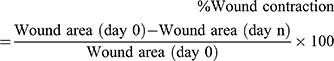
Where, day 0 = day of wound creation, day n = number of days ie, second, fourth, sixth, etc. the day until the wound healing is observed.
Incision Wound Model
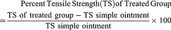
The animals were anesthetized as described in the excision model section and then, a 3 cm length, a linear- paravertebral incision was made through the full thickness of the skin on either side of the vertebral column at a distance of 1 cm from the midline. The incised skin was stitched with black braided silk (no. 00) at a 1cm interval.27,29 After 24 hours of wound creation, the extract ointments were applied and continued daily for 9 days. The suture was removed on the 8th day, and the tensile strength was measured by using a constant water flow method on the 10th day using diethyl ether anesthesia.30 Then, the percent tensile strength was computed as follows:
Infected Wound Model
The mice were anesthetized and the wounds were created in a similar fashion to the excision model. The wounds were inoculated with 100 µL of a standard strain of S. aureus suspension containing approximately 5.0 × 10⁷ Colony-forming units (CFU) and left contaminated over 24 hrs. Then, application of extract ointments commenced on the 2nd day after confirmation of infection. Likewise, wound contraction and epithelialization time were determined. In addition, the presence or absence of the signs and symptoms of established infection such as infiltration, edema, abscess, and exudates were assessed daily throughout the experiment.31,32
Free Radical Scavenging Activity in DPPH Assay
The antioxidant activity of the crude extract was determined by using diphenyl-2- picrylhydrazyl (DPPH) assay based on the methods described by Braca et al.33 Different concentrations of 1 mL of the crude extract ranging from 12.5 to 200µg/mL were added to 3 mL of a 0.004% methanolic solution of DPPH. The absorbance was measured at 517 nm after 30 minutes, and the percent inhibition of DPPH was calculated. The percentage of free radical scavenging potential was determined by the formula: (A0- A1)/A0* 100, where A0 is the absorbance of the control and A1 is the absorbance of the extract/standard.
Data Management, and Analysis
The data were analyzed using Statistical Package for the Social Sciences (SPSS) version 23 and then presented as mean ± standard error of the mean (SEM). To determine variability across the groups, a one-way analysis of variance (ANOVA) followed by Tukey post hoc test was performed. Finally, the results were considered statistically significant at a 95% confidence level in which the p-value was <0.05.
Results
Phytochemical Screening
About 73.6 grams of the crude extract was obtained and the percentage yield value was 36.8%. The preliminary qualitative test revealed the presence of polyphenols, anthraquinones, flavonoids, tannins, glycosides, steroids, and terpenoids in the extract, whereas alkaloids were not detected.
Acute Dermal Toxicity
The application of the 10% (w/w) extract ointment in rats produced no signs of skin toxicity within24 hours. Observation for any skin inflammation and death was continued for 14 days and thus, the safety of the plant for folklore wound healing use seems to be reliable beyond 2000 mg/kg dose.
Excision Model
Wound Contraction and Epithelialization Period
The crude extract ointments promoted wound healing effect substantially from the 6th day to the 16th day. Significant wound contraction effect was observed in mice treated with the 10% (w/w) extract ointment starting from the 6th day (p<0.001), as compared to the negative control group. Besides, a significant contraction effect was also noticed in mice treated with the 5% extract (p<0.05) and the standard drug (p<0.01), as compared to the negative control group. Mice treated with the 10% (w/w) and 5% (w/w) extract ointments showed complete healing effect at the 14th and 16th days, respectively (Table 1). Furthermore, the study revealed that the 10% (w/w) extract ointment produced maximum wound closure at the 10th (93.52%), 12th (98.99) and 14th (100%) post wounding days whereas the 5% (w/w) extract ointment produced maximum wound closures at the 12th (92.07%), 14th (97.68%) and 16th (99.43%) days notifying the dose-dependent effect of the extract ointments. Conversely, maximum wound closure of the standard drug was found to be 92.09% (on 12th day), 98.35% (14th day) and 100% (16th day) (Figure 1).
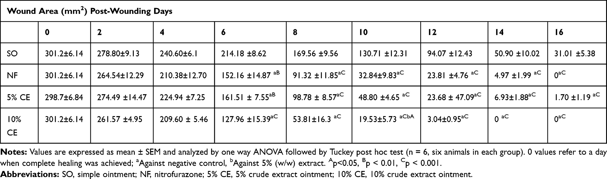 |
Table 1 Wound Healing Activity of 80% Methanolic Crude Extracts Ointments of Leaves of A. schimperi in Terms of Woundarea Contraction in Excision Model |
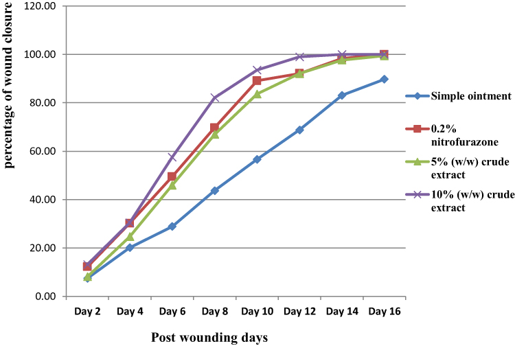 |
Figure 1 Progress of wound contraction of crude extract of leaves of A. schimperi in mice in an excision wound model. |
In addition, a significant reduction (p< 0.001) in the epithelization period was shown in mice treated with both the 10% (w/w) and 5% (w/w) extract ointments, in comparison with their negative controls. Interestingly, the 10% (w/w) extract ointment showed a statistically significant shortening of epithelization periods when compared to the 5% (w/w) extract as well as the standard drug (p<0.05). Moreover, the animals treated with 10% (w/w) extract, nitrofurazone, and 5% (w/w) extract ointments showed a reduction in the epithelialization periods by 34.98%, 25.22%, and 22.78%, respectively when compared to their negative control groups (Table 2).
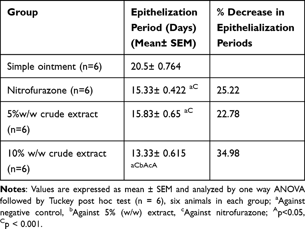 |
Table 2 Wound Healing Activity of 80% Methanolic Crude Extracts Ointments of Leaves of A. schimperi on Period of Epithelialization in Excision Model |
Incision Wound Model
Wound Breaking Strength (Tensile Strength)
The tensile strength was significantly increased (p< 0.001) in mice treated with the 10% (w/w) extract ointment, standard drug, and 5% (w/w) extract ointment by 36.80, 33.67, and 32.23%, respectively, when compared to the negative controls. Moreover, the 10% w/w extract ointment showed the highest increase in tensile strength as compared to the standard drug and the 5% (w/w) extract ointment, though it was not found to be statistically significant (Table 3).
 |
Table 3 Wound Healing Activity of 80% Methanolic Crude Extracts Ointments of Leaves of A. schimperi on Tensile Strength of Incision Model |
Infected Wound Model
Wound Contraction and Epithelialization Period
In the infected wound model, mice treated with negative control remained to exhibit different signs of infection for more than one week. However, mice treated with the test extract ointments started to decrease phlogistic characteristics after 3 days of treatment initiation. Significant wound contraction effect was exhibited among the 10% (w/w) extract ointment treated group starting from day 4 (p< 0.01) and onwards (p< 0.001), in comparison to the negative control group. Conversely, animals treated with the 5% (w/w) extract ointment and the standard drug displayed considerable wound contraction effects at the 4th and 6th days (p< 0.01) and onwards (p< 0.001), as compared to the negative control. However, a statistically significant difference effect was not seen among the treatment and the positive control groups (Table 4). Moreover, the highest wound closure effect was seen for10% extract ointment from the 12th day (97.70%), 14th day (99.65%), and 16th day (100%) (Figure 2). In addition, complete wound healing was recorded at the 16th and18th days for the 10% w/w and 5% w/w extract ointment -treated groups, respectively.
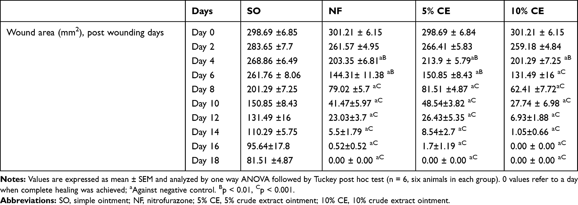 |
Table 4 Wound Healing Activity of 80% Methanolic Crude Extracts Ointments of Leaves of A. schimperi in Terms of Wound Area Contraction in Infected Model |
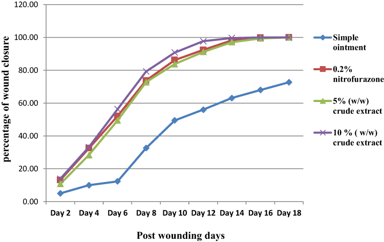 |
Figure 2 Progress of wound contraction of crude extract of leaves of A. schimperi in mice in an infected wound model. |
Furthermore, the 10% extract ointment treated mice displayed rapid healing as evidenced by a faster epithelialization period (p< 0.001), in comparison to the negative control group. The average period of epithelialization was 17.5, 16.3, and 14.3 days for the 5% (w/w), standard drug and 10% (w/w) extract ointments, respectively. However, negative control groups failed to re-epithelize during the course of treatment (Table 5).
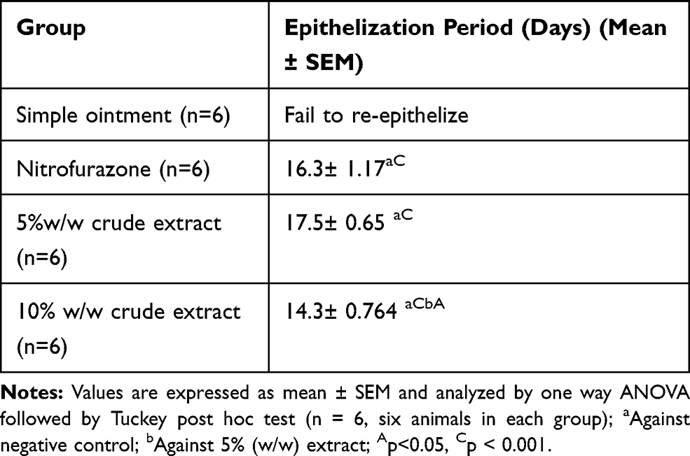 |
Table 5 Wound Healing Activity of 80% Methanolic Crude Extracts Ointments of Leaves of A. schimperi on Period of Epithelialization in Infected Model |
Anti-Oxidant Activity of the Crude Extract
The antioxidant activity of the crude extract of leaves of A. schimperi was concentration-dependent with a higher activity (74.85%) was observed at the 200 µg/mL (Table 6).
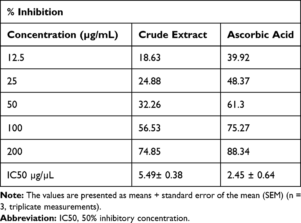 |
Table 6 Anti-Oxidant Activities of the Crude Extract of Leaves of A. schimperi at Various Concentrations in DPPH Assay Model |
Discussion
Traditionally, the fresh or dry leaves of A. schimperi are crushed and tied onto the wound for its wound healing effect. Since the direct application of the extract into the affected part does not have a significant purpose for the intended time, ointment preparation of the extracts is essential to produce a prolonged wound healing effect. Besides, the addition of hard and white soft paraffin during the formulation of ointment base provides an occlusive barrier for moisture whereas wool fat and cetostearyl alcohol (thickeners) are used for the ointment stabilization effect.34,35 This study, therefore, investigated the antioxidant and wound healing activity of A. schimperi leaf extract. Accordingly, the crude extract produced a promising antioxidant activity in a concentration-dependent fashion and the maximum DPPH scavenging activity (74.85%) was seen at 200 µg/mL concentration.
Regarding the wound healing effect, the study revealed that the crude extract ointments of the leaves of A. schimperi promoted wound healing at both doses, in the three models used during the experiment. In the excision model, the 10% (w/w) extract ointment treated groups displayed a faster wound healing effect from day 6 to day 14 when compared to the 5% (w/w) extract ointment treated groups that started from the 8th day and continued to day 16. In addition, a significant reduction of the epithelialization period was shown in both ointment preparations ie 16 days for 5% w/w extract ointment and 13 days for 10% w/w extract ointment. It is noteworthy that the major biological mechanisms behind the wound-healing effects of medicinal plants involve through their antibacterial, antioxidant, anti-inflammatory and mitogenic activities of bioactive constituents.7 For instance, scavenging of excess free radicals and protection of macromolecules like lipids and proteins against oxidative stress is an important biological mechanism for enhanced collagen synthesis and facilitated epithelial cell proliferation, thereby promoting wound healing.36 Therefore, a rapid rate of wound area contraction and a shorter epithelialization period of the extract ointments may be partly explained by its good free scavenging properties as investigated in the present study as well as its antibacterial activity as reported in the previous studies.13,14,37
In the incision model, a significant increase in tensile-strength was revealed in both 10% (by 36.80%) and 5% (by 32.23%) w/w extract ointments, as compared to their negative controls. The increase in tensile strength is associated with an increase in collagen production and maturation, matrix deposition, and cell migration, which are attributable to the potent antioxidant, antibacterial, and anti-inflammatory activities of the bioactive constituents as explained in various studies.32,38,39 This finding is further supported by studies done on other Acokanthera species. As evidence, the leaf extract of Acokanthera oppositifolia showed good anti-inflammatory, antioxidant, and analgesic activities.40,41 Likewise, the leaf extract of Acokanthera oblongifolia also displayed strong antimicrobial, antioxidant, and wound healing activities.42
Moreover, the crude extract ointments of this plant revealed a statistically significant wound healing effect in S. aurous-infected mice, in an infected model. A faster wound contraction and epithelialization effects were noticed in mice treated with the 10% w/w extract ointment than the standard drug and 5% w/w extract ointment-treated mice. Likewise, the phlogistic characteristics of the wound that occurred before the initiation of treatment progressively and significantly reduced in all mice treated with the extract ointments in comparison with the negative-control treated groups. The antibacterial properties of triterpenoids isolated from the leaves of A. schimperi could support this finding.43
Furthermore, secondary metabolites like polyphenols, anthraquinones, flavonoids, tannins, glycosides, steroids, and terpenoids found in the extract could contribute to the wound healing effect. For example, terpenoids facilitate wound healing primarily due to their antimicrobial, antioxidant and anti-inflammatory properties;44,45 flavonoids, and tannins are potent antioxidants;46 polyphenols and flavonoids have anti-inflammatory and antimicrobial activities;47 glycosides have antioxidant, antimicrobial, analgesic, antitumor, and anti-inflammatory effects.48 Hence, the observed wound healing and antioxidant effects of the crude extract may be due to the individual or synergistic effects of active phytochemicals present in the plant. In general, faster rates of wound contraction and epithelialization, and increased tensile strength of the extract ointments were suggestive to support the folklore wound healing claims of the plant and consequently, the study recommends further isolation and characterization, structural elucidation and the study of the structure-activity relationship of the active constituents responsible for this activity.
Conclusion
The present study investigated that the 80% hydromethanolic extract ointments of leaves of A. schimperi possess potential wound healing effects in all three models. The study also suggested that the significant antibacterial and anti-oxidant activities of the extracts are the mechanisms for its wound-healing effects. Thus, the current study strongly supported the traditional claims of the plant for use in the treatment of wounds.
Abbreviations
ANOVA, one way analysis of variance; OECD, Organization for Economic Cooperation and Development; SEM, standard error of the mean; SPSS, Statistical Package for Social Sciences.
Data Sharing Statement
All the data generated or analyzed during this study are included in the manuscript.
Ethics Approval and Consent to Participate
The study was reviewed, presented to and approved by Ethical Review Committee of College of Medicine and Health Sciences, Wollo University with ethical approval number WU CMHS/701/19. All the experiments were conducted in accordance with a guide for the care and use of laboratory animals: Eighth Edition by National Research Council. At the end of the experiment, the animals were killed by Phenobarbital sodium at a dose of 150 mg/kg (euthanasia, mercy killing).
Acknowledgment
The authors would like to thank Wollo University for the financial support extended to complete the research work.
Author Contributions
All authors made a significant contribution to the work reported, whether that is in the conception, study design, execution, acquisition of data, analysis and interpretation, or in all these areas; took part in drafting, revising or critically reviewing the article; gave final approval of the version to be published; have agreed on the journal to which the article has been submitted; and agree to be accountable for all aspects of the work.
Funding
Wollo University covered the costs of chemicals, reagents and equipments used.
Disclosure
The authors declare that they have no competing interests in this work.
References
1. Cutting KF, Tong A. Wound Physiology & Moist Wound Healing: Medical Communications UK. 2003.
2. Bowler PG. Wound pathophysiology, infection and therapeutic options. Ann Med. 2002;34(6):419–427.
3. Zafar A, Anwar N, Ejaz H. Bacteriology of infected wounds a study conducted at children hospital Lahore. Biomedica. 2007;23(8):1–4.
4. Menke NB, Ward KR, Witten TM, Bonchev DG, Diegelmann RF. Impaired wound healing. Clin Dermatol. 2007;25(1):19–25.
5. Mohanty A, Sahu P, Das C. Wound healing activities of methanolic extract of Cissus quadrangularis on albino rat. Int J Drug Formul Res. 2010;1:176–184.
6. Kokane DD, More RY, Kale MB, Nehete MN, Mehendale PC, Gadgoli CH. Evaluation of wound healing activity of root of Mimosa pudica. J Ethnopharmacol. 2009;124(2):311–315.
7. Soni H, Singhai AK. A recent update of botanicals for wound healing activity. Int Res J Pharm. 2012;3(7):1–7.
8. Ghosh PK, Gaba A. Phyto-extracts in wound healing. Int J Pharm Pharm Sci. 2013;16(5):760–820.
9. Teklehaymanot T, Giday M. Ethnobotanical study of medicinal plants used by people in Zegie Peninsula, Northwestern Ethiopia. J Ethnobiol Ethnomed. 2007;3(1):12.
10. Bitew H, Gebregergs H, Tuem KB, Yeshak MY. Ethiopian medicinal plants traditionally used for wound treatment: a systematic review. Ethiop J Health Dev. 2019;33(2).
11. Abebe D, Ayehu A. Medicinal Plants and Enigmatic Health Practices of Northern Ethiopia. 1993.
12. Omino E, Kokwaro J. Ethnobotany of apocynaceae species in Kenya. J Ethnopharmacol. 1993;40(3):167–180.
13. Taye B, Giday M, Animut A, Seid J. Antibacterial activities of selected medicinal plants in traditional treatment of human wounds in Ethiopia. Asian Pac J Trop Biomed. 2011;1(5):370–375.
14. Tadeg H, Mohammed E, Asres K, Gebre-Mariam T. Antimicrobial activities of some selected traditional Ethiopian medicinal plants used in the treatment of skin disorders. J Ethnopharmacol. 2005;100(1–2):168–175.
15. Chaithanya K, Abrha B, Gopalakrishnan V, Hagos Z, Hiruy M, Devaki K. Phytochemical screening and in vitro antioxidant activities of ethanolic extract of Acokanthera schimperi leaves. J Pharm Res. 2018.
16. Gebre-Mariam T, Neubert R, Schmidt P, Wutzler P, Schmidtke M. Antiviral activities of some Ethiopian medicinal plants used for the treatment of dermatological disorders. J Ethnopharmacol. 2006;104(1–2):182–187.
17. Mohammed T, Erko B, Giday M. Evaluation of antimalarial activity of leaves of acokanthera schimperi and croton macrostachyus against plasmodium berghei in swiss albino mice. BMC Complement Altern Med. 2014;14(1):314.
18. National Research Council. Guide for the Care and Use of Laboratory Animals. National Academies Press; 2010.
19. Festing MF, Altman DG. Guidelines for the design and statistical analysis of experiments using laboratory animals. ILAR J. 2002;43(4):244–258.
20. Karapolat S, Karapolat B, Buran A, et al. The effects of nitrofurazone on wound healing in thoracoabdominal full-thickness skin defects. Wounds. 2020;32(5):134–141.
21. British Pharmacopoeia. Department of Health and Social Security Scottish Home and Health Department. Vol. 2. UK: Office of the British Pharmacopoeia Commission; 1988:713.
22. Trease G, Evans M. Text Book of Pharmacognosy.
23. Debella A. Manual for Phytochemical Screening of Medicinal Plants, Department of Drug Research, Ethiopian Nutrition and Research Institute. Addis Ababa, Ethiopia; 2002:45–71.
24. Amcoff P. Appendix 5: OECD guidelines for the testing of chemicals. Altern Lab Anim. 2005;33(1_suppl):223–228.
25. Wilhelm K-P, Maibach HI. 33 OECD guidelines for testing of chemicals. Dermatotoxicology. 2007;303.
26. Morton J, Malone M. Evaluation of vulneray activity by an open wound procedure in rats. Arch Int Pharmacodyn Ther. 1972;196(1):117.
27. Mulisa E, Asres K, Engidawork E. Evaluation of wound healing and anti-inflammatory activity of the rhizomes of rumex abyssinicus J. (polygonaceae) in mice. BMC Complement Altern Med. 2015;15(1):341.
28. Yesuf A, Asres K. Wound healing and antiinflammatory properties of Allophylus abyssinicus (Hochst.) Radlk. Phytopharmacology. 2013;4(2):442–453.
29. Ehrich H. Effect of cortisone and anabolic steroids on tensile strength of healing wound. Ann Surg. 1969;170(2):203–206.
30. Lee K. Studies on the mechanism of action of salicylates III. Effect of vitamin A on the wound healing retardation action of aspirin. J Pharm Sci. 1968;57(7):1238–1240.
31. Demilew W, Adinew GM, Asrade S. Evaluation of the wound healing activity of the crude extract of leaves of acanthus polystachyus delile (acanthaceae). Evid Based Complement Alternat Med. 2018;2018.
32. Bastos MLA, Houly RLS, Conserva LM, Andrade VS, Rocha EMM, Lyra Lemos R. Antimicrobial and wound healing activities of Piper hayneanum. J Chem Pharm Res. 2011;3:213–222.
33. Braca A, De Tommasi N, Di Bari L, Pizza C, Politi M, Morelli I. Antioxidant principles from bauhinia t arapotensis. J Nat Prod. 2001;64(7):892–895.
34. Ansel HC. Introduction to Pharmaceutical Dosage Forms. 1969.
35. Manjunatha B, Vidya S, Rashmi K, Mankani K, Shilpa H, Singh SJ. Evaluation of wound-healing potency of Vernonia arborea Hk. Indian J Pharmacol. 2005;37(4):223.
36. Arunachalam KD, Subhashini S, Annamalai S. Wound healing and antigenotoxic activities of Aegle marmelos with relation to its antioxidant properties. J Pharm Res. 2012;5(3):1492–1502.
37. Le N T, Ho D V, Quoc Doan T, et al. Biological activities of essential oils from leaves of Paramignya trimera (Oliv.) Guillaum and Limnocitrus littoralis (Miq.) swingle. Antibiotics. 2020;9(4):207.
38. Donadu MG, Trong Le N, Viet Ho D, et al. Phytochemical compositions and biological activities of essential oils from the leaves, rhizomes and whole plant of Hornstedtia bella Škorničk. Antibiotics. 2020;9(6):334.
39. Getie M, Gebre-Mariam T, Rietz R, et al. Evaluation of the anti-microbial and anti-inflammatory activities of the medicinal plants Dodonaea viscosa, Rumex nervosus and Rumex abyssinicus. Fitoterapia. 2003;74(1–2):139–143.
40. Ondua M, Adebayo S, Shai L, Lebelo S. The Anti-Inflammatory and Anti-Nociceptive Activities of Some Medicinal Plant Species Used to Treat Inflammatory Pain Conditions in Southern Africa. 2016.
41. Adebayo S, Ondua M, Shai L, Lebelo S. Inhibition of nitric oxide production and free radical scavenging activities of four South African medicinal plants. J Inflamm Res. 2019;12:195.
42. Otang-Mbeng W, Afolayan AJ. Antimicrobial and antioxidant efficacy of Acokanthera oblongifolia Hochst (apocynaceae). Int J Pharmacol. 2017;13(8):1086–1091.
43. Matebie WA, Zhang W, Zhang S, Xie G. Triterpenoids from Acokanthera schimperi in Ethiopia. Rec Nat Prod. 2019;13(3):182–188.
44. Bodenstein J, Du Toit K. The susceptibility of Staphylococcus aureus and Klebsiella pneumoniae to naturally derived selected classes of flavonoids. Antimicrob Agents. 2012;73–84.
45. Petr D, Marian H, David V, et al. Anti-inflammatory and antitumor-promoting effects of the triterpene acids from the leaves of Eriobotrya japonica. Nat Prod Rep. 2006;23:394–411.
46. Getie M, Gebre-Mariam T, Rietz R, Neubert R. Evaluation of the release profiles of flavonoids from topical formulations of the crude extract of the leaves of Dodonea viscosa (Sapindaceae). Pharmazie. 2002;57(5):320–322.
47. Armstrong DG, Jude EB. The role of matrix metalloproteinases in wound healing. J Am Podiatr Med Assoc. 2002;92(1):12–18.
48. Ling S-K, Tanaka T, Kouno I. Effects of iridoids on lipoxygenase and hyaluronidase activities and their activation by β-glucosidase in the presence of amino acids. Biol Pharm Bull. 2003;26(3):352–356.
 © 2020 The Author(s). This work is published and licensed by Dove Medical Press Limited. The full terms of this license are available at https://www.dovepress.com/terms.php and incorporate the Creative Commons Attribution - Non Commercial (unported, v3.0) License.
By accessing the work you hereby accept the Terms. Non-commercial uses of the work are permitted without any further permission from Dove Medical Press Limited, provided the work is properly attributed. For permission for commercial use of this work, please see paragraphs 4.2 and 5 of our Terms.
© 2020 The Author(s). This work is published and licensed by Dove Medical Press Limited. The full terms of this license are available at https://www.dovepress.com/terms.php and incorporate the Creative Commons Attribution - Non Commercial (unported, v3.0) License.
By accessing the work you hereby accept the Terms. Non-commercial uses of the work are permitted without any further permission from Dove Medical Press Limited, provided the work is properly attributed. For permission for commercial use of this work, please see paragraphs 4.2 and 5 of our Terms.