")
Back to Journals » International Medical Case Reports Journal » Volume 12
Transient Inverse Bell’s Phenomenon Following Frontalis Sling–Suspension Ptosis Surgery: A Rare Ophthalmic Phenomenon
Authors Pandey TR , Limbu B, Rajkarnikar Sthapit P, Gurung HB , Saiju R
Received 23 May 2019
Accepted for publication 23 September 2019
Published 30 October 2019 Volume 2019:12 Pages 325—327
DOI https://doi.org/10.2147/IMCRJ.S216805
Checked for plagiarism Yes
Review by Single anonymous peer review
Peer reviewer comments 4
Editor who approved publication: Dr Scott Fraser
Triptesh Raj Pandey,1 Ben Limbu,2 Purnima Rajkarnikar Sthapit,2 Hom Bahadur Gurung,2 Rohit Saiju2
1Mechi Netralaya & Ophthalmic Research Center (P) Ltd., Jhapa, Nepal; 2Tilganga Institute of Ophthalmology, Kathmandu, Nepal
Correspondence: Triptesh Raj Pandey
Mechi Netralaya & Ophthalmic Research Center (P) Ltd., Jhapa, Nepal
Tel +977 984 175 2811
Email [email protected]
Introduction: Inverse Bell’s phenomenon is a rare ophthalmic phenomenon where downward instead of upward movement of the eyeball occurs during eyelid closure. It may be associated with peripheral facial nerve palsy, conjunctival scarring, and ptosis surgery.
Case report: A 9-year-old male patient with right upper–lid congenital ptosis developed inverse Bell’s phenomenon 2 days after frontalis sling–suspension ptosis surgery. At the 3-week postoperative visit, there had been spontaneous resolution of the inversion of Bell’s phenomenon without any corneal complication.
Conclusion: Inverse Bell’s phenomenon, more often reported to be associated with levator-resection surgery, may develop following frontalis sling–suspension ptosis surgery. Close monitoring and frequent instillation of topical lubricants are necessary to prevent exposure keratopathy until the resolution of inverse Bell’s phenomenon in patients with lagophthalmos after ptosis surgery.
Keywords: frontalis sling, inverse Bell’s phenomenon, keratopathy, levator resection, ptosis
Introduction
Bell’s phenomenon is a protective mechanism and an essential preoperative assessment in every case of ptosis surgery. Normal Bell’s phenomenon consists of an upward and outward movement of the eyes associated with eyelid closure. However, in inverse Bell’s phenomenon, downward movement of the eyes occurs during closure of the eyelids and is an uncommon phenomenon.1 There have been many case reports of inverse Bell’s phenomenon following levator-resection surgery, but few following frontalis sling–suspension ptosis surgery.2–5 The authors herein report a very rare inverse Bell’s phenomenon case following frontalis sling–suspension ptosis surgery. The patient’s parents provided written informed consent to have the case details and accompanying images published. The present case report was approved by the Institutional Review Committee of Tilganga Institute of Ophthalmology.
Case Report
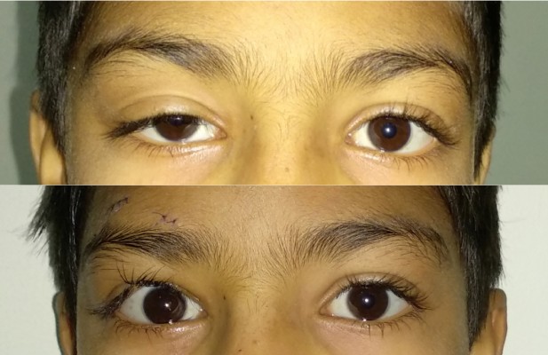
A 9-year old male presented with a history of drooping of the right upper lid since childhood with visual acuity of 6/6 (Snellen chart) in both eyes (Figure 1, top). Levator function was 4 mm and 15 mm in the right and left eye, respectively. The marginal reflex distance 1 of the upper lid from corneal light reflex was 1 mm for the right eye and 5 mm for the left. Extraocular movements were normal and Bell’s phenomenon was normal (upward and outward). There was no evidence of Marcus Gunn phenomenon, and corneal sensations were normal. No other ocular abnormalities were noted on anterior- or posterior-segment evaluation. He underwent right-eye frontalis sling–suspension ptosis surgery (Fox pentagon technique) using a silicone sling under general anesthesia. Frost suture was applied. No intraoperative complications were noted.
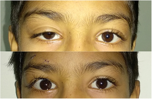 |
Figure 1 Preoperative photograph (top) and postoperative (day 2) photograph after frontalis sling–suspension surgery (bottom). |
Frost suture was removed on the second postoperative day. There was overcorrection of the ptosis by 1 mm in primary gaze with mild lid edema and prominent lagophthalmos (Figure 1, bottom). Inverse Bell’s phenomenon (downward rolling of the eyeball) was noted during eyelid closure (Figure 2). There was no restriction of eye movement on any gaze. The patient was advised to instill lubricating eyedrops frequently and apply eye ointment to prevent exposure keratopathy. At the 3-week postoperative visit, vertical palpebral fissure height was 11 mm in both eyes, with minimal lagophthalmos in the right eye (Figure 3, top). There was spontaneous resolution of inverse Bell’s phenomenon. It had became normal (upward and outward), without any evidence of exposure keratopathy (Figure 3, bottom).
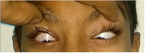 |
Figure 2 Postoperative (day 2) photograph showing inversion of Bell’s phenomenon in both eyes following right frontalis sling–suspension surgery. |
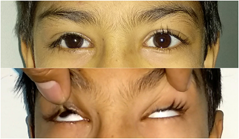 |
Figure 3 Postoperative (day 22) photograph of the patient (top) and showing reversal to normal Bell’s phenomenon in both eyes (bottom). |
Discussion
Inverse Bell’s phenomenon, described as downward movement of eyes during closure of the eyelids, is rather uncommon, but may be present in 2% of otherwise-normal cases.1 However, it may occur in peripheral facial nerve palsies, tabes dorsalis, lid swelling, conjunctival scarring, and following ptosis surgery.1 It has been often been observed following levator-resection surgery.2–4
Possible explanations for inverse Bell’s phenomenon are intraoperative injury to the oculomotor nerve in the superior rectus muscle, leading to alteration of trigemino-oculomotor projection and severe edema and hyperemia of the superior fornix, altering the relationship between eyelid, superior rectus movement, and normal Bell’s phenomenon.3 However, inverse Bell’s phenomenon may be affected by edema of soft tissue, rather than aberrant connections of the nervous system.3
Such extensive intraoperative tissue manipulation and nerve injury is uncommon in frontalis-sling surgery compared to levator-resection surgery. Nevertheless, direct and indirect trauma to tissue may occur during silicone-sling guidance and retrieval, causing edema and hemorrhage in muscular and subcutaneous planes and leading to inversion of Bell’s phenomenon.
The likelihood of exposure keratopathy increases following inversion of Bell’s phenomenon, as the lower lid may not provide adequate coverage to the globe in the presence of postoperative lagophthalmos. Also, there are alterations in tear film and the ocular surface after frontalis sling–suspension surgery.6 In such situations, postoperative lubricants and ointment application become of utmost importance to prevent exposure keratitis. As reported in other cases, there was spontaneous resolution of inverse Bell’s phenomenon in this case 3 weeks after surgery without any complications, probably coinciding with the resolution of tissue edema and inflammation.2–5
Conclusion
Preoperative and postoperative evaluation of Bell’s phenomenon is essential in all ptosis surgeries. Though described more often in association with levator-resection surgery, inverse Bell’s phenomenon may develop following frontalis sling–suspension ptosis surgery. Close monitoring and frequent instillation of topical lubricants are necessary to prevent exposure keratopathy until resolution of inverse Bell’s phenomenon.
Disclosure
The authors report no conflicts of interest in this work.
References
1. Gupta JS, Chatterjee A, Kumar K. Inverse Bell’s phenomenon as a protective mechanism. Am J Ophthalmol. 1965;59:931–933. doi:10.1016/0002-9394(65)93035-7
2. Betharia SM, Sharma V. Inverse Bell’s phenomenon observed following levator resection for blepharoptosis. Graefes Arch Clin Exp Ophthalmol. 2006;244(7):868–870. doi:10.1007/s00417-005-0122-4
3. Na KS, Yang SW. Two cases of inverse Bell’s phenomenon following levator resection: a contemplation of the mechanism. Eur J Ophthalmol. 2009;19(2):285–287. doi:10.1177/112067210901900218
4. Shitole S, Jakkal T, Khaire B. Inverse Bell’s phenomenon: rare ophthalmic finding following ptosis surgery. J Clin Diagn Res. 2015;9(3):ND01–2. doi:10.7860/JCDR/2015/11340.5686
5. Dhivya AK, Agarwal A.Transient inverse bells Phenomenon following frontalis sling surgery. Int J Ocul Oncol Oculoplasty. 2016;2(4):258–259. October-December.
6. Yoon JS, Lew H, Lee SY. Bell’s phenomenon protects the tear film and ocular surface after frontalis suspension surgery for congenital ptosis. J Pediatr Ophthalmol Strabismus. 2008;45(6):350–355.
 © 2019 The Author(s). This work is published and licensed by Dove Medical Press Limited. The full terms of this license are available at https://www.dovepress.com/terms.php and incorporate the Creative Commons Attribution - Non Commercial (unported, v3.0) License.
By accessing the work you hereby accept the Terms. Non-commercial uses of the work are permitted without any further permission from Dove Medical Press Limited, provided the work is properly attributed. For permission for commercial use of this work, please see paragraphs 4.2 and 5 of our Terms.
© 2019 The Author(s). This work is published and licensed by Dove Medical Press Limited. The full terms of this license are available at https://www.dovepress.com/terms.php and incorporate the Creative Commons Attribution - Non Commercial (unported, v3.0) License.
By accessing the work you hereby accept the Terms. Non-commercial uses of the work are permitted without any further permission from Dove Medical Press Limited, provided the work is properly attributed. For permission for commercial use of this work, please see paragraphs 4.2 and 5 of our Terms.