")
Back to Journals » Cancer Management and Research » Volume 11
The Significance of Adjuvant Therapy for Extrahepatic Cholangiocarcinoma After Surgery
Authors Wang G, Wang Q, Fan X, Ding L, Dong L
Received 24 July 2019
Accepted for publication 3 December 2019
Published 30 December 2019 Volume 2019:11 Pages 10871—10882
DOI https://doi.org/10.2147/CMAR.S224583
Checked for plagiarism Yes
Review by Single anonymous peer review
Peer reviewer comments 2
Editor who approved publication: Dr Sanjeev K. Srivastava
Gaoyuan Wang, Qiang Wang, Xia Fan, Lijuan Ding,* Lihua Dong*
Department of Radiation Oncology, The First Hospital of Jilin University, Changchun, People’s Republic of China
*These authors contributed equally to this work
Correspondence: Lijuan Ding
Department of Radiation Oncology, The First Hospital of Jilin University, Changchun, People’s Republic of China
Tel +8615843073216
Email [email protected]
Abstract: Extrahepatic cholangiocarcinoma (EHCC) is a rare malignant tumor, and current treatment methods are also relatively limited. Radical surgery is the only potentially curative method for the long survival time. However, despite undergoing radical resection, prognosis remained poor due to the high recurrence rate and distant metastasis. Therefore, adjuvant chemotherapy and radiotherapy should be offered to patients who have undergone surgery. Unfortunately, the low incidence of this disease has resulted in a lack of high-level evidence to confirm the importance of adjuvant chemotherapy or radiotherapy. At present, it is still controversial whether adjuvant therapy can prolong the survival of patients after operation, especially patients with negative margins or lymph nodes. Furthermore, standard regimens of adjuvant have not been identified. This review summarizes the currently available evidence of the effect of adjuvant therapy in the management of EHCC. Ultimately, we concluded that adjuvant therapy may improve survival in high-risk (positive margin or lymph node or advanced stage) patients and adjuvant concurrent chemoradiotherapy followed by chemotherapy may be the optimum selection for them. This needs to be verified by randomized prospective clinical trials.
Keywords: extrahepatic cholangiocarcinoma, adjuvant chemotherapy, adjuvant radiotherapy, adjuvant chemoradiotherapy
Introduction
Cholangiocarcinoma (CC) is a rare malignancy which arises from the epithelium of bile ducts, and it has a tendency to extend along the biliary tract.1 It accounts for about 3% of gastroenteric tumors and is the second most common primary hepatic neoplasm.2,3 CC is generally categorized as either intrahepatic or extrahepatic based on the anatomic location with respect to the second-order bile ducts. This article mainly discusses the effect of postoperative treatment in EHCC which is further divided into hilar cholangiocarcinoma (HCC) (Klastskin tumors) and distal cholangiocarcinoma (DCC).4 The HCC which arises from malignant proliferation of epithelium of bile ducts is the most common type of CC (50–60%). It is located between the second-degree bile ducts and insertion of cystic duct into the common bile duct.5,6 DCC is defined as bile duct tumors arising between the cystic duct and the ampulla of Vater (except Klastskin tumors and ampulla of Vater cancer).7 Radical resection with negative surgical margins is the most effective and only potential curative treatment.8 However, only approximately 20% of biliary tract tumor could be considered resectable, and the prognosis of this disease is even poor.9 Furthermore, only 5–10% of patients would be alive 5 years after surgery.2 Despite undergoing a complete resection, the reported 5-year survival rate of patients with HCC and DCC is in the range of 20% to 42% and 16% to 52%, respectively.10,11 This may be attributable to the high rate of recurrence (60–75%).12 Due to these poor outcomes, adjuvant therapy (AT), including chemotherapy, radiotherapy and chemoradiotherapy, should be considered for postoperative patients. To our knowledge, the role of adjuvant chemotherapy or radiotherapy after resection of EHCC is just uncertain for the lack of high-level evidence. The current data are mainly based on several retrospective and prospective studies with certain limitations. The purpose of this review is to summarize the effect of adjuvant chemotherapy and radiation therapy in EHCC, and to seek the best postoperative treatment regulations. Furthermore, we try to point out possible future research directions. Some studies of AT in EHCC and details about ongoing clinical trials can be seen in Tables 1 and 2, respectively.
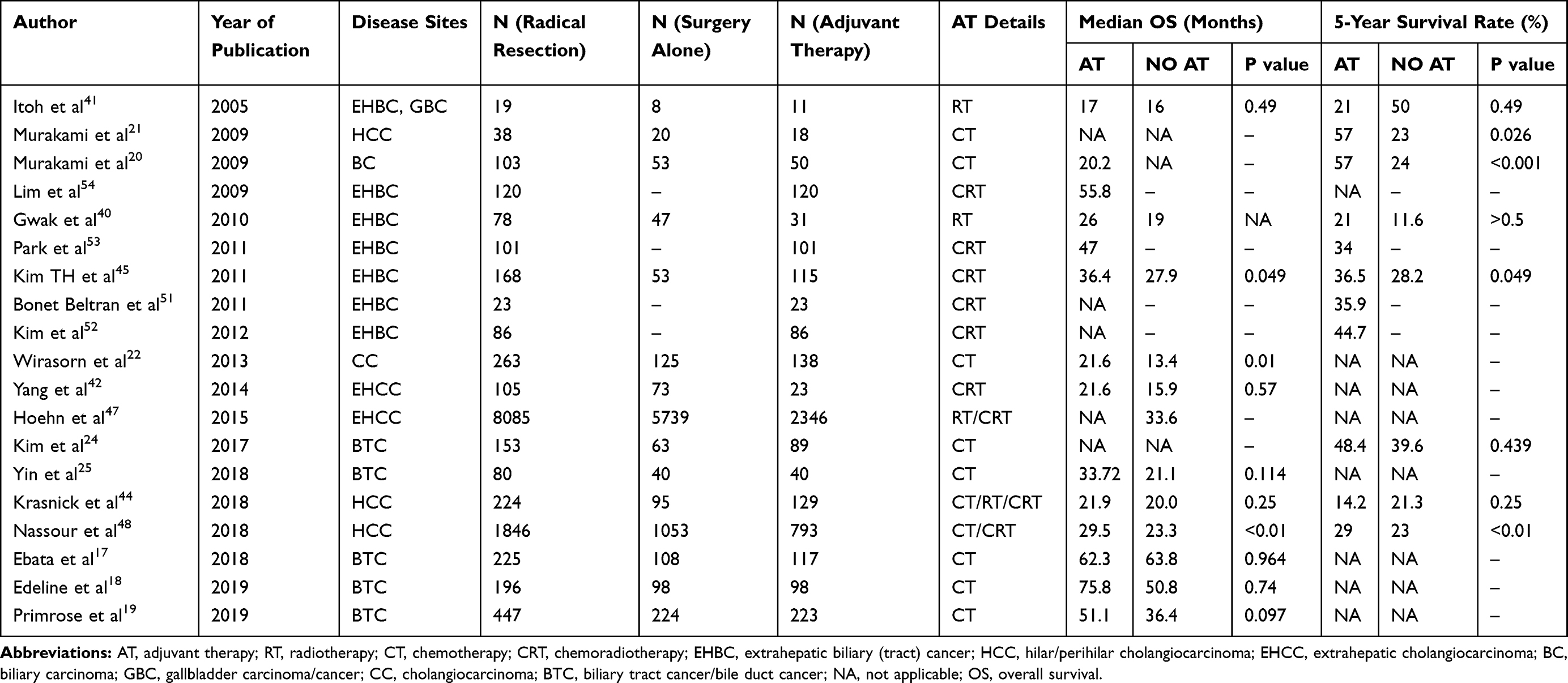 |
Table 1 Results from Studies of Adjuvant Therapy in Biliary Tract Cancer |
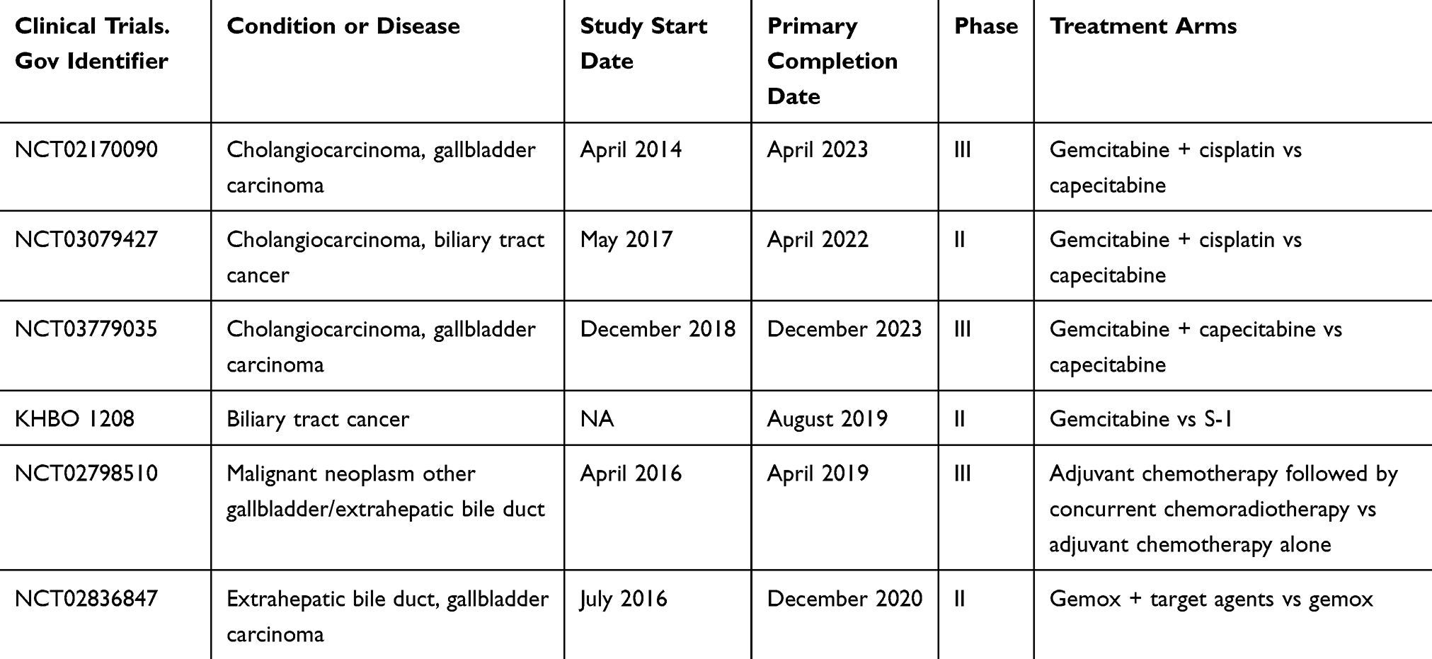 |
Table 2 Ongoing Studies of Adjuvant Therapy in Biliary Tract Cancer |
Adjuvant Chemotherapy
EHCC is intrinsically a chemo-resistant malignancy.13 The purpose of adjuvant chemotherapy is to improve the poor prognosis of this disease after radical surgery. According to National Comprehensive Cancer Network (NCCN) guidelines, the chemotherapy protocol mainly includes fluoropyrimidine-based or gemcitabine-based chemotherapy, but they are based on the extrapolation of data from studies of patients with advanced disease.14 There are a few data evaluating the role of adjuvant chemotherapy in EHCC,15,16 and only three prospective studies could be found so far.17–19
A retrospective study in a Japanese center was published in 2009.20 The study included 103 patients with HCC, DCC, ampullary carcinoma and gallbladder cancer (GBC) who underwent aggressive surgical resection. Of these, 50 patients received 10 cycles of adjuvant gemcitabine plus s-1 chemotherapy and the other 53 patients had postoperative observation only. In the report, the patients who received adjuvant chemotherapy showed a better 5-year survival rate than the observation group (57% vs 24%, P<0.001). And the toxicity during chemotherapy was controllable.
Similarly, another Japanese study published in 2009 concluded that postoperative adjuvant gemcitabine-based chemotherapy may improve survival after surgical resection for HCC.21 This study appraised 38 eligible patients with HCC who completed surgical resection. In this study, 18 patients received postoperative adjuvant chemotherapy (gemcitabine or gemcitabine plus S-1), while the other 20 patients were treated with surgery alone. The 5-year survival rates of these two groups of patients were 57% and 23%, respectively (P=0.026).
A third retrospective study, published on the use of adjuvant chemotherapy by Wirasorn et al, was a cohort from Thailand.22 The study included 263 postoperative CC patients with a clear margin (R0) or microscopic margin (R1). Of these, 138 patients underwent gemcitabine-based or 5-fluorouracil (FU)-based adjuvant chemotherapy, and the other 125 patients were treated with surgery alone. The result showed that patients who received postoperative chemotherapy had significantly longer median overall survival (OS) (21.6 vs 13.4 months, P=0.01). And the report indicated that the combination of gemcitabine and capecitabine made the longest survival time (median OS time of gemcitabine and capecitabine 31.5 months; 5-FU and mitomycin C 17.3 months; 5-FU alone 22.2 months; capecitabine alone 21.6 months; gemcitabine alone 7.9 months, respectively, P=0.02). But, the baseline characteristics between the two groups may be an imbalance. The group that received adjuvant chemotherapy was younger than the surgery alone group (mean age 57.7 vs 60.4 years, P=0.01), and fewer had serum albumin<3g/dl (11.6% vs 20.8%, P=0.04). Furthermore, this study concluded that the patients who had high-risk features, including high level of CA19-9, T4 stage, lymph node involvement, and R1 margin, were more likely to get benefits from adjuvant chemotherapy.
In contrast, a retrospective study authored by Bergeat et al from a French center in 2018 did not draw the same conclusion23 The study included 178 patients with DCC who underwent pancreaticoduodenectomy, of whom 56 patients were treated with adjuvant chemotherapy (mostly based on gemcitabine regimen). After propensity score (PS) matching, the median time of OS and disease-free survival (DFS) in the adjuvant chemotherapy group and surgery alone group were comparable (26.27 months vs 43.33 months, P=0.34; and 15.47 vs 14.70 months, P=0.79, respectively). In the entire cohort, there was no evidence of improvement of the OS of those treated with adjuvant chemotherapy. On univariate analysis, adjacent organ resection (P<0.01), perineural infiltration (P=0.01), microvascular invasion (P=0.01), tumor size >2cm (P=0.04), positive margin resection (P=0.01), AJCC classification ≥II (P=0.01), and number of involved nodes ≥4 (P=0.07) were associated with a poor OS. The multivariate analysis indicated that adjacent organ resection (HR=4.17, 95% CI 1.87–9.29, P<0.01) and AJCC classification ≥ II (HR=3.34, 95% CI 1.50–7.43, P < 0.01) was independent risk factors for OS.
Another retrospective study from South Korea included a series of 153 patients with gallbladder, intrahepatic, hilar, and distal bile ducts cancer between March 1999 and December 2013.24 All of them were treated with microscopically margin negative (R0) resection. Compared with the observation group, the cohort of patients (N=89) that received fluoropyrimidine-based adjuvant chemotherapy showed no significant differences in 5-year OS rates (48.4% vs 39.6%, P=0.439) or 3-year recurrence-free survival (RFS) rates (49.1% vs 39.5%, P=0.299). In the subgroup analysis, the patients with stage I and IV disease who received adjuvant chemotherapy did not show any survival benefit compared with observation group, whereas the patients with stage II and III disease who received chemotherapy had a higher 5-year OS rate (52.4% vs 35.6%, P=0.002) and 3-year RFS (55.5% vs 39.1%, P=0.021) than observation group.
Recently, a retrospective cohort study from a Chinese center evaluated the efficacy of adjuvant chemotherapy and discussed the regimen choice in biliary tract cancer. This report analyzed 80 patients after curative-intent R0 resection from 2008 to 2016.25 Among them, 40 patients received adjuvant chemotherapy and the others in the observation group were 1:1 matched by clinical characteristics including gender, age, tumor stage, and ECOG performance status score. Mean survival time showed no significant difference between AT group and the observation group (33.72±5.02 months vs 21.05±4.12 months, respectively, P=0.114). But the mean DFS time of patients with chemotherapy was longer than the observation group (18.63±3.63 months vs 10.36±1.67 months, respectively, P=0.029). On multivariate analysis, adjuvant chemotherapy and N factor were significant factors for OS. The chemotherapy protocol included gemcitabine or S-1 or capecitabine only or combination. Subgroup analysis illustrated that the combination of chemotherapy regimens containing oral drugs prolonged DFS and improved OS of patients.
In the last 2 years, three randomized clinical trials were published. The first of them is BCAT trial. In this study, 225 patients were divided into gemcitabine group and observation group with well-balanced baseline characteristics.17 Both median OS (HR=1.01, 95% CI 0.70–1.45, P=0.964) and relapse-free survival (HR=0.93, 95% CI 0.66–1.32, P=0.693) were not significantly different. Another randomized Phase III study (PRODIGE 12-ACCORD) compared the effect of gemcitabine and oxaliplatin (Gemox) chemotherapy with observation group.18 Similarly, there were no benefits of adjuvant Gemox in OS (HR=1.08, 95% CI 0.70–1.66, P=0.74) and RFS (HR=0.88, 95% CI 0.62–1.25, P=0.48). In the BILCAP study, which is from England in 2019, 447 patients with completely excisional CC or GBC were randomized to receive either adjuvant capecitabine or observation.19 In the intent-to-treat analysis, the difference between the study arms for median OS was not statistically significant (51.1 months vs 36.4 months, respectively, HR=0.81, 95% CI 0.63–1.04, P=0.097). In contrast, the difference was statistically significant in the per-protocol analysis (HR=0.75, 95% CI, 0.58–0.97, P=0.028) and protocol-specified sensitivity analysis (HR=0.71, 95% CI, 0.55–0.92, P=0.010).
From the above discussion, there are limited clinical trial data to define a standard regimen or definitive benefit. The present studies mostly concentrate on gemcitabine or 5-FU or capecitabine or S-1 alone or combination. Adjuvant chemotherapy may be advantageous to patients with positive resection margin or advanced stage tumor, when combination chemotherapy regimens, which contain oral medication, are used. Some other protocols, such as gemcitabine/cisplatin, gemcitabine/oxaliplatin (Gemox), gemcitabine/albumin-bound paclitaxel and fluoropyrimidine/oxaliplatin or cisplatin, were supported in some clinical trials for patients with advanced biliary tract cancers.26–31 Besides, capecitabine should be considered as first-line standard of adjuvant chemotherapy. As a result, it is essential to conduct more clinical trials to find out which is the best adjuvant chemotherapy regimens for postoperative patients.
Adjuvant Radiotherapy
As mentioned above, complete resection of EHCC is the only potentially curative treatment. Nevertheless, the excision rate of the disease is low, with a range from 10% to 47%.32 Even though the patients have been treated with radical surgery, the local recurrence rate is very high, and many cases of close resection margins may remain.33,34 A study reviewed a series of 189 patients who underwent resection for primary CC but did not receive radiation therapy between 1999 and 2014.35 Of these 145 had enough follow-up, with 86 (59%) documented recurrence, and 44 (51%) had loco-regional component. The major areas of locoregional recurrence are the biliary anastomosis/cut liver surface, portal lymph nodes, and retroperitoneal lymph nodes. Based on this, adjuvant radiotherapy, as a local treatment, should be considered as a useful therapeutic modality for residual tumor, ultimately to lower the risk of loco-regional recurrence and maximize the survival rate.36 Unfortunately, the role of radiotherapy as a sole adjuvant modality has not been published in any prospective clinical trials. The studies about relative effectiveness of adjuvant radiotherapy are found only in a few retrospective series and population-based registry analyses with conflicting results.37–41
A retrospective series of patients treated with adjuvant radiotherapy for HCC from a Japanese center was reported in 2005.38 The study reviewed 69 patients who underwent surgery for HCC between June 1980 and April 1998. Of the 69 patients, 39 received adjuvant external beam radiotherapy (EBRT) with or without intraluminal beam radiotherapy (ILRT), and the remaining 30 patients were treated with surgery alone. Ages, sex, pathological stage, and the presence of residual tumors had no statistically significant difference between these two groups. The 3-year survival time of adjuvant radiotherapy group and surgery alone group were 40.9% and 33.3% (HR=0.847, 98% CI, 0.488–1.470, P=0.554), respectively. The result did not show an increase in OS for patients who underwent adjuvant radiotherapy. On subgroup analysis, however, we found a longer survival time for patients who received curative resection for p-stage III or IVa disease followed by radiotherapy (HR=0.081, 95% CI, 0.007–0.914, P=0.042). But the data of the study were insufficient owing to the small number of patients included.
The Surveillance Epidemiology and End Results (SEER) analysis formed the basis of a large series, in which patients were examined for the effect of adjuvant radiotherapy in resected EHCC, reported by Vern-Gross et al in 2011.39 The records for 2322 patients were obtained between 1973 and 2003, and 1491 patients were eligible for analysis. Of these 473 patients had adjuvant radiotherapy. The patients were divided into two groups due to changes in the database during the period. One group had localized disease (stage T1-T2), and the other group had regional disease (stage T3 or greater with or without node positive). Margin status and adjuvant chemotherapy were not mentioned. In the localized disease group, the patients treated with and without adjuvant radiotherapy had a median OS time of 28 months and 36 months (P=0.038), respectively. The cause-specific survival time were 46 months and 33 months (P=0.057), respectively. In the regional disease group, both the median OS and the cause-specific survival time had no significant difference among the patients who underwent postoperative radiotherapy and those who did not. The value of adjuvant radiotherapy was not shown through univariate analysis (HR=0.89, 95% CI, 0.785–1.010, P=0.074). On multivariate analysis, adjuvant radiotherapy made a positive effect on survival in the short term (P<0.001), whereas it suggested a negative effect in the long term (P<0.001).
A third retrospective study compared the effect of postoperative radiation therapy with surgery alone in 78 patients with extrahepatic bile ducts cancers (EHBDC) from 1997 to 2005.40 These patients were stratified by the absence of adjuvant radiotherapy (n=47, group I) versus adjuvant radiotherapy (n=31, group II) after surgery. The primary endpoints were OS, DFS, and prognostic factors. There were no statistically significant differences in 5-year survival rates between group I and group II (11.6% vs 21.0%, respectively, P>0.5). However, a significant difference in DFS could be seen in incomplete resection case in subgroup analysis (4.1% vs 13.9%, respectively, P=0.042). On multivariate analysis, resection margin status, tumor differentiation, and lymph node status were significant prognostic factors for DFS (P=0.039, P=0.005, P=0.018, respectively). Based on the prognostic factor analysis, 5-year DFS of the patients with a positive resection margin and lymph node metastasis who underwent adjuvant radiation therapy might be higher than those without postoperative radiotherapy (12.5% vs 6.3%, respectively). Finally, the report concluded that postoperative adjuvant radiotherapy might be useful in patients with EHBDC to decrease local failure, especially for those with residual tumors and positive lymph nodes.
To sum up, the correlative data are insufficient conclusive evidence to favor the use of adjuvant radiotherapy for all patients who underwent surgery. Postoperative radiation therapy is more likely to ameliorate the local control rate for patients with advanced stage, residual tumor, or positive lymph nodes. On the other hand, the disparity of radiation techniques and quality control level, the different dose fraction, the discrepancy of target range, the distinction of patients’ characteristics and the heterogeneity of tumors from current studies led to the controversial conclusions. So, we need multicenter cooperation to explore appropriate standards of adjuvant radiotherapy.
Adjuvant Chemoradiotherapy
There are several studies investigating the effect of systemic chemotherapy combined with local radiotherapy as a modality of AT for patients after radical surgery.42–46 Most studies are retrospective and remained controversial, because of the small size, the lack of correction for multiple comparisons, heterogeneity in terms of patients’ characteristics and so on.14 Furthermore, the combined patterns of chemoradiotherapy, the technologies of radiotherapy, and the regimens of chemotherapy have not been confirmed.
One of those studies was reported from a Korean center in 2011 with 168 patients who had extrahepatic biliary tract cancer (EHBTC) and curative resection between 2001 and 2009.45 Of these, 115 received adjuvant chemoradiotherapy (CRT group) and 53 did not (no-CRT group). All the CRT group patients received four fields (anteroposterior, posteroanterior, and both lateral fields) EBRT and concurrent 5-FU-based chemotherapy. The median radiation dose was 45Gy (range 45–55.8Gy) in 25 fractions at 1.8Gy/fraction daily. The chemotherapy drugs were 5-FU and leucovorin for 3 days in the first and fifth week of radiotherapy or capecitabine twice daily during radiotherapy. The 5-year local recurrence control (LRC) and 5-year DFS rates in the CRT group were significantly better than those in the no-CRT group (LRC: 58.5% vs 44.4%, P=0.007; DFS: 32.1% vs 26.1%, P=0.041, respectively). Also, the 5-year OS rate of the CRT was higher than that of the no-CRT group (36.5% vs 28.2%, P=0.049, respectively), even though the stages were more advanced (P=0.041) and the ages were much younger (P=0.007) in the CRT group. On multivariate analysis, N stage, perineural invasion, and the use of adjuvant CRT were significantly associated with LRC, while N stage, histologic differentiation, resection margin, vascular invasion, perineural invasion, and the use of adjuvant CRT were significantly associated with OS and DFS (P<0.05).
Conversely, a retrospective series of 105 patients with EHCC following surgical resection between March 2008 and December 2013 indicated that AT was not significantly associated with improved OS (HR=0.87, 95% CI, 0.52–1.44, P=0.57).42 In the study, a total of 32 patients received AT (18 patients chemotherapy, 11 patients radiotherapy, and 3 patients chemoradiotherapy). On subgroup analysis, the patients with pathological lymphatic metastasis demonstrated AT group had a better survival than non-adjuvant therapy group (median OS, 21.6 months vs 10.4 months; and 3-year OS, 16.6% vs 0%, respectively, P=0.02). The different outcomes are attributable to the imbalance in baseline characteristics, such as age, AJCC stage, lymphatic metastasis, surgical margin, and treatment modality. On multivariate analysis, lymph node metastasis (HR=2.185, 95% CI 1.215–3.931, P=0.009), positive surgery margin (HR=1.893, 95% CI 1.131–3.171, P=0.015), and AT (HR=0.451, 95% CI 0.244–0.834, P=0.011) remained independently associated with OS.
A similar conclusion reported in another retrospective study which examined the role of AT in HCC.44 It included 224 patients after resection between 2000 and 2015 from ten institutions in the US There were 129 patients receiving AT (89 patients [chemoradiotherapy], 35 patients [chemotherapy], and 5 patients [radiation therapy]). The results suggested no statistical difference between AT group and non-AT group in OS (median OS, 21.9 months vs 20.0 months; 5-year OS rate, 14.2% vs 21.3%, respectively, P=0.25). The improvement of OS for AT group could only be seen in the lymph node-positive patients. In addition, after propensity matching of AT and non-AT groups, the patients who received AT had significantly improved OS (21.5 months vs 13.5 months, HR=0.660, P=0.033). However, we could not confirm which combination of AT offers more benefits from the above reports.
In 2015, Hoehn et al made comparisons between a large series of patients who received adjuvant chemoradiotherapy, adjuvant chemotherapy and surgery alone.47 This registry analysis identified patients with EHCC between 1998 and 2006 from the American College of Surgeons National Cancer Data Base (NCDB). These patients were classified into three cohorts: surgery alone (S) (n=5739), surgery plus adjuvant chemotherapy (AC) (n=444), and surgery plus adjuvant chemotherapy and radiation therapy (ACR) (n=1902). The patients of AC-group and ACR-group were more likely to have positive lymph nodes and positive surgical margins. In that case, the median survival of these three groups were 2.80 years (S), 2.07 years (AC), 2.76 years (ACR) (P=0.011). Patients receiving adjuvant chemotherapy showed the worst survival. Nevertheless, considering the patients with positive lymph nodes or positive surgical margins, ACR group had better survival than AC group or S group (node-positive median: S 1.56 years, AC 1.85 years, ACR 2.28 years, respectively, P<0.001; positive-margin survival: S 1.11 years, AC 1.86 years, ACR 1.86 years, respectively, P<0.001).
A more recent propensity-matched study published in 2018 further analyzed the effect of adjuvant chemotherapy and chemoradiotherapy.48 The study investigated 1846 patients with HCC from NCDB, of whom 1053 patients were postoperative observation (OB) group and 793 were in the AT group. The OB and AT groups were matched by propensity score in a 1:1 ratio to balance baseline characteristics. The AT group had a better OS than the OB group (HR=0.73, 95% CI 0.64–0.83, P<0.01). On subgroup analysis, AT demonstrated a special benefit to patients with positive margin (HR=0.53, 95% CI 0.42–0.67, p<0.01). When comparing adjuvant chemotherapy (ACT) with adjuvant chemoradiotherapy (ACRT), ACRT group also showed a longer median survival time than ACT group (31months vs 25 months, respectively, HR=0.80, 95% CI 0.64–0.99, P=0.04). As a result, adjuvant chemoradiotherapy was associated with survival benefit when compared to chemotherapy and observation, especially in positive-margin patients.
In another retrospective research the role of adjuvant chemoradiotherapy and radiotherapy was further clarified,49 with a total of 84 EHCC patients who underwent radical resection. Of these, 52 patients with negative resection margins were put in observation after surgery (R0+S group), while the other 32 patients with microscopically positive resection margins were treated with either adjuvant concurrent chemoradiation therapy (R1+CCRT group, n=19) or adjuvant radiation therapy alone (R1+RT group, n=13). Regardless of 2-year DFS and 2-year OS, the R1+CCRT group did not show any benefits compared with R0+S group (2-year DFS: 57.8% vs 62.6%, respectively, P=0.139; 2-year OS: 57.9% vs 61.5%, respectively, P=0.148). The 2-year DFS and 2-year OS for R1+RT group was significantly lower than both R0+S group (2-year DFS: 9.6% vs 62.6%, respectively, P=0.002; 2-year OS: 15.4% vs 61.5%, respectively, P<0.001) and R1+CCRT group (2-year DFS: P=0.005; 2-year OS: P=0.017). Therefore, it was obvious that adjuvant concurrent chemoradiotherapy may have a greater advantage for controlling microscopic residual tumor and thus is superior to adjuvant radiotherapy alone.
The most influential reports regarding adjuvant chemoradiotherapy were from a meta-analysis authored by Horgan et al in 2012.50 The study included 20 eligible studies between 1960 and November 2010 and analyzed 6712 patients with gallbladder, and intrahepatic, perihilar, and distal bile ducts. There were 4915 patients undergoing surgery alone and 1797 patients receiving AT. On overall analysis, AT failed to show a significant enhancement in survival compared with surgery alone (OR=0.74, 95% CI 0.55–1.01, P= 0.06). When studies that reported nodal or margin positive only were analyzed, significant benefits of AT in node-positive disease (OR=0.49, 95% CI 0.3–0.8, P=0.004) and R1 disease (OR=0.36, 95% CI 0.19–0.68, P=0.002) was seen. Besides, the evidence from R1 studies supports radiotherapy as an adjuvant approach (OR=0.33, 95% CI 0.14–0.81, P=0.01), but the role in R0 disease was uncertain (OR=1.26, 95% CI 0.88–1.79, P=0.20). This report drew the conclusions that AT was beneficial to patients with LN-positive or R1 disease and adjuvant chemotherapy or adjuvant chemoradiotherapy derived greater benefit than adjuvant radiotherapy alone. Unfortunately, the best treatment for low-risk patients was not to be found for the lack of data.
As has been noted, adjuvant chemoradiotherapy should be recommended to node-positive or margin-positive EHCC patients, and adjuvant chemotherapy combined with radiotherapy may show even better survival than chemotherapy or radiotherapy alone. Yet now the detailed rules for adjuvant chemoradiotherapy have not been constituted. Some studies examined patients with EHCC that received AT, without comparison to groups treated with surgery alone.51–53 Based on these studies, concurrent chemoradiotherapy with or without following chemotherapy was used in the patients after radical resection. Total dose of radiotherapy ranged from 40Gy to 60Gy, and concomitant chemotherapy regimens were 5FU, or 5FU-leucovorin, or 5FU-cisplatin, or cisplatin. A nonrandomized study published in 2009 that comes from a Korean center explored which modality of adjuvant chemoradiotherapy can be more beneficial to patients with radically resected EHBTC.54 A total of 120 patients, divided into two groups, were reported with 30 of them receiving concurrent chemoradiation therapy (CCRT) alone. The remaining 90 patients underwent CCRT followed by adjuvant chemotherapy. The CCRT followed by adjuvant chemotherapy group had not only a longer 3-year median DFS than CCRT group (29.9 months vs 17.7 months, respectively, P=0.04) but also a higher 3-year OS (62.6% vs 30.8%, respectively, P<0.01). Thus, comparing with CCRT alone, adjuvant CCRT followed by adjuvant chemotherapy may prolong DFS and OS for patients with curative resection.
So far, the highest level of evidence for adjuvant chemoradiotherapy has come from the Phase II SWOG 0809 which was published in 2015.55 It was eligible for patients with pathological diagnosis of EHCC or GBC after complete resection. Treatment consisted of four cycles of chemotherapy with gemcitabine (1000mg/m2 intravenously on day 1 and 8) and capecitabine (1500mg/m2 per day on days 1 to 14, in divided doses twice daily) every 21 days. And then, patients without progression were treated with capecitabine (1330mg/m2 per day, in divided doses twice daily, 7 days per week) concurrent with EBRT (once daily, 5 days per week). The clinical target volumes included regional lymph nodes (retropancreaticoduodenal, celiac, and portal vein nodes) and preoperative tumor bed, which were based on review of preoperative scans, postoperative scans, markers placed by the surgeon, and surgery summary notes. The dose of regional lymph was 45Gy, while preoperative tumor bed received irradiation with a range of 54–59.4Gy. Both three-dimensional radiotherapy (3D-CRT) and intensity-modulated radiotherapy (IMRT) were allowed. A series of 79 eligible patients (EHCC 68%, GBC 32%) were involved in the analysis. 2-year survival rate of all patients was 65% (95% CI, 53–74%), and median OS was 35 months. Grade 3 and grade 4 adverse effects were observed in 52% and 11%, respectively. Despite lack of a control group, this study offered a safe and feasible AT regimen to patients with CC or GBC after surgery.
As elaborated above, we can reach the conclusions that patients with positive margins or lymph nodes should be recommended to receive AT, and concurrent chemoradiotherapy followed by adjuvant chemotherapy may be the best treatment pattern for them. The role of AT in R0 or negative lymph node patients needs to be further explored through more multicenter prospective randomized clinical trials.
Prognostic Factors
Complete surgical resection with histologically negative (R0) margins is currently considered to be the most important prognostic factor.56 Other prognostic factors including age, lymph node status, TNM or tumor stage, tumor differentiation, perineural and vascular invasion, and hepatic resection have also been pointed out in many reports.7,15,57–61 Negative histologic margins, concomitant partial hepatectomy and portal vein resection, and well-differentiated tumor histology are associated with improved outcome after surgery, while increasing T-stage significantly correlated with reduced R0 resection rate, higher distant metastasis rate, and lower median survival. In addition, advanced age, lymph node involvement microvascular or perineural invasion and poor differentiation are poor prognosis factors.62,63 The prognostic factors for survival are summarized in Table 3.
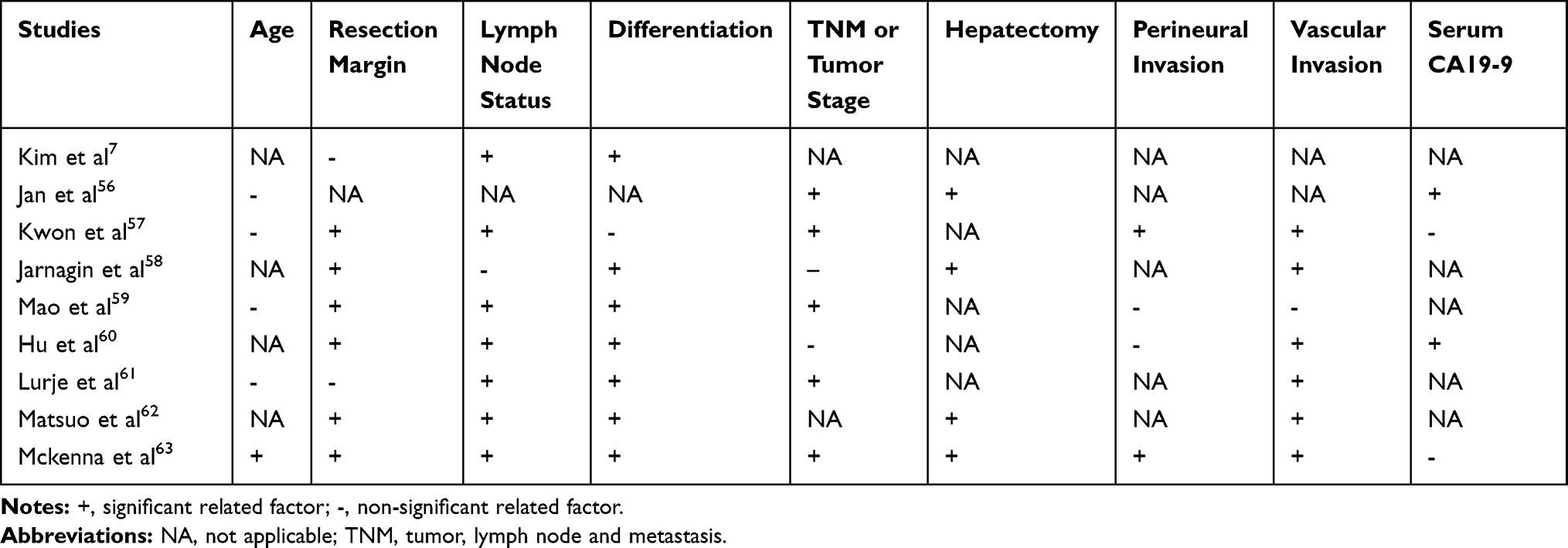 |
Table 3 Prognostic Factors for Survival |
Guidelines and Future Direction
The NCCN guideline for EHCC suggests that patients with R0 margins, negative regional nodes or carcinoma in situ at margin should only be observed or be offered fluoropyrimidine chemoradiation or fluoropyrimidine- or gemcitabine-based chemotherapy, while fluoropyrimidine- or gemcitabine-based chemotherapy or adjuvant fluoropyrimidine-based chemoradiation followed or not by additional fluoropyrimidine- or gemcitabine-based chemotherapy is recommended for patients with R1 margins or positive regional nodes.14 From the American Society of Clinical Oncology (ASCO) Clinical Practice Guidelines, patients who have been treated with resection should receive adjuvant capecitabine chemotherapy for 6 months and a microscopically positive surgical margin resection (R1) may be offered adjuvant chemoradiotherapy.64 The regimens of chemoradiotherapy were based on the SWOG trial. In the absence of data from randomized trials, prospective clinical trials participation should be encouraged. There are several ongoing clinical trials further appraising the effect of AT in CC. Two prospective randomized trials (NCT02170090 and NCT03079427) aim to compare adjuvant chemotherapy with gemcitabine plus cisplatin vs capecitabine in EHCC and GBC. A phase III trial (NCT03779035) is ongoing with the purpose of examining the effect of adjuvant chemotherapy between gemcitabine plus capecitabine and capecitabine alone in CC and GBC. Another phase II trial (KHBO 1208) is exploring gemcitabine versus S-1 after major hepatectomy for biliary tract cancer.65 In addition, a randomized phase III trial (NCT 02798510) has been designed to determine whether adjuvant concurrent chemoradiotherapy improves OS in EHBTC/GBC. The study randomized patients to adjuvant chemotherapy (gemcitabine plus capecitabine) followed by concurrent chemoradiotherapy (capecitabine alone) or adjuvant chemotherapy (gemcitabine plus capecitabine) alone. The primary outcome is 2-year over survival, and the result is promising.
In recent years, immunotherapy has transformed the treatment paradigm for tumors such as melanoma, lung cancer, and renal cell carcinoma. The efforts are ongoing to explore whether a benefit can also be seen in other solid tumors. There are studies indicating that dMMR tumors are sensitive to PD-1 blockade, and the outcomes have been published from a study of patients with dMMR tumors of twelve different cancer types.66,67 In the study, four patients with dMMR CC received pembrolizumab, and one of them had a complete response while the other three patients had stable disease. Based on this background, pembrolizumab (for MSI-H/dMMR tumors) as an alternative treatment option was recommended for patients with advanced or metastatic disease. Furthermore, the role of target therapy is being considered. A phase II trial (NCT02836847) randomized patients with advanced or recurrent EHCC and GBC into two arms to evaluate the feasibility, efficacy and safety of this target therapy. The arm-1 were treated with chemotherapy (gemcitabine and oxaliplatin) combined with target agents (cetuximab, trastuzumab, gefitinib, lapatinib, everolimus, sorafenib, crizotinib). The arm-2 only received conventional chemotherapy (Gemox). The results of the experiment are also noteworthy. Therefore, we consider that the use of adjuvant target therapy should be explored.
Summary
EHCC is associated with a poor prognosis even after undergoing radical surgery. Based on the present evidences, AT demonstrates a tendency to prolong survival in high-risk (positive margin or lymph node or advanced stage) patients. Adjuvant concurrent chemoradiotherapy followed by chemotherapy is probably the optimum choice of postoperative treatment. Capecitabine should be considered as standardized adjuvant drug, and the radiotherapy scheme could refer to the SWOG trial. However, owing to the low incidence of the disease and the heterogeneity of tumors, many studies have a mixture of intrahepatic cholangiocarcinoma (ICC) or GBC and the baselines between each group of patients are usually imbalance. They are unfavorable factors to define a standard regulation or definitive benefit. Consequently, it is integral to spare every effort to conduct well-designed multicenter clinical trials. In that way, standard regimens of AT for EHCC will be found in the future.
Disclosure
The authors report no conflicts of interest in this work.
References
1. Longmire WP, Mcarthur MS, Bastounis EA, et al. Carcinoma of the extrahepatic biliary tract. Ann Surg. 1973;178(3):333–345.
2. de Groen PC, Gores GJ, LaRusso NF, et al. Biliary tract cancers. N Engl J Med. 1999;341(18):1368–1378. doi:10.1056/NEJM199910283411807
3. Kumar JR. Advances in diagnosis and treatment of hilar cholangiocarcinoma – a review. Med Sci Monit. 2013;19:648–656. doi:10.12659/MSM.889379
4. Sahai P, Kumar S. External radiotherapy and brachytherapy in the management of extrahepatic and intrahepatic cholangiocarcinoma: available evidence. Br J Radiol. 2017;90(1076):20170061. doi:10.1259/bjr.20170061
5. Deoliveira ML, Cunningham SC, Cameron JL, et al. Cholangiocarcinoma: thirty-one-year experience with 564 patients at a single institution. Ann Surg. 2007;245(5):755–762. doi:10.1097/01.sla.0000251366.62632.d3
6. Nakeeb A. Cholangiocarcinoma: a spectrum of intrahepatic, perihilar, and distal tumors. Ann Surg. 1996;224(4):473–475. doi:10.1097/00000658-199610000-00005
7. Kim BH, Kim K, Chie EK, et al. Long-term outcome of distal cholangiocarcinoma after pancreaticoduodenectomy followed by adjuvant chemoradiotherapy: a 15-year experience in a single institution. Cancer Res Treat. 2016;49(2):473–483. doi:10.4143/crt.2016.166
8. Khan SA, Davidson BR, Goldin R, et al. Guidelines for the diagnosis and treatment of cholangiocarcinoma: consensus document. Gut. 2002;51(Supplement 6):vi1–vi9. doi:10.1136/gut.51.suppl_6.vi1
9. Nyberg K. Gemcitabine hydrochloride and oxaliplatin or observation in treating patients with biliary tract cancer that has been removed by surgery. Clinical Trials. gov. 2017.
10. Akamatsu N, Sugawara Y, Hashimoto D. Surgical strategy for bile duct cancer: advances and current limitations. World J Clin Oncol. 2011;2(2):94–107. doi:10.5306/wjco.v2.i2.94
11. Nagino M, Ebata T, Yokoyama Y, et al. Evolution of surgical treatment for perihilar cholangiocarcinoma: a single-center 34-year review of 574 consecutive resections. Ann Surg. 2012;258(1):129–140. doi:10.1097/SLA.0b013e3182708b57
12. Park SW, Park YS, Chung JB, et al. Patterns and relevant factors of tumor recurrence for extrahepatic bile duct carcinoma after radical resection. Hepato-Gastroenterol. 2004;51(60):1612–1618.
13. Vogel A, Saborowski A. Cholangiocellular carcinoma. Digestion. 2017;95(3):181–185. doi:10.1159/000454763
14. Benson AB
15. Cereda S, Belli C, Reni M. Adjuvant treatment in biliary tract cancer: to treat or not to treat? World J Gastroenterol. 2012;18(21):2591–2596. doi:10.3748/wjg.v18.i21.2591
16. Brunner TB, Seufferlein T. Radiation therapy in cholangiocellular carcinomas. Best Pract Res Clin Gastroenterol. 2016;30(4):593–602. doi:10.1016/j.bpg.2016.08.003
17. Ebata T, Hirano S, Konishi M, et al. Randomized clinical trial of adjuvant gemcitabine chemotherapy versus observation in resected bile duct cancer. Br J Surg. 2018;105(3):192–202. doi:10.1002/bjs.2018.105.issue-3
18. Edeline J, Benabdelghani M, Bertaut A, et al. Gemcitabine and oxaliplatin chemotherapy or surveillance in resected biliary tract cancer (PRODIGE 12-ACCORD 18-UNICANCER GI): a randomized Phase III study. J Clin Oncol. 2019;37(8):658–667. doi:10.1200/JCO.18.00050
19. Primrose JN, Fox RP, Palmer DH, et al. Capecitabine compared with observation in resected biliary tract cancer (BILCAP): a randomised, controlled, multicentre, Phase 3 study. Lancet Oncol. 2019;20(5):663–673. doi:10.1016/S1470-2045(18)30915-X
20. Murakami Y, Uemura K, Sudo T, et al. Adjuvant gemcitabine plus S-1 chemotherapy improves survival after aggressive surgical resection for advanced biliary carcinoma. Ann Surg. 2009;250(6):950–956. doi:10.1097/SLA.0b013e3181b0fc8b
21. Murakami Y, Uemura K, Sudo T, et al. Gemcitabine-based adjuvant chemotherapy improves survival after aggressive surgery for hilar cholangiocarcinoma. J Gastrointest Surg. 2009;13(8):1470–1479. doi:10.1007/s11605-009-0900-0
22. Wirasorn K, Ngamprasertchai T, Khuntikeo N, et al. Adjuvant chemotherapy in resectable cholangiocarcinoma patients. J Gastroenterol Hepatol. 2013;28(12):1885–1891. doi:10.1111/jgh.2013.28.issue-12
23. Bergeat D, Turrini O, Courtin-Tanguy L, et al. Impact of adjuvant chemotherapy after pancreaticoduodenectomy for distal cholangiocarcinoma: a propensity score analysis from a French multicentric cohort. Langenbecks Arch Surg. 2018;403(6):701–709. doi:10.1007/s00423-018-1702-1
24. Kim YS, Jeong CY, Song HN, et al. The efficacy of fluoropyrimidine-based adjuvant chemotherapy on biliary tract cancer after R0 resection. Chin J Cancer. 2017;36(1):9. doi:10.1186/s40880-017-0182-y
25. Yin L, Xu Q, Li J, et al. The efficiency and regimen choice of adjuvant chemotherapy in biliary tract cancer A STROBE-compliant retrospective cohort study. Med. 2018;97(50):13570. doi:10.1097/MD.0000000000013570
26. Valle JW, Wasan HS, Palmer DD, et al. Cisplatin plus gemcitabine versus gemcitabine for biliary tract cancer. N Eng J Med. 2010;362:1273–1281. doi:10.1056/NEJMoa0908721
27. Sharma A, Dwary AD, Mohanti BK, et al. Best supportive care compared with chemotherapy for unresectable gall bladder cancer: a randomized controlled study. J Clin Oncol. 2010;28(30):4581–4586. doi:10.1200/JCO.2010.29.3605
28. Nehls O, Klump B, Arkenau HT, et al. Oxaliplatin, fluorouracil and leucovorin for advanced biliary system adenocarcinomas: a prospective phase II trial. Br J Cancer. 2002;87(7):702–704. doi:10.1038/sj.bjc.6600543
29. Nehls O, Oettle H, Hartmann JT, et al. Capecitabine plus oxaliplatin as first-line treatment in patients with advanced biliary system adenocarcinoma: a prospective multicentre phase II trial. Br J Cancer. 2008;98(2):309–315. doi:10.1038/sj.bjc.6604178
30. Kim TW. Phase II study of capecitabine plus cisplatin as first-line chemotherapy in advanced biliary cancer. Ann Oncol. 2003;14(7):1115–1120. doi:10.1093/annonc/mdg281
31. Kobayashi K, Tsuji A, Morita S, et al. A phase II study of LFP therapy (5-FU (5-fluorourasil) continuous infusion (CVI) and low-dose consecutive (Cisplatin) CDDP) in advanced biliary tract carcinoma. BMC Cancer. 2006;6(1):121. doi:10.1186/1471-2407-6-121
32. Schoenthaler R, Phillips TL, Castro J, et al. Carcinoma of the extrahepatic bile ducts the University of California at San Francisco experience. Ann Surg. 1994;219(3):267–274. doi:10.1097/00000658-199403000-00006
33. Washburn WK, Lewis WD, Jenkins RL. Aggressive surgical resection for cholangiocarcinoma. Arch Surg. 1995;130(3):270–276. doi:10.1001/archsurg.1995.01430030040006
34. Kopelson G, Galdabini J, Warshaw AL, et al. Patterns of failure after curative surgery for extra-hepatic biliary tract carcinoma: implications for adjuvant therapy. Int J Radiat Oncol Biol Phys. 1981;7(3):413–417. doi:10.1016/0360-3016(81)90118-8
35. Ghiassi-Nejad Z, Tarchi P, Moshier E, et al. Prognostic factors and patterns of locoregional failure after surgical resection in patients with cholangiocarcinoma without adjuvant radiation therapy: optimal field design for adjuvant radiation therapy. Int J Radiat Oncol Biol Phys. 2017;99(4):805–811. doi:10.1016/j.ijrobp.2017.06.2467
36. Oh D, Lim DH, Heo JS, et al. The role of adjuvant radiotherapy in microscopic tumor control after extrahepatic bile duct cancer surgery. Am J Clin Oncol. 2007;30(1):21–25. doi:10.1097/01.coc.0000245467.97180.78
37. Doherty MK, Knox JJ. Adjuvant therapy for resected biliary tract cancer: a review. Chin Clin Oncol. 2016;5(5):64. doi:10.21037/cco
38. Sagawa N, Kondo S, Morikawa T, et al. Effectiveness of radiation therapy after surgery for hilar cholangiocarcinoma. Surg Today. 2005;35(7):548–552. doi:10.1007/s00595-005-2989-4
39. Vern-Gross TZ, Shivnani AT, Chen K, et al. Survival outcomes in resected extrahepatic cholangiocarcinoma: effect of adjuvant radiotherapy in a surveillance, epidemiology, and end results analysis. Int J Radiat Oncol Biol Phys. 2011;81(1):189–198. doi:10.1016/j.ijrobp.2010.05.001
40. Gwak HK, Kim WC, Kim HJ, et al. Extrahepatic bile duct cancers: surgery alone versus surgery plus postoperative radiation therapy. Int J Radiat Oncol Biol Phys. 2010;78(1):194–198. doi:10.1016/j.ijrobp.2009.07.003
41. Itoh H, Nishijima K, Kurosaka Y, et al. Magnitude of combination therapy of radical resection and external beam radiotherapy for patients with carcinomas of the extrahepatic bile duct and gallbladder. Dig Dis Sci. 2005;50(12):2231–2242. doi:10.1007/s10620-005-3040-8
42. Yang H, Zhou J, Wei X, et al. Survival outcomes and prognostic factors of extrahepatic cholangiocarcinoma patients following surgical resection: adjuvant therapy is a favorable prognostic factor. Mol Clin Oncol. 2014;2(6):1069–1075. doi:10.3892/mco.2014.377
43. Kim YJ, Kim K, Min SK, et al. Role of adjuvant radiotherapy for localized extrahepatic bile duct cancer. Br J Radiol. 2017;90(1071):20160807. doi:10.1259/bjr.20160807
44. Krasnick BA, Jin LX, Davidson JT, et al. Adjuvant therapy is associated with improved survival after curative resection for hilar cholangiocarcinoma: a multi-institution analysis from the U.S. extrahepatic biliary malignancy consortium. J Surg Oncol. 2017;117(3):363–371. doi:10.1002/jso.24836
45. Kim TH, Han SS, Park SJ, et al. Role of adjuvant chemoradiotherapy for resected extrahepatic biliary tract cancer. Int J Radiat Oncol Biol Phys. 2011;81(5):E853—E859. doi:10.1016/j.ijrobp.2010.12.019
46. Han IW, Jang JY, Lee KB, et al. Clinicopathological analysis and prognosis of extrahepatic bile duct cancer with a microscopic positive ductal margin. HPB. 2014;16(6):575–581. doi:10.1111/hpb.12193
47. Hoehn RS, Wima K, Ertel AE, et al. Adjuvant chemotherapy and radiation therapy is associated with improved survival for patients with extrahepatic cholangiocarcinoma. Ann Surg Oncol. 2015;22(3):1133–1139. doi:10.1245/s10434-015-4599-8
48. Nassour I, Mokdad A, Porembka MR, et al. Adjuvant therapy is associated with improved survival in resected perihilar cholangiocarcinoma: a propensity matched study. J Am Coll Surg. 2018;25(5):1193–1201.
49. Lee J, Kang SH, Noh O, et al. Adjuvant concurrent chemoradiation therapy in patients with microscopic residual tumor after curative resection for extrahepatic cholangiocarcinoma. Clin Transl Oncol. 2018;20(8):1011–1017. doi:10.1007/s12094-017-1815-y
50. Horgan AM, Amir E, Walter T, et al. Adjuvant therapy in the treatment of biliary tract cancer: a systematic review and meta-analysis. J Clin Oncol. 2012;30(16):1934–1940. doi:10.1200/JCO.2011.40.5381
51. Beltrán MB, Roth AD, Mentha G, et al. Adjuvant radio-chemotherapy for extrahepatic biliary tract cancers. BMC Cancer. 2011;11(1):267. doi:10.1186/1471-2407-11-267
52. Kim K, Chie EK, Jang JY, et al. Adjuvant chemoradiotherapy after curative resection for extrahepatic bile duct cancer: a long-term single center experience. Am J Clin Oncol. 2012;35(2):136. doi:10.1097/COC.0b013e318209aa29
53. Park JH, Choi EK, Ahn SD, et al. Postoperative chemoradiotherapy for extrahepatic bile duct cancer. Int J Radiat Oncol BiolPhys. 2011;79(3):696–704. doi:10.1016/j.ijrobp.2009.12.031
54. Lim KH, Oh DY, Chie EK, et al. Adjuvant concurrent chemoradiation therapy (CCRT) alone versus CCRT followed by adjuvant chemotherapy: which is better in patients with radically resected extrahepatic biliary tract cancer? A nonrandomized, single center study. BMC Cancer. 2009;9(1):345. doi:10.1186/1471-2407-9-345
55. Ben-Josef E, Guthrie KA, El-Khoueiry AB, et al. SWOG S0809: a phase II Intergroup Trial of adjuvant capecitabine and gemcitabine followed by radiotherapy and concurrent capecitabine in extrahepatic cholangiocarcinoma and gallbladder carcinoma. J Clin Oncol. 2015;33(24):2617–2622. doi:10.1200/JCO.2014.60.2219
56. Jan YY, Yeh CN, Yeh TS, Chen TC. Prognostic analysis of surgical treatment of peripheral cholangiocarcinoma: two decades of experience at Chang Gung Memorial Hospital. World J Gastroenterol. 2005;11:1779–1784. doi:10.3748/wjg.v11.i12.1779
57. Kwon HJ, Kim SG, Chun JM, et al. Prognostic factors in patients with middle and distal bile duct cancers. World J Gastroenterol. 2014;20(21):6658–6665. doi:10.3748/wjg.v20.i21.6658
58. Jarnagin WR, Fong Y, Dematteo RP, et al. Staging, resectability, and outcome in 225 patients with hilar cholangiocarcinoma. Ann Surg. 2001;234(4):507–519. doi:10.1097/00000658-200110000-00010
59. Mao Z, Guo X, Su D, et al. Prognostic factors of cholangiocarcinoma after surgical resection: a retrospective study of 293 patients. Med Sci Monit. 2015;21:2375–2381. doi:10.12659/MSM.893586
60. Hu HJ, Mao H, Shrestha A, et al. Prognostic factors and long-term outcomes of hilar cholangiocarcinoma: a single-institution experience in China. World J Gastroenterol. 2016;22(8):2601–2610. doi:10.3748/wjg.v22.i8.2601
61. Lurje G, Bednarsch J, Czigany Z, et al. The prognostic role of lymphovascular invasion and lymph node metastasis in perihilar and intrahepatic cholangiocarcinoma. Eur J Surg Oncol. 2019;30411–1(19):S0748–7983.
62. Matsuo K, Rocha FG, Ito K, et al. The blumgart preoperative staging system for hilar cholangiocarcinoma: analysis of resectability and outcomes in 380 patients. J Am Coll Surg. 2012;215(3):343–355. doi:10.1016/j.jamcollsurg.2012.05.025
63. Bird NTE, Mckenna A, Dodd J, et al. Meta-analysis of prognostic factors for overall survival in patients with resected hilar cholangiocarcinoma. Br J Surg. 2018;105(11):1408–1416. doi:10.1002/bjs.10921
64. Shroff RT, Kennedy EB, Bachini M, et al. Adjuvant therapy for resected biliary tract cancer: ASCO clinical practice guideline. J Clin Oncol. 2019;37(12):1015–1027. doi:10.1200/JCO.18.02178
65. Kobayashi S, Nagano H, Tomokuni A, et al. A prospective, randomized Phase II study of adjuvant gemcitabine versus S-1 after major hepatectomy for biliary tract cancer (KHBO 1208): Kansai hepato-biliary oncology group. Ann Surg. 2019;270(2):230–237. doi:10.1097/SLA.0000000000002865
66. Le DT, Uram JN, Wang H, et al. PD-1 blockade in tumors with mismatch-repair deficiency. N Engl J Med. 2015;372(26):2509–2520. doi:10.1056/NEJMoa1500596
67. Le DT, Durham JN, Smith KN, et al. Mismatch-repair deficiency predicts response of solid tumors to PD-1 blockade. Sci. 2017;357(6349):409–441. doi:10.1126/science.aan6733
 © 2019 The Author(s). This work is published and licensed by Dove Medical Press Limited. The full terms of this license are available at https://www.dovepress.com/terms.php and incorporate the Creative Commons Attribution - Non Commercial (unported, v3.0) License.
By accessing the work you hereby accept the Terms. Non-commercial uses of the work are permitted without any further permission from Dove Medical Press Limited, provided the work is properly attributed. For permission for commercial use of this work, please see paragraphs 4.2 and 5 of our Terms.
© 2019 The Author(s). This work is published and licensed by Dove Medical Press Limited. The full terms of this license are available at https://www.dovepress.com/terms.php and incorporate the Creative Commons Attribution - Non Commercial (unported, v3.0) License.
By accessing the work you hereby accept the Terms. Non-commercial uses of the work are permitted without any further permission from Dove Medical Press Limited, provided the work is properly attributed. For permission for commercial use of this work, please see paragraphs 4.2 and 5 of our Terms.