")
Back to Journals » Advances in Medical Education and Practice » Volume 11
The Role of Technology in Anatomy Teaching: Striking the Right Balance
Authors Zargaran A, Turki MA , Bhaskar J, Spiers HVM, Zargaran D
Received 26 November 2019
Accepted for publication 17 March 2020
Published 31 March 2020 Volume 2020:11 Pages 259—266
DOI https://doi.org/10.2147/AMEP.S240150
Checked for plagiarism Yes
Review by Single anonymous peer review
Peer reviewer comments 3
Editor who approved publication: Dr Md Anwarul Azim Majumder
Alexander Zargaran, 1 Mohammed Adil Turki, 2 Jared Bhaskar, 3 Harry Victor Michael Spiers, 4 David Zargaran 3
1King’s College London, Department of Medicine, London, UK; 2St George’s, University of London, Department of Medicine, London, UK; 3Imperial College London, Department of Medicine, London, UK; 4Manchester Royal Infirmary, Department of Medicine, London, UK
Correspondence: David Zargaran
Imperial College London Email [email protected]
Background: This study assesses the scope for using technology to supplement the undergraduate anatomy curriculum at medical school.
Methods: A narrative literature review explored the current landscape of anatomy learning. Medical student usage and preferences of technological interventions for anatomy learning were then explored through a cross-sectional survey.
Results: The literature review revealed the current teaching strategies for anatomy learning, exploring recent multimedia innovations. The survey demonstrated that technology usage was ubiquitous among medical students with 98% of medical students owning smartphones. Medical education apps were used by 64.3% of medical students, with 61.9% of these apps covering anatomy, and 60.4% of students preferring traditional cadaveric teaching to other technological interventions.
Conclusion: Novel technological innovations present the opportunity to deliver accessible and standardised teaching of anatomy to medical students. Many students already use smartphone applications as part of their anatomy learning. Uptake of smartphones and other devices provides opportunities to reach larger target audiences. However, traditional cadaveric teaching remains the learning resource of choice for medical students, and technological interventions are best designed as adjuncts or supplements to cadaveric teaching.
Keywords: technology, anatomy, medical education, smartphone
A Letter to the Editor has been published for this article.
Introduction
Comprehensive knowledge of human anatomy underpins the understanding and practice of medicine. Anatomy teaching is an established part of the undergraduate curriculum. However, with a variety of tools and techniques employed to teach anatomy, there is uncertainty regarding methods to deliver optimal teaching. With rapid advancement of technology and e-learning, it is important to explore the potential of technology in delivering tailored anatomical teaching. This paper seeks to answer the question: what is the best method to teach anatomy to undergraduate medical students, including striking the right balance between use of novel technologies and traditional methods. A narrative literature review was conducted to explore established methods and their efficacy, which informed a cross-sectional survey at a medical school in the United Kingdom.
Method of Literature Search
The search was conducted using Ovid MEDLINE® and Embase databases, and was structured using a PICO framework (Population→medical students, Intervention→technology, Comparison→Anatomy teaching methods, Outcomes→student satisfaction/improved student performance). A PRISMA flow chart and search string are provided in Appendix 1. The inclusion and exclusion criteria are detailed in Table 1
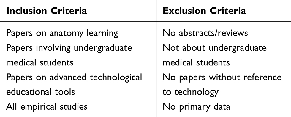 |
Table 1 Inclusion and Exclusion Criteria for Narrative Review |
The application of inclusion and exclusion criteria yielded 25 papers for review. Abstract screening and the literature review were conducted by two people, and the Kappa coefficient for interrater reliability was calculated at 0.9. Papers were structured into five main themes (3D & Virtual technology, Cadaveric materials, Web-based applications, Imaging tools and Video learning tools), which are explored in this literature review. An additional section on the views of students and teachers has been included due to the importance of end-user perceptions in implementing any teaching strategy.
3D and Virtual Technology
Computer-assisted and 3D printing technologies have been used to complement or replace traditional anatomy teaching. Studies have evaluated the effectiveness of 3D printed models compared to cadavers. These studies have been summarised in Table 2.
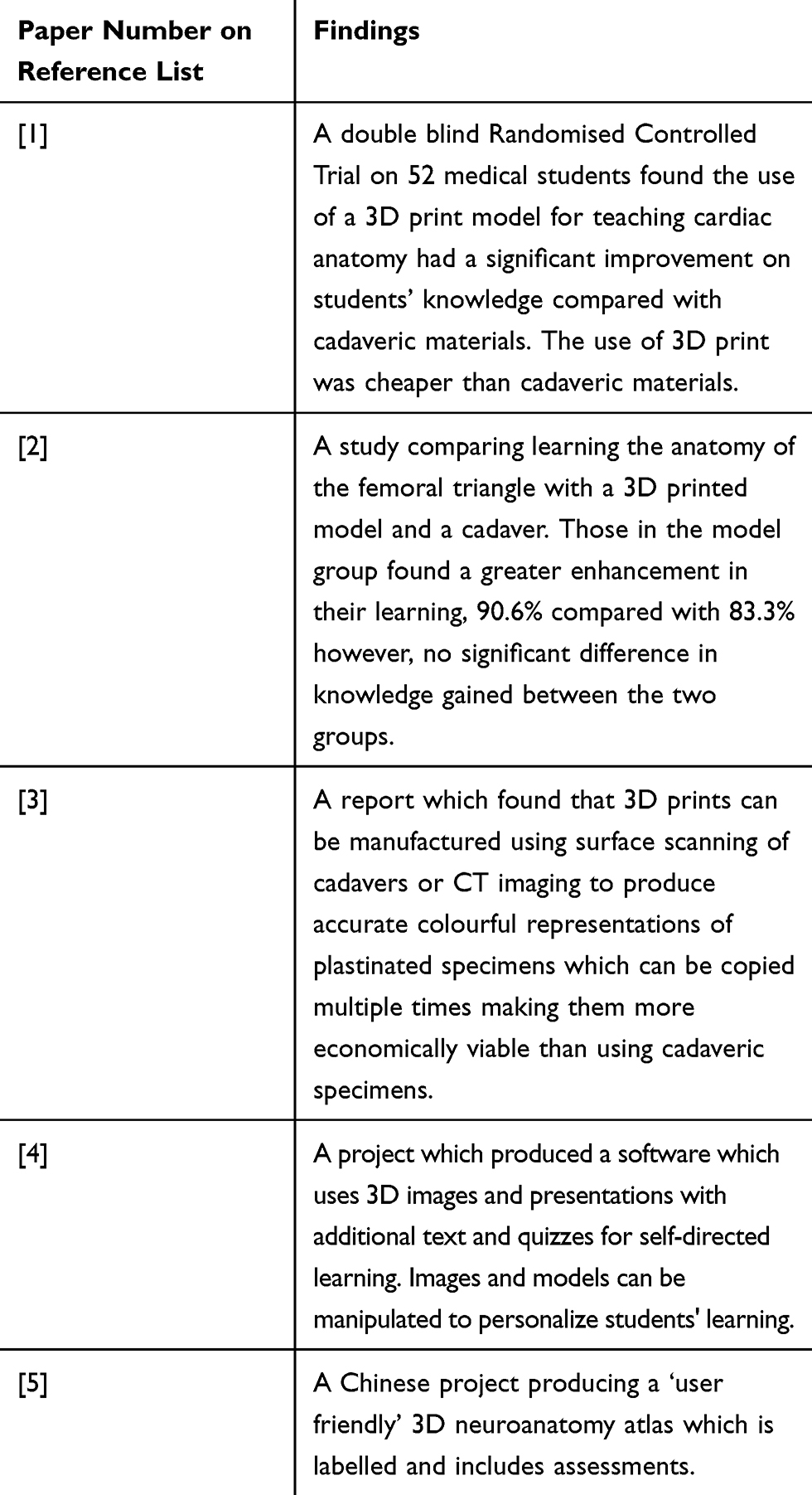 |
Table 2 Summary of Key Findings for 3D and Virtual Technology |
There is consensus among the studies that 3D and virtual technology represent accessible and cost-effective options for delivering anatomy teaching, without compromising the quality of learning opportunities for students. Furthermore, studies4,5 demonstrate how this technology can be designed for personalized use, which could provide an effective adjunct for self-directed learning, supplementing traditional methods in the classroom.
Despite the clear potential for these technologies to enhance teaching, there remain challenges, as these innovative technologies are in their infancy, and uncertainty surrounds the best way of commercialising and integrating them into the current curriculum.
Cadaveric Materials
Cadaveric materials have traditionally been regarded as the most efficacious teaching method for learning anatomy. However, it is important to determine whether this is still the case. Findings for this section are summarized in Table 3.
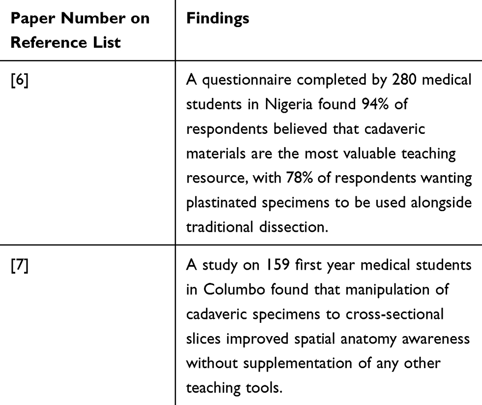 |
Table 3 Key Findings About Use of Cadaveric Materials |
Traditional methods can be transformed to enhance anatomical learning, as demonstrated in the study.7 This is important for educators to acknowledge when considering the best strategy for teaching anatomy. More efficient anatomical learning can be achieved by modifying existing methods; not necessarily requiring advanced technology. In certain instances, technology can be used to mimic and upscale traditional methods of teaching.3
Web-Based Applications
Web-based applications are beginning to integrate into the curriculum due to their increased development and availability, yet their efficacy remains unclear. Two papers were studied, which involved the design of a new Web-based application, and an investigation into its success when incorporated into the undergraduate curriculum. Key findings are summarised in Table 4.
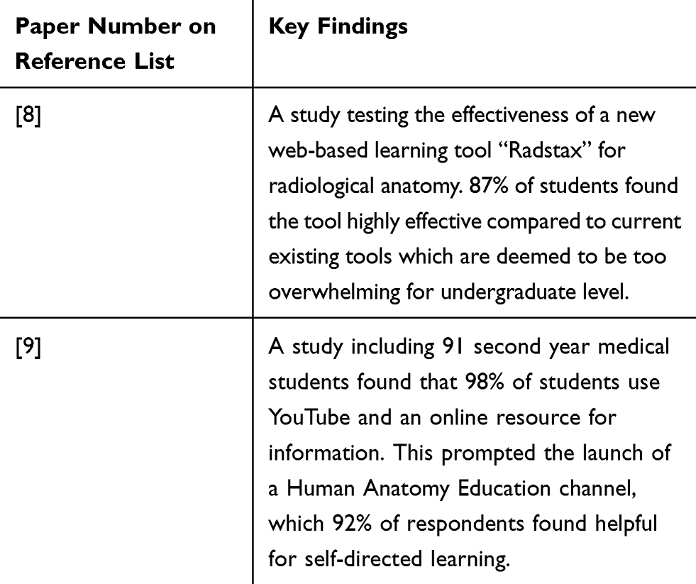 |
Table 4 Key Findings About Use of Web-Based Applications |
Many existing Web-Based anatomy programmes are designed for postgraduate learning and provide an overwhelming level of detail for medical students.8 The high levels of self-reported satisfaction in both interventions demonstrate the positive potential impact of online applications. Where facilitated correctly and aimed at an appropriate level of understanding, they can be a useful self-directed learning tool for students. However, it is important to note that high satisfaction does not necessarily correlate to improved performance by students and further work needs to be done to assess whether such tools have an impact on increasing knowledge levels.
Integration of Imaging Tools
Slideshow presentations, live drawing illustrations and radiological imaging techniques such as computer tomography (CT), magnetic resonance imaging (MRI) and ultrasound scanning (USS) can be used to supplement traditional cadaveric anatomical teaching. Medical schools have explored the impact of integrating medical imaging components into the anatomy curriculum. Table 5 summarises the key findings:
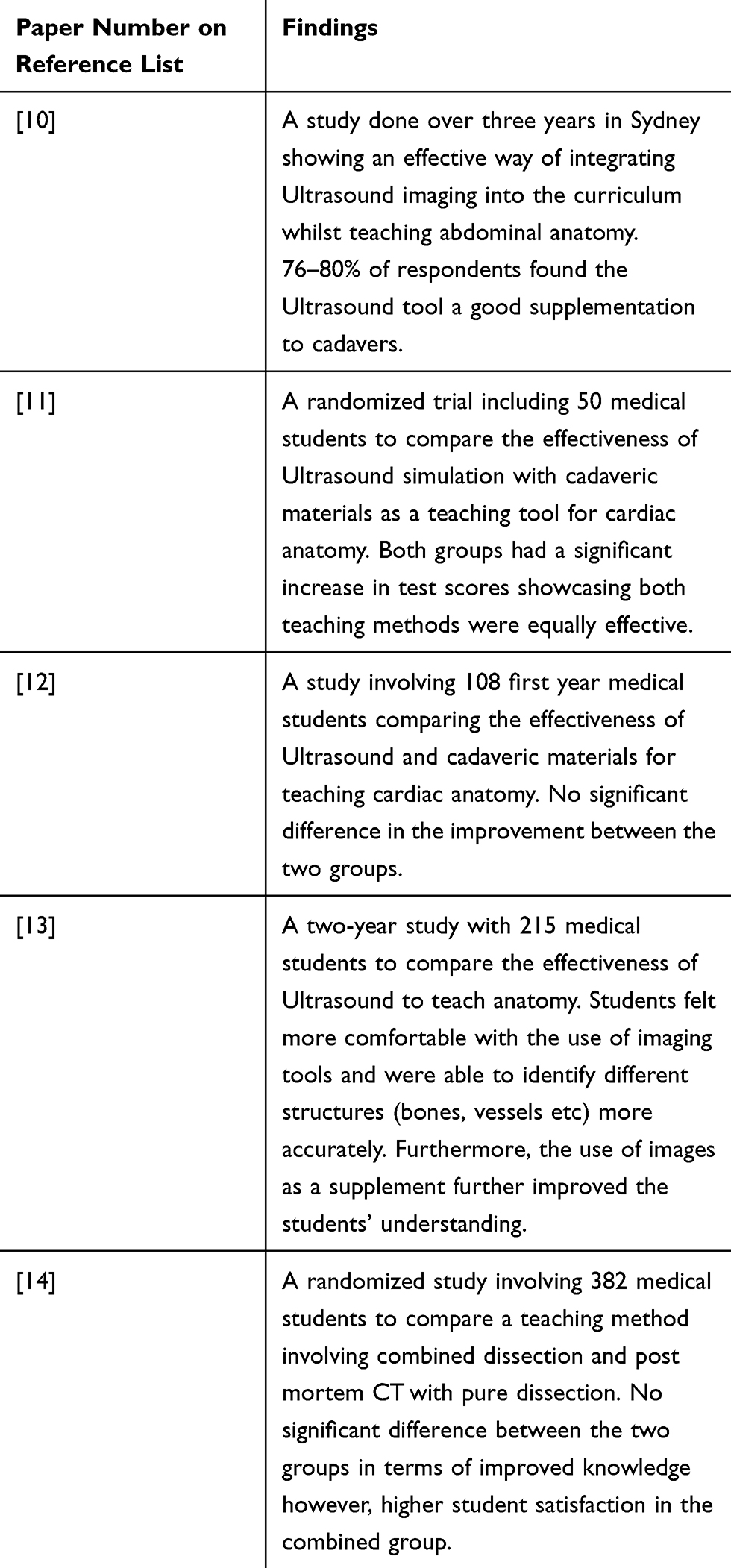 |
Table 5 Key Findings About Integration of Imaging Tools |
The results from these studies point towards imaging tools being a useful adjunct to anatomical learning in a clinical context. Another benefit of integrating imaging technology with anatomy is that this exposure can prime students for later encounters with imaging devices in clinical practice, often considered a difficult technique to grasp. Nonetheless, study14 indicates that when used in isolation, imaging tools are not potent teaching tools for anatomy learning. However, when combined with the existing curriculum, such tools may promote student interest and improve understanding.13
Video Learning Tools
Pre-recorded or live videos offer a realistic visual experience of anatomy, easily accessible to reference when required. There was one study including multimedia tools, with the findings summarised in Table 6.
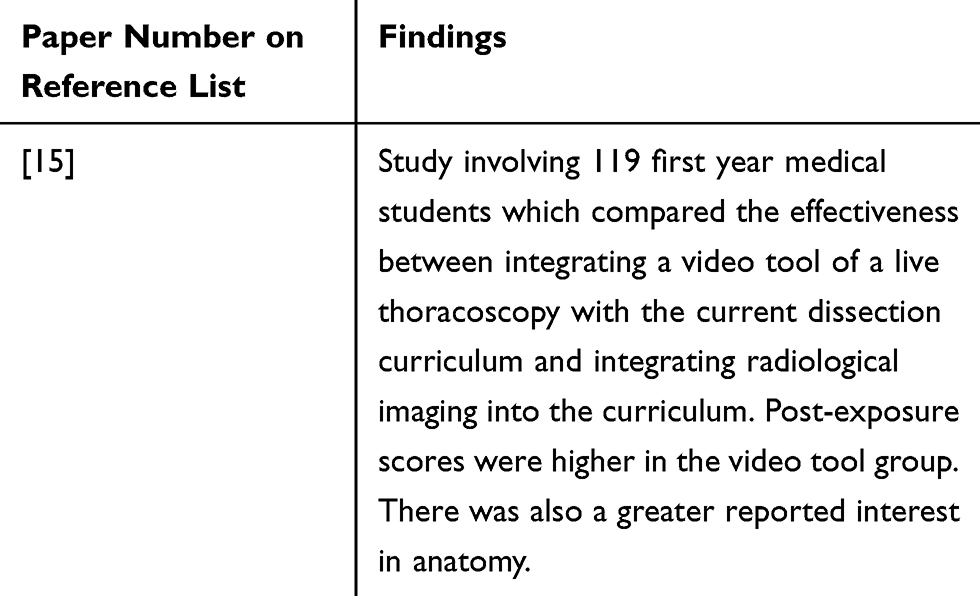 |
Table 6 Key Findings About Use of Video Learning Tools |
This study proposes that the integration of video tools into the current curriculum would be more effective than the use of radiological imaging, since they have a greater positive impact on student performance as well as attentiveness to the subject. However, time taken to record the videos as well as ethical issues of recording live patients must be considered.
Views of Students and Teachers
For a learning method to be considered successful, it must satisfy both educators and learners. Consequently, the views of these personnel are extremely important when debating the efficacy of the current curriculum with new possibilities in technology. A summary of the findings is included in Table 7.
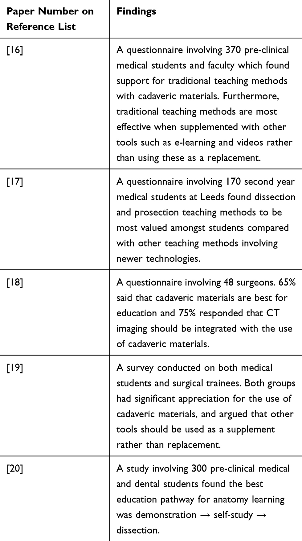 |
Table 7 Key Findings About Teacher and Student Perceptions |
The papers included in this section demonstrate that the current learning methods for anatomy are highly valued, and that any changes would seek to improve rather than replace cadaveric teaching.
The format in which teaching is delivered is also an important consideration and it is investigated in the study.20 By looking at which methods of anatomical learning are preferred by students, a medical school can concentrate their resources to provide appropriate tools and enhance self-directed learning. This may be the area of learning where advanced technological teaching tools are most useful, rather than as a replacement in the classroom where standardised learning occurs.
Although in many ways the current anatomy curriculum is competent and serves its purpose, access to cadavers is not always feasible. Furthermore, accessible advancements in technology such as web-based applications have transformed education, with an increasing trend towards long-distance learning. It is likely that technology will drive the future of anatomy teaching in medical schools and it is therefore important to determine how best to incorporate technology in any teaching strategy for undergraduate medical students.
In this cross-sectional study, we aim to evaluate how medical students learn anatomy. However, it is also important to outline technological uptake among undergraduate medical students to assess how feasible any new strategy would be. We hope to outline which methods and tools students find most useful for the assimilation and application of anatomical knowledge, helping to improve the teaching of anatomy and provide students with more appropriate material and methods to grasp the core anatomy needed at undergraduate level.
Methods
A cross-sectional survey of a convenience sample of 120 undergraduate medical students was taken on 2nd of March 2017 at the annual anatomy competition in a medical school in the UK. Students in the first 2 years of their Biomedical Sciences degree were also included, as anatomy teaching in the first 2 years is the same for Biomedical Sciences students and medical students. The survey was designed by medical students and doctors who teach as anatomy demonstrators. The question that arose from the literature review was how best to incorporate technology in teaching undergraduate anatomy education. There was a paucity of literature on technological use among undergraduate medical students, therefore the survey explored the following:
- Technological use including smartphones, tablets, medical education apps and anatomy related apps. In terms of usage, it also assessed how often any technologies were used.
- Current anatomy learning including the type of resource used such as books, lectures, online videos, cadaveric teaching and dissection.
Questions were laid out as a mixture of multiple-choice and a Guttmann scale, requiring delegates to provide a “yes” or “no” answer. The second section used a modified Likert scale to understand views on the range, type and nature of resources from a scale of 1–5. All responses were anonymised and collated for analysis.
Statistical analysis was performed using IBM SPSS statistical software. Levene’s Test for Equality of Variances and an independent samples t-test were used to compare means. Significance was set at p<0.05.
Results
A total of 102 delegates completed surveys, giving a response rate of 85%; 85.3% of respondents were medical students and 14.7% studied biomedical sciences. The mean age of respondents was 20.89 years. A breakdown of distribution of year groups is shown in Table 8.
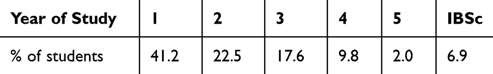 |
Table 8 Demographic Characteristics of Survey Participants by Year Group. IBSc Is an Intercalated Bachelor of Science (BSc) Which Can Be Taken at Any Stage After Year 2 and Before Year 5 |
Interest in areas of medicine included Surgery (56.9%), Medicine (36.3%), Psychiatry (4.9%) and General Practice (2%). Smartphone ownership was 98% among students, with 60.2% owning an iPhone, 38.8% owning Android smartphones and 1% owning a different type of smartphone. Tablet ownership was 79.4%, with 82.7% iPad and 17.3% Android. Medical Education apps were used by 64.3%, with Biomedical Sciences students more likely to use Medical Education apps than Medical Students (p<0.01). Table 9 illustrates how the types of Medical Education apps used changes depending on the stage of medical training.
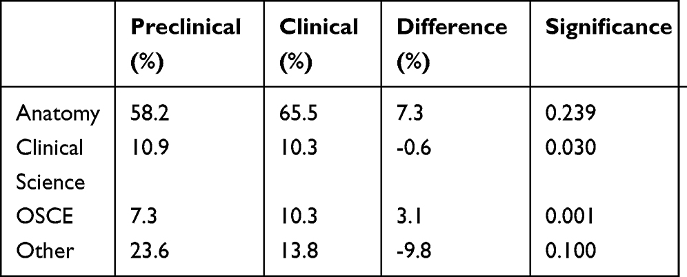 |
Table 9 Content of Medical Education Apps by Stage of Education |
Virtual Simulation for learning anatomy was preferred by 39.4% of students, whilst 60.4% preferred traditional cadaveric teaching (p<0.01). Surgery-related apps were used by 63.7% of students, with 68.3% using these apps at least once a week. Table 10 demonstrates the range of resources used by students for extracurricular anatomy learning.
 |
Table 10 Extracurricular Learning Resources Used by Survey Participants |
Discussion
Students from all years of medical school took part in the survey. Pre-clinical students (years one and two) accounted for the largest proportion of responses (63.7%). The distribution of students can be explained by the focus of the medical curriculum, with more emphasis on core concepts including anatomy being placed during pre-clinical years. The proportion of students in their final 2 years was low (11.8%), with only 2% of those who took part being final years.
Most students who took part were interested in a career in surgery (56.9%), perhaps reflecting the importance of anatomy to surgery. The findings of this study concur with the consensus that a career in general practice is less appealing to undergraduate medical students compared to other fields,21 with only 2% of those who took part interested in a career in general practice. Whilst it is important to acknowledge that respondents attended a Surgical Society event, only 2% of medical students being interested in general practice as a career despite government policy encouraging a figure closer to 50%,22 is very low. However, the low number of respondents interested in becoming GPs is potentially due to the misconception that anatomy will not be important for their future daily practice. More needs to be done to emphasise the importance of a comprehensive knowledge and understanding of anatomy across different specialities within medicine. The high levels of smartphone and tablet ownership demonstrate the high penetration of electronic devices among students and the potential for technologies to be harnessed to facilitate educational objectives. Biomedical science students were more likely to use medical education applications than medical students. However, only 12% of respondents studied Biomedical Sciences. This makes it difficult to make reliable inferences, but highlights the wide scope of resources that cover anatomy and its appeal to wider audiences than medical students.
There was no significant difference in the proportion of pre-clinical students compared to clinical, who used anatomical education applications (p=0.239). This is despite anatomy making up a smaller percentage of assessment in clinical years, suggesting that the subjective importance of anatomy pervades all years of medical school and extends beyond the boundaries of the curriculum. This highlights the need for anatomical resources to be available, outside of the medical school curriculum and easily accessible to students to help drive self-directed study. This, combined with the high proportion of those owning a smart device, provides an insight into why the number of students relying on e-resources and applications for self-directed learning is rising. Applications that cover anatomy had the highest proportion of students using them, 61.9% across all those who took part.
The number of those using applications concerned with clinical sciences was similar between pre-clinical and clinical year students (10.9 to 10.3). This is to be expected, as medicine is the study of clinical sciences, whether theoretical (pre-clinical) or practical (clinical). However, across the cohort, only 10.6% of respondents were using clinical sciences applications compared to 61.9% for anatomy. This could either suggest that students feel that clinical sciences are adequately covered by other teaching methods or that there is a lack of valuable technologies delivering clinical sciences learning.
Resources regarding Objective Structured Clinical Examinations (OSCE) saw a notable change from pre-clinical to clinical years. This finding is in line with the different focus between preclinical and clinical years, as there is a greater focus on patient interaction and ward-based learning in clinical years. Overall, the use of anatomical applications was higher than those for other aspects of the curriculum. This reflects the large variety of electronic resources available to assist the study of anatomy, from videos to virtual reality. This study demonstrates strong interest in the use of electronic resources and applications to supplement and augment anatomy learning. It is likely that if more innovative resources to learn clinical science or OSCE were provided, the proportion of students learning via such methods will also rise.
Student Perspectives on Anatomical Teaching Methods
Anatomical educational resources vary in design and content. Students identified various methods of learning anatomy that they found most useful. As previous studies have suggested, traditional cadaveric teaching is most favoured.16 In this study, only 39.4% of students preferred virtual simulation, whilst 60.4% preferred cadaveric teaching, which corroborates with existing literature.
Cadaveric teaching can take two forms – dissection and prosection. Respondents of this study least preferred dissection as a method for learning anatomy (Table 3). Whilst it offers surgical experience and an opportunity to familiarize with surgical instruments, it can be overwhelming to dissect out anatomical structures with little guidance. This is advantageous to medical schools, as dissection is more resource intensive, as the same structure in human tissue cannot be dissected twice and requires closer supervision. By contrast, cadaveric teaching (Table 3) was the most popular method of learning anatomy. Cadaveric prosections and plastinations offer students the opportunity to familiarise themselves with the human body and tissue, without the responsibility of dissection. This principle allows undergraduate teaching to be focused on prosections and plastinations with an option to explore dissection in self-selected modules.
Traditional resources used to consolidate learning in the dissecting room such as books (52.9%) and lectures (52.9%) outscored more novel methods such as online videos (51.0%) and applications (35.3%). However, videos scored almost as high as traditional methods. Videos range in variety and quality from simple animations to thorough guided walk-throughs of the human body. Video atlases have also been produced, such as Acland’s,23 providing high-quality videos with an instructor talking through the structures on cadavers, offering a dissecting room-like experience at the student’s desk. With improved video technologies, this resource will continue to rise in popularity and overtake more traditional methods.
Frequency of Application Use
Of those who used applications with surgical resources (63.7%), 68.3% used them once a week. This reflects the growing importance of non-traditional methods to augment undergraduate learning.
Conclusion
Our findings reinforce that students appreciate the traditional methods of learning, such as cadaveric teaching.19 Technological penetration amongst medical students, combined with the climate of evolving resources to reinforce and revise the content in the student’s own time, provides an opportunity for educational providers and innovators to develop novel teaching methods to improve the delivery of the medical curriculum to undergraduate students. This in turn can help develop an interest in anatomy in medical students and better equip them to apply anatomical knowledge in their clinical practice.13
The frequency of use of e-learning methods as an adjunct to supplement classroom learning was high among participants. The influence of this on modern learning can already be observed, with universities augmenting traditional teaching with electronic resources and components. This should be considered in medical curriculum performance reviews and a way to incorporate these novel methods of learning into the traditional curriculum should be found.
The knowledge acquired in medical school influences performance as a doctor.24 Both clinicians and students acknowledge anatomy knowledge to be vital for good clinical practice.25 Therefore, the decline of time dedicated to anatomy within the medical curriculum26 is concerning. It is vital that resources tailored to student preferences are available to address this issue.
Our study demonstrates that students readily make use of electronic resources to support their learning of anatomy. We feel that medical schools adopting and officially supporting innovative methods of anatomical learning will empower students in their study of anatomy.
Declarations
This research was presented at the Association of Surgeons in Training International Surgical Conference, 22nd – 24th March 2019, Belfast, UK as an abstract with interim findings. The abstract was published in “ASiT Oral Presentations” in the British Journal of Surgery.
Data Sharing Statement
The datasets used and/or analysed during the current study are available from the corresponding author on reasonable request.
Author Contributions
All authors contributed to data analysis, drafting and revising the article, gave final approval of the version to be published, and agree to be accountable for all aspects of the work.
Disclosure
The authors declare that they have no competing interests.
References
1. Lim KH, Loo ZY, Goldie SJ, et al. Use of 3D printed models in medical education: a randomized control trial comparing 3D prints versus cadaveric materials for learning external cardiac anatomy. Anat Sci Educ. 2016;9(3):213–221. doi:10.1002/ase.1573
2. O’Reilly MK, Reese S, Herlihy T, et al. Fabrication and assessment of 3D printed anatomical models of the lower limb for anatomical teaching and femoral vessel access training in medicine. Anat Sci Educ. 2016;9(1):71–79. doi:10.1002/ase.v9.1
3. McMenamin PG, Quayle MR, McHenry CR, et al. The production of anatomical teaching resources using three-dimensional (3D) printing technology. Anat Sci Educ. 2014;7(6):479–486. doi:10.1002/ase.v7.6
4. Tworek. JK, Jamniczky HA, Jacob C, et al. The LINDSAY virtual human project: an immersive approach to anatomy and physiology’. Anat Sci Educ. 2013;6(1):19–28. doi:10.1002/ase.v6.1
5. Li. Q, Ran X, Zhang S, et al. A digital interactive human brain atlas based on Chinese visible human datasets for anatomy teaching. J Craniofac Surg. 2014;25(1):303–307. doi:10.1097/SCS.0b013e3182a4c54a
6. Azu. OO, Peter AI, Etuknwa BT, et al. The awareness of medical students in Nigerian Universities about the use of plastinated specimens for anatomical studies. Maced J Med Sci. 2012;5(1):5–9. doi:10.3889/MJMS.1857-5773.2011.0202
7. Samarakoon L, Vithoosan S, Kokulan S, et al. Anatomy of teaching anatomy: do prosected cross sections improve students understanding of spatial and radiological anatomy? Anat Res Int. 2016;2016:1–4. doi:10.1155/2016/8984704
8. Colucci. PG, Kostandy P, Shrauner WR, et al. Development and utilization of a web-based application as a robust radiology teaching tool (RadStax) for medical student anatomy teaching. Acad Radiol. 2015;22(2):247–255. doi:10.1016/j.acra.2014.09.014
9. Jaffar. AA. YouTube: an emerging tool in anatomy education. Anat Sci Educ. 2012;5(3):158–164. doi:10.1002/ase.v5.3
10. Moscova M, Bryce DA, Sindhusake D, Young N. Integration of medical imaging including ultrasound into a new clinical anatomy curriculum. Anat Sci Educ. 2015;8(3):205–220. doi:10.1002/ase.v8.3
11. Canty DJ, Hayes JA, Story DA, Royse CF. Ultrasound simulator-assisted teaching of cardiac anatomy to preclinical anatomy students: a pilot randomized trial of a three-hour learning exposure. Anat Sci Educ. 2015;8(1):21–30. doi:10.1002/ase.v8.1
12. Griksaitis MJ, Sawdon MA, Finn GM. Ultrasound and cadaveric prosections as methods for teaching cardiac anatomy: a comparative study. Anat Sci Educ. 2012;5(1):20–26. doi:10.1002/ase.v5.1
13. Swamy M, Searle RF. Anatomy teaching with portable ultrasound to medical students. BMC Med Educ. 2012;12(1):99. doi:10.1186/1472-6920-12-99
14. Buenting M, Mueller T, Raupach T, et al. Post mortem CT scans as a supplementary teaching method in gross anatomy. Ann Anat. 2016;208:165–169. doi:10.1016/j.aanat.2016.05.003
15. AlNassar S, Hajjar W, Rahal S, Clifton J, Finley R, Sidhu R. The use of thoracoscopy to enhance medical students′ interest and understanding of thoracic anatomy. Ann Thorac Med. 2012;7(3):145. doi:10.4103/1817-1737.98847
16. Davis CR, Bates AS, Ellis H, Roberts AM. Human anatomy: let the students tell us how to teach. Anat Sci Educ. 2014;7(4):262–272. doi:10.1002/ase.v7.4
17. Prasad KR, Marangoni G, Hakeem AR, Stephen J. Anatomy in medical education: perceptions of undergraduate medical students. Ann Anat. 2013;195(5):409–414. doi:10.1016/j.aanat.2013.03.005
18. Sheikh AH, Barry DS, Gutierrez H, Cryan JF, O’Keeffe GW. Cadaveric anatomy in the future of medical education: what is the surgeons view? Anat Sci Educ. 2016;9(2):203–208. doi:10.1002/ase.v9.2
19. Marom A, Tarrasch R. On behalf of tradition: an analysis of medical student and physician beliefs on how anatomy should be taught. Clin Anat. 2015;28(8):980–984. doi:10.1002/ca.22621
20. Jarrral SA, Afzal MF. Preferred instructional strategies for gross anatomy -a student perspective. Pak J Med Health Sci. 2016;10(2).
21. Rimmer A. BMJ careers - less than 70% of GP training places are filled in some areas. BMJ online; 2014. Available from: http://careers.bmj.com/careers/advice/view-article.html?id=20018042.
22. Department of Health. Delivering high quality, effective, compassionate care: developing the right people with the right skills and the right values; 2013. Available from: https://hee.nhs.uk/sites/default/files/documents/WES_DH_HEE_Mandate.pdf.
23. Acland’s Video Atlas of Human Anatomy [Internet]. Aclandanatomy.com; 2018 [
24. Carr S, Celenza A, Puddey I, Lake F. Relationships between academic performance of medical students and their workplace performance as junior doctors. BMC Med Educ. 2014;14(1). doi:10.1186/1472-6920-14-157
25. Sbayeh A, Qaedi Choo M, Quane K, et al. Relevance of anatomy to medical education and clinical practice: perspectives of medical students, clinicians, and educators. Perspect Med Educ. 2016;5(6):338–346. doi:10.1007/s40037-016-0310-4
26. Turney B. Anatomy in a modern medical curriculum. Ann R Coll Surg Engl. 2007;89(2):104–107. doi:10.1308/003588407X168244
 © 2020 The Author(s). This work is published and licensed by Dove Medical Press Limited. The full terms of this license are available at https://www.dovepress.com/terms.php and incorporate the Creative Commons Attribution - Non Commercial (unported, v3.0) License.
By accessing the work you hereby accept the Terms. Non-commercial uses of the work are permitted without any further permission from Dove Medical Press Limited, provided the work is properly attributed. For permission for commercial use of this work, please see paragraphs 4.2 and 5 of our Terms.
© 2020 The Author(s). This work is published and licensed by Dove Medical Press Limited. The full terms of this license are available at https://www.dovepress.com/terms.php and incorporate the Creative Commons Attribution - Non Commercial (unported, v3.0) License.
By accessing the work you hereby accept the Terms. Non-commercial uses of the work are permitted without any further permission from Dove Medical Press Limited, provided the work is properly attributed. For permission for commercial use of this work, please see paragraphs 4.2 and 5 of our Terms.