")
Back to Journals » International Journal of Nanomedicine » Volume 18
The Dual Effect of 3D-Printed Biological Scaffolds Composed of Diverse Biomaterials in the Treatment of Bone Tumors
Authors Ma Y , Zhang B, Sun H, Liu D, Zhu Y, Zhu Q, Liu X
Received 21 September 2022
Accepted for publication 3 January 2023
Published 15 January 2023 Volume 2023:18 Pages 293—305
DOI https://doi.org/10.2147/IJN.S390500
Checked for plagiarism Yes
Review by Single anonymous peer review
Peer reviewer comments 2
Editor who approved publication: Dr Yan Shen
Yihang Ma,1,* Boyin Zhang,1,* Huifeng Sun,2 Dandan Liu,1 Yuhang Zhu,1 Qingsan Zhu,1 Xiangji Liu3
1Department of Spine Surgery, China-Japan Union Hospital of Jilin University, Changchun, People’s Republic of China; 2Department of Respiratory Medicine, No.964 Hospital of People’s Liberation Army, Changchun, People’s Republic of China; 3Department of Spine Surgery, The Second Hospital of Dalian Medical University, Dalian, People’s Republic of China
*These authors contributed equally to this work
Correspondence: Xiangji Liu, Email [email protected]
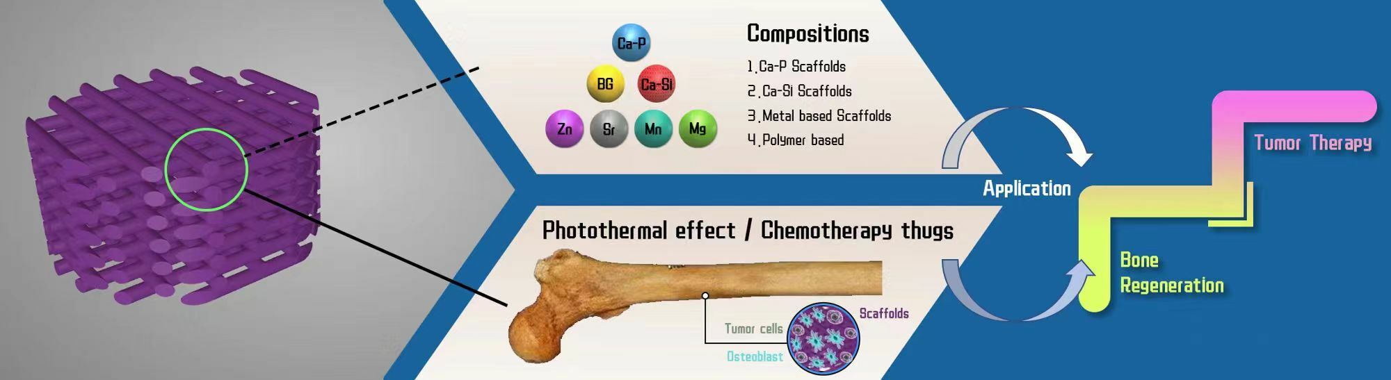
Abstract: Bone tumors, including primary bone tumors, invasive bone tumors, metastatic bone tumors, and others, are one of the most clinical difficulties in orthopedics. Once these tumors have grown and developed in the bone system, they will interact with osteocytes and other environmental cells in the bone system’s microenvironment, leading to the eventual damage of the bone’s physical structure. Surgical procedures for bone tumors may result in permanent defects. The dual-efficacy of tissue regeneration and tumor treatment has made biomaterial scaffolds frequently used in treating bone tumors. 3D printing technology, also known as additive manufacturing or rapid printing prototype, is the transformation of 3D computer models into physical models through deposition, curing, and material fusion of successive layers. Adjustable shape, porosity/pore size, and other mechanical properties are an advantage of 3D-printed objects, unlike natural and synthetic material with fixed qualities. Researchers have demonstrated the significant role of diverse 3D-printed biological scaffolds in the treatment for bone tumors and the regeneration of bone tissue, and that they enhanced various performance of the products. Based on the characteristics of bone tumors, this review synthesized the findings of current researchers on the application of various 3D-printed biological scaffolds including bioceramic scaffold, metal alloy scaffold and nano-scaffold, in bone tumors and discussed the advantages, disadvantages, and future application prospects of various types of 3D-printed biological scaffolds. Finally, the future development trend of 3D-printed biological scaffolds in bone tumor is summarized, providing a theoretical foundation and a larger outlook for the use of biological scaffolds in the treatment of patients with bone tumors.
Graphic Abstract:
Keywords: 3D-printed, scaffolds, bone tumors, nanomaterials
Graphical Abstract:
Introduction
Bone is a natural composite material that has yet to be properly replicated by man-made materials. As a result, the development of biological materials capable of repairing bone defects has become a significant focus of study in clinical orthopedics. In addition to tumors damaging the bone’s physical structure, the surgery for bone tumors will also result in bone defects.1 Bone tumors are categorized into primary bone tumors (osteosarcoma), invasive bone tumors (melanoma), and metastatic bone tumors (eg, lung cancer, prostate cancer, and others).2–5 Without exception, these tumors can impair the physical connections between bones by interacting with normal bone cells. In order to assure complete resection, surgical resection, the gold standard for the treatment of bone tumor disorders, will also cause harm to normal bone tissue.6–8 Nevertheless, tumor cells will persist in the bone tissue after surgery.9,10 Therefore, we require a substance that is not only capable of promoting bone tissue regeneration and repairing bone defect, but also of destructing residual bone tumor cells. In bone tissue engineering, scaffolds serve a critical role by creating a three-dimensional (3D) environment for cell adhesion and proliferation.11–13 A number of biological scaffolds, including bioceramics and metal nanoparticles, have been developed by simulating the structures and physical characteristics of normal bone tissue. In addition to effectively filling bone defects, these scaffolds can also kill residual tumor cells through their own physical and chemical properties or loaded drugs, thereby reducing postoperative complications of bone tumors and enhancing the life quality and survival time of patients.14–16 3D printing offers a rapid transition from product design or conceptual models to rapid prototyping and functional part manufacturing, allowing for the rapid fabrication of diverse complicated structural components without specialized tools.17,18 3D printing techniques make it possible to precisely adjust the geometry, connectivity, and porosity of biological scaffolds, allowing them to better adapt to cell growth and tissue regeneration.19–22 Based on an understanding of the current clinical difficulties of treating patients with bone tumors, such as irreparable bone defects, and tumor recurrence, this review summarized the current role of 3D-printed biological scaffolds in treating the patients with bone tumors, as well as the differences, advantages, and disadvantages of various types of scaffolds. In addition to offering a reference scheme for the current treatment of bone tumors with biological scaffolds, it is anticipated that the future trend of 3D printing biological scaffolds will give researchers with new ideas.
Destruction of Bone Structure by Bone Tumors
According to its macroscopic structure, bone is composed of cortical bone on the outside and trabecular bone on the inside. The cortical bone structure is extraordinarily dense (6% porosity and 130–190mpa compressive strength). Trabeculae cross-arrange in the internal bone to generate cancellous bone, which has a loose structure, but its arrangement and thickness are crucial for the bone to withstand pressure and weight.23–26 Bone tissue is made up of bone matrix and other cells at the microscopic structure. The bone matrix is composed of both inorganic and organic substances. Theses inorganic substances are predominantly made of hydroxyapatite crystals (calcium and phosphorus), compounds (sodium, potassium, magnesium, and fluorine), salts (chloride), and some trace elements (silicon, zinc, and copper). The majority of these organic substances consists of collagen fibers, osteocalcin, osteophosphoprotein, and other compounds.27–30
Most primary bone tumors are amenable to surgical treatment. The tumor resection will also result in bone defects at the resection site. Due to the presence of leftover tumor cells at the defect site, recurrence of bone tumors is often a significant source of distress for patients.31 In addition, although some invasive and metastatic bone tumors do not undergo surgical excision, the tumor cells themselves interact with bone cells and inorganic mineralized bone matrix, and can produce substances like parathyroid hormone-related protein (PTHrP) and other factors; these factors stimulate osteoclast aggregation and generates a cascade reaction, ultimately resulting in the transition of bone from the pre-osteolytic to the osteolytic phase.32–34 In addition, research on the relationship between breast cancer cells and bone matrix reveals that breast cancer cells inhibit bone formation by inhibiting the proliferation of osteoblasts and mesenchymal stem cells (MSC) and the activity of alkaline phosphatase (ALP), which leads to bone loss caused by metastatic bone tumors.35 It is evident that many forms of bone tumors can result in bone defects for various reasons. The introduction of biological scaffolds for such bone deficiency has inspired optimism. Biological scaffolds can not only mimic the chemical composition and layered structure of bone so that their physical and chemical properties are similar to those of human bone, but they can also transport cells, nutrients, oxygen and growth factors to promote tissue regeneration.36,37 Besides, the transporting of medications or the loading of particular metal ions and nanoparticles can provide biological scaffolds the power to inhibit or even kill tumor cells, which is a significant step forward for the application of biological scaffolds in bone tumors.38 Due to the various site and shape of bone defects, however, biological scaffolds are frequently unable to suit the needs of specific sites and shapes. The advent of 3D printing can compensate for this flaw.
Introduction of 3D Printing Technology, Different 3D Printing Methods and Their Advantages and Disadvantages
The conventional biological scaffolds are prepared by methods of gas flocculation, particle rinsing, and phase isolation.39–41
However, these preparation methods are incapable of customizing product properties like as porosity, permeability, and connection. Professor Sachs of the Massachusetts Institute of Technology innovatively created 3D printing technology, often known as additive manufacturing or fast printing prototype, to overcome these restrictions.42 Through layer-by-layer deposition, curing, and material fusion, the 3D digital model is finally transformed into a physical model. This manufacturing process not only successfully controls costs, but also allows for the modification of the 3D digital model based on specific requirements, the rapid and efficient realization of the design scheme, and the ultimate creation of customized products. Over time, the 3D printing technology has evolved from photosculpture and geomorphology to selective laser sintering (SLS), fused deposition modeling (FDM), and stereolithography (SLA), ink jet 3D printing, adhesive droplet and powder bed-based AM, digital laser processing, continuous liquid interface production.43–46 Using powder bed melting technology, SLS fabricates scaffolds in a directional layering manner by the repetitive deposition of powder particle materials and laser or electron beam melting or sintering, which is mostly used for metallic printing.FDM is the most common technique for printing polymers, which entails injecting thermoplastic polymers into a heating chamber, melting them, and extruding them through a nozzle under pressure.44 For printing, the tiny filament-like material was layered from bottom to top, row by row, while the heated nozzle moved over the layer contour. Currently, FDM is economic and frequently used.47 SLA printing process is similar to SLS, with the exception that powder is deposited on the print bed instead of a liquid photosensitive resin, and a laser (DLP utilizes a photomask instead of a laser) is used to track the product layer by layer until it is produced. Both SLA and DLP have the capacity to print high-resolution, structurally complicated items. However, the implementation of these two technologies is limited by the toxicity of the photosensitive resins employed as raw materials and the expense of printing equipment. Inkjet printing has limited use, and its application concept is comparable to that of 2D inkjet printing, which is often used to create product molds rather than the actual product.47 The benefit of 3D-printing printing is the ability to alter the product’s shape, porosity/pore size, and other mechanical properties as needed, instead of having fixed qualities like natural and manufactured substances. In addition, 3D printing allows for the rapid production of products and the reduction of experimental repetition time.48 (Table 1) In recent years, since 3D printing has garnered broad interest in the field of biomedicine, it has been regarded as the most optimal technical method for the advancement of scaffold-based tissue engineering.
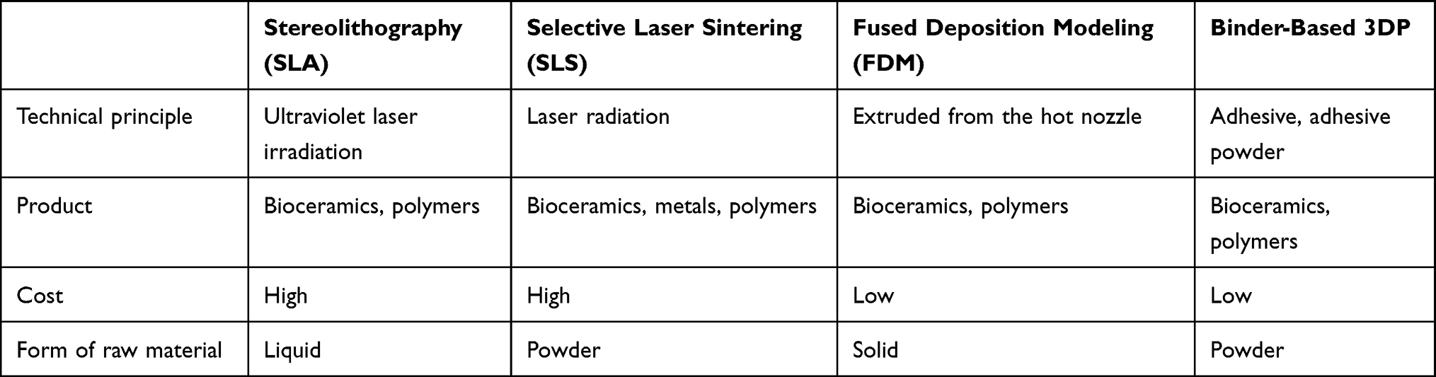 |
Table 1 Different Technical Means Applied to 3D Printing |
Application of Different Types of 3D Printed Biological Scaffolds in Bone Tumors
The biological scaffolds utilized in the treatment of bone tumors should have the following characteristics: 1) a porous structure, which facilitates the transport of nutrients, medicines, and cell migration. 2) Biocompatibility and biodegradability: the biocompatibility ensure the scaffolds are not rejected by the immune system in vivo and decreases the inflammatory response; the biodegradability assures that the scaffolds do not create substances that can harm the human body. 3) Diversified mechanical strength, allowing the scaffolds to accommodate various defect sites. 4) Biological activity, which causes biochemical reactions with bodily tissues, resulting in the establishment of unique chemical linkages between organisms and substances.49–51 The mechanical strength of 3D-printed biological scaffolds can be precisely adjusted to accommodate a variety of clinical scenarios, and their pore sizes can be tuned to meet specific functional requirements. Currently, bioceramics, metal alloy scaffolds, polymer scaffolds, nanoscaffolds, and composite scaffolds comprise the majority of 3D-printed biological scaffolds applied to patients with bone tumors52,53 (Table 2).
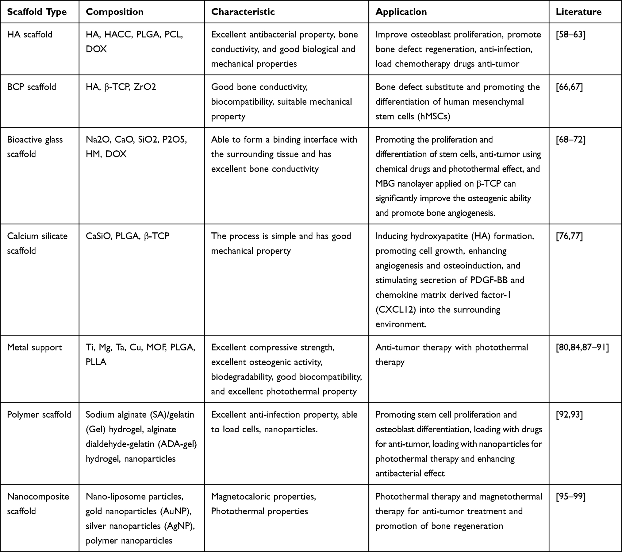 |
Table 2 Application of 3D-Printed Scaffold of Different Materials in Bone Tumors |
3D-Printed Bioceramic Scaffolds
Due to their similarity to inorganic bone components, bioceramic scaffolds are now the most commonly utilized 3D-printed scaffolds for bone defects. Compositionally, bioceramics are categorized primarily as calcium-phosphate-based bioactive materials and calcium-silica-based bioactive materials.40 Calcium-phosphorus-based bioactive materials include hydroxyapatite (HA), β-tricalcium phosphate (β-TCP), and biphasic calcium phosphate (BCP), whereas calcium-silica-based bioactive materials include bioactive glass, calcium silicate, tricalcium silicate, akermanite, and calcium silicate. These materials offer good bone conductivity, biocompatibility, and bioactivity, as well as robust compression qualities.54–56 However, conventional 3D-printed bioceramics scaffolds also exhibit disadvantages such as single function, difficulty inducing angiogenesis, low bone formation efficiency, inability to treat tumors, and ease of bacterial reproduction, which hinder the development of bioceramics scaffolds.57 Due to the advancement of research, bioceramic scaffolds composed of a single material are rarely employed. Based on traditional calcium phosphate-based and calcium silicon-based materials, 3D-printed bioceramics scaffolds are further optimized by methods such as apparent function change, trace element doping, and nanostructure construction, making them an ideal alternative material in bone tissue engineering.8,58
Calcium-Phosphorus-Based Bioceramic Scaffolds
Hydroxyapatite(HA)
HA bioceramics are the most prevalent calcium-phosphate-based bioceramics, which have positive effects on osteoblast proliferation and adhesion, due to their comparable chemical structure to human bone.59–61 However, scaffolds supported by HA degrade slowly and have poor mechanical strength, which inhibits bone repair and raises the risk of infection. Infection is one of the leading reasons of nonunion after trauma or tumor removal.62,63 Through 3D printing technology, Ying Yang et al prepared quaternized chitosan (HACC)-Grafted Polylactide-co-glycolide (PLGA)/ (HA) scaffolds, and their good antibacterial characteristics and bone conductivity were validated in vitro.HACC exhibits excellent antibacterial properties by electrostatically binding to bacterial membranes, thereby reducing the risk of bacterial resistance by a substantial margin. The biological properties of Hacc-coated bone scaffolds, titanium implants, and absorbable sutures are satisfactory both in vitro and in vivo. By varying the substitution degree (DS) of quaternary ammonium salt, the cytocompatibility and antibacterial activity of HACC were enhanced.64 They also demonstrated that PLGA/HA scaffolds transplanted with HACC exhibited significantly increased anti-infection and bone regeneration properties on rat and rabbit cortical bone defects. They also hypothesized that the degradation velocity of the scaffolds was strongly tied to its antibacterial effect, which would explain why many scaffolds with poor biodegradability had a greater risk of infection. This is a typical example of constructing composite scaffolds using polymers and HA.65 Likewise, Fan Liu et al fabricated biodegradable PCL/HA composites using poly-ε-caprolactone (PCL) and hydroxyapatite (HA) nanoparticles. Using melt deposition formation, PCL/HA scaffolds were prepared from PCL/HA composites. A series of in vitro assessments revealed that PCL/HA composites had strong biodegradability, low cytotoxicity, and good biocompatibility, hence enhancing the osteoblasts MC3T3-E1 cell proliferation. Moreover, multifunctional 3D-printed PCL/HA/DOX scaffolds with a composite design loaded with anticancer pharmaceuticals (doxorubicin, DOX) and attaining sustained drug release qualities might accelerate bone healing and are anticipated to inhibit tumor cells following removal of malignant bone tumors.66,67 Consequently, the development of suitable 3D-printed scaffolds can not only repair bone defects produced by bone tumors, but also inhibit tumor cells, but also delay tumor development, playing a role in preventing the recurrence and metastasis of bone tumors. As a form of additive manufacturing, 3D printing permits the customization of product parameters. For example, the 3D high-resolution (<210 μm) printing at room temperature was used to create scaffolds with regulated HA components. Yoontae Kim et al manipulated the formation rate and amount of HA by adjusting the concentration of Na2HPO4 in order to produce bioceramic scaffolds with varying amounts of HA in order to optimize the scaffolds’ mechanical characteristics, porosity, and osteoclast activity.68,69
Biphasic Calcium Phosphate (BCP)
BCP bioceramics are a blend of hydroxyapatite (HA) and β-tricalcium phosphate (β-TCP) in varying proportions, achieving good chemical bone bonding capacity and biological absorption qualities.70 TCP’s utility cannot be questioned. By continuously coating tricalcium phosphate (TCP) scaffolds with TiN microparticles and doxorubicin (DOX), Dang et al were able to achieve the combined effect of photothermal therapy and chemotherapy on osteosarcoma. By immersing the scaffold in a solution containing varied concentrations of TiN and DOX, it was possible to alter the quantity of TiN and DOX present in the scaffold. In vitro and in vivo, both precision photothermal treatment and local controlled release chemotherapy have shown positive therapeutic results.71 However, it has been demonstrated in vitro, in vivo, and in clinical models that BCP has superior biocompatibility, bone conductivity, and safety.72 Moreover, investigations have demonstrated that the bioceramic mixture of 60%HA and 40% β-TCP has a superior capacity for bone tissue regeneration and can be employed as a filler for bone defects produced by bone tumors or other factors.73 Using fused deposition modeling (FDM), Min-woo Sa et al effectively created 3D slurry foams including mixes. The microstructure and shape of BCP/ZrO2 scaffolds were validated by scanning electron microscopy (SEM), and the biocompatibility of BCP and BCP/ZrO2 scaffolds was assessed by measuring the mechanical strength using a compression testing machine. We examined their impact on MG63 cells by measuring cell proliferation. Human mesenchymal stem cells (hMSCs) were seeded onto BCP or BCP/ZrO2 to show the efficacy of BCP/ZrO2 for bone tissue engineering applications.By removing the combined material following a high-temperature sintering procedure, they obtained BCP/ZrO2 composite scaffolds with particular pore characteristics, porosity, and size. Not only did the inclusion of ZrO2 increased the mechanical strength of BCP scaffolds, but it was also shown that BCP/ZrO2scaffolds were biocompatible for the proliferation of MG63 cells for 7 days and were favourable to the differentiation of human mesenchymal stem cells (hMSCs).74
Calcium Silicon-Based Bioceramic Scaffolds
Bioactive Glass Scaffold
The most prevalent calcium-silica-based bioceramic scaffolds are bioactive glass scaffolds, whose primary components are Na2O, CaO, SiO2 and P2O5. Bioactive glasses need to be bone conductive and bone potent in order to promote stem cell proliferation and differentiation.75,76 It has also been established that bioactive glass degradation products could impact the cell cycle of osteoblasts and promote bone tissue formation. The production of a layer of bioactive hydroxyapatite (HA) and hydroxy-carbonate apatite (HCA) on the surface in contact with bodily fluids, which offers a binding interface between bioactive glass and surrounding tissue is a common denominator of bioactive glass function.77 Wentao Dang et al investigated the use of bioactive scaffolds to fill bone defect, sustain bone tissue, and stimulate bone formation, as well as to reduce drug resistance and tumor recurrence following continuous chemotherapy following surgical excision of osteosarcoma.78 They prepared multifunctional scaffolds by applying heme and doxorubicin (DOX) to the surface of 3D-printed bioactive glass (BGC-HM-DOX). BGC-HM-DOX scaffolds had an exceptional photothermal sensitivity to NIR laser irradiation because to the heme particles on their surface. Combining photothermal therapy with DOX chemotherapy enabled the BGC-HM-DOX scaffolds with to kill osteosarcoma cells in vitro and inhibit tumor growth in vivo. This 3D-printed bioactive glass scaffold with multifunction is a significant improvement over the conventional 3D-printed scaffolds, the latter of which is only utilized to fill bone defects caused by bone tumors. Patients with bone tumors benefit greatly from it.
In addition, research on mesoporous bioactive glass (MBG) is ongoing. Due to its organized mesoporous (nanoporous) structure, MBG has much improved surface area and pore volume than conventional bioactive glasses. The presence of silanol groups on the surface of MBG makes it simple to functionalize submicroparticles for a variety of drug delivery applications. M. Ravanbakhsh et al created submicron MBG particles of uniform size and shape for local drug delivery. Due to the negatively charged surface of MBG, the researchers introduced amine groups to change the surface charge to a positive charge to improve drug attachment and loading efficiency.79. Considering the unusual mesoporous structure and biological activity of MBG, its application to modify porous bioceramic scaffolds is of major importance.80,81 Yali Zhang et al constructed β-TCP porous scaffolds using 3D printing. The MBG nanolayers were then uniformly coated on the β-TCP scaffolds using a spin coating technique. MBG-β-TCP ultimately demonstrated more osteogenic and angiogenesis-related gene expression than BG-TCP. Compared to the β-TCP scaffold alone, the osteogenic capacity of the MBG-β-TCP scaffold was dramatically enhanced.82
Calcium Silicate Scaffold
Calcium silicate ceramics are a particular bioactive compounds that have been demonstrated to promote bone regeneration. Using 3D printing to construct CS scaffolds is a straightforward method with good mechanical properties.83 In 2005, Wenyuan Zhao et al investigated the in vitro biological activity of CaSiO by soaking it in simulated bodily fluid (SBF) for various durations.84 CaSiO slurry was found to stimulate hydroxyapatite (HA) production and dissolve slowly in SBF. CaSiO paste enhanced cell proliferation within a specific concentration range, as determined by indirect cytotoxicity screening. In a co-culture system of human bone marrow stromal cells (hBMSC) and human umbilical vein endothelial cells (HUVECs), the (PLGA)/calcium silicate scaffold raised the level of VEGF, resulting in a significant osteogenic and angiogenic synergy. Compared to β-TCP alone, 5% CaSiO3-β-TCP scaffolds increased angiogenesis and osteoinduction and encouraged the release of PDGF-BB and chemokine matrix derived factor-1 (CXCL12) into the surrounding environment.85
3D Printed Metal Composite Scaffolding
Metal implants have traditionally been a crucial material for repairing bone defects. The markers that define the suitability of metal materials are mechanical stability and biological reactivity, which may be done more effectively through 3D printing’s customization.86
Using 3D printing technologies to make metal implants may lessen the stiffness of metal implants by producing pores, hence decreasing overall stress shielding to obtain a closer resemblance to human bone and providing space for bone regeneration. In addition, metal ceramic materials, which are made by mixing metal materials with bioceramic materials, may complement the two substantially.87 Titanium (Ti), magnesium (Mg), and tantalum (Ta)are commonly used metals.88–90 The combining of metallic materials and bioceramic materials to prepare a ceramic-metal composition could significantly complement both substances. Xin Wang et al91 used 3D printing technology based on digital laser processing (DLP) to fabricate CaTiO3 bioceramical scaffolds and discovered that the color of the scaffolds became darker as the sintering temperature increased; when the color reached pink exposed to NIR laser, CaTi scaffolds exhibited the highest compressive strength and satisfactory photothermal properties. This permits the use of CaTi scaffolds as photothermal biomaterials for the effective in vitro and in vivo treatment of bone tumors. (Figure 1) In addition, both in vitro and in vivo bone formation was observed in the pink CaTi scaffolds. Therefore, the use of this biphasic metal-ceramic material to cure bone defects induced by malignancies is a potential method.92
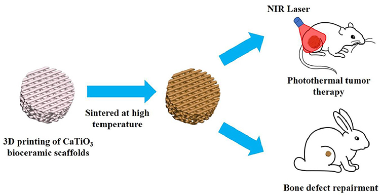 |
Figure 1 DLP-based 3D printed bifunctional calcium titanate scaffolds for photothermal therapy and bone regeneration. Notes: Reprinted from Journal of Materials Chemistry, 22(24), Wu C, Fan W, Zhou Y, Luo Y. 3D-printing of highly uniform CaSiO3 ceramic scaffolds: preparation, characterization and in vivo osteogenesis. 12288–12295, Copyright 2012, with permission from Elsevier.83 |
Due to its appropriate mechanical qualities, biodegradability, and good osteogenic activity, magnesium metal is also regarded a viable bioactive material for bone regeneration. However, pure magnesium scaffolds deteriorate too rapidly, and the enormous amount of hydrogen created would harm the human body’s normal tissue structure.93,94 To create composite scaffolds, the researchers decided to include Mg particles into a biodegradable polymer matrix (such as poly (Lactide-co-Glycolide), PLGA; Poly-l-lactide, PLLA). Recent investigations95 have demonstrated that the developed PLGA/Mg composite scaffold has outstanding osteogenic activity, biodegradability, and biocompatibility, and that its photothermal properties make it an essential antitumor medium for photothermal therapy. (Figure 2) These characteristics satisfy the prerequisites for future clinical translation. Similarly, porous tantalum (Ta) scaffold is a novel form of metallic scaffolding material. Utilizing selective laser melting (SLM) technology, researchers have created porous Ta scaffolds. In comparison to conventional Ta scaffolds, 3D-printed Ta scaffolds not only have regular pore structure and connectivity, but they also have a tunable elastic modulus and compressive strength, and their osteogenic impact in vitro and in vivo is greatly enhanced.96
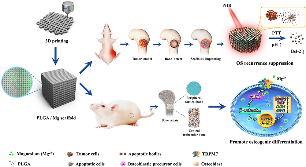 |
Figure 2 Schematic illustration of the 3D printed PLGA/Mg scaffold used to inhibit postoperative osteosarcoma recurrence and promote bone regeneration. Notes: Reprinted from Zadpoor AA, J Mech Behav Biomed Mater, 70, Mechanics of additively manufactured biomaterials. 1–6, Copyright 2017, with permission from Elsevier.87 |
Metal-organic frameworks (MOFs) are a recently found type of metal-organic frameworks that are often composed of metal ions or clusters of polytopic organic ligands. MOF-based compounds are frequently utilized as chemical drug carriers, and PTT reagents for tumor therapy are created by converting near-infrared light into thermal energy.97,98 Consequently, bioscaffolds prepared using these MOFs may be beneficial for chemotherapy or photothermal therapy of bone tumors. By means of a solvothermal reaction, Wentao Dang et al coated the surface of 3D-printed TCP scaffolds with Cu-TCPP nanosheets of around 50 nm in thickness. Due to the photothermal characteristics of Cu-TCPP nanosheets, it was experimentally proven that Cu-TCPP-TCP scaffolds effectively kill osteosarcoma cells in vitro and in vivo.99
3D-Printed Polymer Scaffolds
In addition to assisting bioceramic or metallic particles in preparing composite scaffolds, polymers have a promising future.100,101 By means of 3D printing, Ce Zhu et al prepared PEEK/graphene composite scaffolds. The scaffolds were subsequently electrophoretically deposited (EPD) with drug-loaded bioactive coatings based on hydroxyapatite (HA). HA is the predominant inorganic component of bone, and its release of Ca2+ and PO43- may stimulate bone repair. The in-scaffold graphene nanosheets function as potent PT conversion agents, whilst the loaded pharmaceuticals, such as antibacterial stearyl trimethylammonium chloride (STAC) and/or anticancer medication cisplatin (DDP), destroy tumor cells and bacteria through NIR laser-induced heating. This scaffold has been effectively used to the multimodal treatment of bone regeneration, tumor therapy, and antimicrobial therapy.102 Studies have demonstrated that 3D-printed sodium alginate (SA)/gelatin (Gel) hydrogel scaffolds promoted stem cell proliferation and osteoblast differentiation, and an obvious mineralization effect was observable in simulated body fluids after doping with different concentrations of nano-attapulgite, making them ideally suited for bone regeneration and defect repair.103 Nonetheless, a unique 3D-printed composite scaffold including alginate dialdehyde-gelatin (ADA-GEL) hydrogel has been developed, which is enhanced by lysozyme-loaded mesoporous cerium desilica-calcium nanoparticles (Lys-CE-MSN). In a simulated humoral environment, Lys-Ce-MSN could increase the mechanical strength of hydrogel composite scaffolds and trigger surface apatite mineralization. This ADA-GEL/Lys-Ce-MSN nanocomposite inhibits the formation and growth of malignancies in addition to inducing bone regeneration and resolving the infection.104
3D Printing Nano Material Composite Scaffolding
Nanomaterials are frequently employed in tissue engineering because they not only have sizes and shapes that penetrate most physiological barriers, but also imitate the functions of many spherical biological macromolecules.105 In bone tissue engineering, nanoparticles are thus often used as drug delivery vehicles, or play roles depending on their inherent electromagnetic and optical capabilities. In addition to mesoporous biological scaffolds previously mentioned, researchers frequently incorporate nanoparticles, such as nano-liposome particles,106 gold nanoparticles (AuNP),107 silver nanoparticles (AgNP),108 and polymer nanoparticles,109 into 3D-printed scaffolds to promote bone regeneration and inhibit the tumor cells growth.A set of investigations on the creation of nanocomposites for bone tissue engineering applications, including the cost-effective preparation of akermanite (CaMgSiO) nano-bioceramic, have provided considerable insight. Coating this nanoceramic solution onto a 3D filament-printed poly-lactic acid (EC-PLA) scaffold significantly increased its chemical stability and stress tolerance. The scaffold was then infused with Magnetic Nanoparticles (MNPs), and its porosity was remarkably comparable to that of regular bone tissue. In addition, the heat and magnetic properties of MNPS might attract cancer cells to separate and kill them, as well as restrict their proliferation.110–114 Sarkar et al integrated curcumin-encapsulated nano-liposomes into calcium phosphate 3D-printed scaffolds, which not only stimulated the bone formation but also shown antitumor effect.106 Moreover, it has been shown that both AuNPs and AgNPs produce anti-tumor effects in vitro owing to their superior features, such as their multi-functionalization capacity, high surface area-to-volume ratio, and high permeability. AgNP-prepared Ag-GO composite nanoparticles were incorporated into 3D-printed β-TCP scaffolds. The composite scaffolds not only had excellent osteogenic activity, but also induced oxidative stress in tumor cells after the generation of reactive oxygen species (ROS) by AgNP and promoted tumor cell apoptosis.107–109
Fe3O4 nanoparticles have magnetic thermal characteristics and are often used in tumor magnetic thermal treatment. Some pieces of research have combined calcium peroxide (CaO2) and iron oxide (Fe3O4) nanoparticles into 3D-printed AKT-Fe3O4-CaO2 scaffolds to achieve H2O2-self-sufficient chemodynamic therapy (CDT) for osteosarcoma, employing CaO2 nanoparticles as a source of hydrogen peroxide (H2O2) and the magnetic heating action of Fe3O4 to accelerate the CDTprocess. Obtaining H2O2-independent CDT osteosarcoma treatment. Adequate calcium ion (Ca2+) also stimulates osteanagenesis. This approach of interdisciplinary research blends nanoparticles with 3D-printed scaffolds to enable the advancement of the present frontier research-CDT for tumor therapy.115
Conclusion and Prospect
Despite the fact that bone tissue has a robust capacity for self-repair, it is difficult for patients with bone tumors to repair themselves due to the ongoing destruction of their own tissue or bone defects induced by surgical resection. The purpose of conventional biological scaffolds is to fill the defects. Due to the constraints of the preparation processes, however, the mechanical and biological qualities (biodegradability and biocompatibility) required to fulfill a variety of clinical circumstances are frequently not obtained. This disadvantage is remedied with the advent of 3D printing, which is able to suit specific needs by carefully regulating parameters and computer-controlled editing of objects with variable parameters. Initially, the most basic bioceramic scaffolds could cover the majority of application cases. It has gradually been obvious that just filling a defect in patients with bone tumors is of limited benefit. The true influence on life expectancy and life quality is the recurrence and progression of the tumor. Consequently, 3D-printed biological scaffolds for the treatment of tumors have increasingly entered the public consciousness. Researchers have mixed bioceramics with metal particles, polymers, and other materials to take advantage of the drug-loading and photothermal capabilities of the materials in the treatment of tumors, and even to overcome the faults of scaffolds that are prone to infection. Researchers have employed 3D printing as a manufacturing method to continually develop biological scaffolds suitable for the patients with bone tumors, compensating for the positive and negative properties of these materials. However, clinical application of these scaffolds is still some time away. In addition to upgrading the current 3D printing technology, the mechanical strength and durability of its products may be continuously modified to suit the actual clinical environment. Researchers should also begin at the microscopic molecular level of bone, while simulating the macroscopic bone structure, and improve the degree of fit between biological scaffolds and bone at the microscopic level, so that biological scaffolds can more accurately simulate the function of bone tissue and regenerative medicine can reach a new plateau.
Acknowledgments
This study was supported by the Natural Science Foundation of Liaoning Province, 2021-MS-291 and Jilin Provincial Science and Technology Department Grant (20200201436JC, ZBY. Yihang Ma, Boyin Zhang are co-first authors for this study.
Disclosure
The authors report no conflicts of interest in this work.
References
1. McGovern JA-O, Griffin M, Hutmacher DA-O. Animal models for bone tissue engineering and modelling disease. Dis Mod Mechan. 2018;11(4). doi:10.1242/dmm.033084
2. Kwakwa KA, Vanderburgh JP, Guelcher SA, et al. Engineering 3D models of tumors and bone to understand tumor-induced bone disease and improve treatments. Curr Osteoporos Rep. 2017;15(4):247–254. doi:10.1007/s11914-017-0385-9
3. Aboulafia AJ, Levine AM, Schmidt D, et al. Surgical therapy of bone metastases. Semin Oncol. 2007;34(3):206–214. doi:10.1053/j.seminoncol.2007.03.002
4. Johnson RW, Schipani E, Giaccia AJ. HIF targets in bone remodeling and metastatic disease. Pharmacol Ther. 2015;150:169–177. doi:10.1016/j.pharmthera.2015.02.002
5. Zhao X, Wu Q, Gong X, et al. Osteosarcoma: a review of current and future therapeutic approaches. Biomed Eng Online. 2021;20(1):24. doi:10.1186/s12938-021-00860-0
6. Li Y, Yang Y, Huang Z, et al. Bone defect reconstruction with autologous bone inactivated with liquid nitrogen after resection of primary limb malignant tumors: an observational study. Medicine. 2020;99(24):e20442. doi:10.1097/MD.0000000000020442
7. Liu W, Wang B, Zhang S, Li Y, Hu B, Shao Z. Wrist reconstruction after En bloc resection of bone tumors of the distal radius. Orthop Surg. 2021;13(2):376–383. doi:10.1111/os.12737
8. Huang Y, Yu X, He L, et al. 3D porous acellular cartilage matrix scaffold with surface mediated sustainable release of TGF-β3 for cartilage engineering. Chinese Chemical Letters. 2020;31(7):1797–1800.
9. Ma H, Luo J, Sun Z, et al. 3D printing of biomaterials with mussel-inspired nanostructures for tumor therapy and tissue regeneration. Biomaterials. 2016;111:138–148. doi:10.1016/j.biomaterials.2016.10.005
10. Yuan J, Ye Z, Zeng Y, et al. Bifunctional scaffolds for tumor therapy and bone regeneration: synergistic effect and interplay between therapeutic agents and scaffold materials. Mater Today Bio. 2022;15:100318. doi:10.1016/j.mtbio.2022.100318
11. Kim H, Hwangbo H, Koo Y, Kim G. Fabrication of mechanically reinforced gelatin/hydroxyapatite bio-composite scaffolds by core/shell nozzle printing for bone tissue engineering. Int J Mol Sci. 2020;21(9):3401.
12. Daneshmandi S, Dibazar SP, Fateh S. Effects of 3-dimensional culture conditions (collagen-chitosan nano-scaffolds) on maturation of dendritic cells and their capacity to interact with T-lymphocytes. J Immunotoxicol. 2016;13(2):235–242. doi:10.3109/1547691X.2015.1045636
13. Ye M, Mohanty P, Ghosh G. Biomimetic apatite-coated porous PVA scaffolds promote the growth of breast cancer cells. Mater Sci Eng C Mater Biol Appl. 2014;44:310–316. doi:10.1016/j.msec.2014.08.044
14. Woodard JR, Hilldore AJ, Lan SK, et al. The mechanical properties and osteoconductivity of hydroxyapatite bone scaffolds with multi-scale porosity. Biomaterials. 2007;28(1):45–54. doi:10.1016/j.biomaterials.2006.08.021
15. Gu Y, Zhang J, Zhang X, et al. Three-dimensional printed mg-doped β-TCP bone tissue engineering scaffolds: effects of magnesium ion concentration on osteogenesis and angiogenesis in vitro. Tissue Eng Regen Med. 2019;16(4):415–429. doi:10.1007/s13770-019-00192-0
16. Yang C, Ma H, Wang Z, et al. 3D printed wesselsite nanosheets functionalized scaffold facilitates NIR-II photothermal therapy and vascularized bone regeneration. Adv Sci. 2021;8(20):e2100894. doi:10.1002/advs.202100894
17. Ahangar P, Aziz M, Rosenzweig DH, et al. Advances in personalized treatment of metastatic spine disease. Ann Transl Med. 2019;7(10):223. doi:10.21037/atm.2019.04.41
18. Gu BK, Choi DJ, Park SJ, Kim YJ, Kim CH. 3D bioprinting technologies for tissue engineering applications. Adv Exp Med Biol. 2018;1078:15–28.
19. Butscher A, Bohner M, Hofmann S, et al. Structural and material approaches to bone tissue engineering in powder-based three-dimensional printing. Acta Biomater. 2011;7(3):907–920. doi:10.1016/j.actbio.2010.09.039
20. Elviri L, Foresti R, Bergonzi C, et al. Highly defined 3D printed chitosan scaffolds featuring improved cell growth. Biomed Mater. 2017;12(4):045009. doi:10.1088/1748-605X/aa7692
21. Roohani-Esfahani SI, Newman P, Zreiqat H. Design and fabrication of 3D printed scaffolds with a mechanical strength comparable to cortical bone to repair large bone defects. Sci Rep. 2016;6:19468. doi:10.1038/srep19468
22. Zheng P, Ding J. Calcium ion nanomodulators for mitochondria-targeted multimodal cancer therapy. Asian J Pharm Sci. 2022;17(1):1–3. doi:10.1016/j.ajps.2021.10.004
23. Eliaz N, Metoki N. Calcium phosphate bioceramics: a review of their history, structure, properties, coating technologies and biomedical applications. Materials. 2017;10(4). doi:10.3390/ma10040334
24. Barabaschi GD, Manoharan V, Li Q, Bertassoni LE. Engineering pre-vascularized scaffolds for bone regeneration. Adv Exp Med Biol. 2015;881:79–94.
25. Koester KJ, Ager JW, Ritchie RO. The true toughness of human cortical bone measured with realistically short cracks. Nat Mater. 2008;7(8):672–677. doi:10.1038/nmat2221
26. Sandberg OH, Aspenberg P. Inter-trabecular bone formation: a specific mechanism for healing of cancellous bone. Acta Orthop. 2016;87(5):459–465. doi:10.1080/17453674.2016.1205172
27. Mann FA, Payne JT. Bone healing. Semin Vet Med Surg Small Anim. 1989;4(4):312–321.
28. Andrulewicz-Botulińska E, Wiśniewska R, Brzóska MM, et al. Beneficial impact of zinc supplementation on the collagen in the bone tissue of cadmium-exposed rats. J Appl Toxicol. 2018;38(7):996–1007. doi:10.1002/jat.3608
29. Henry JP, Bordoni B. Histology, osteoblasts. In: StatPearls. Treasure Island (FL): StatPearls PublishingCopyright © 2022, StatPearls Publishing LLC; 2022.
30. Young MF. Bone matrix proteins: their function, regulation, and relationship to osteoporosis. Osteoporos Int. 2003;14(Suppl 3):S35–42. doi:10.1007/s00198-002-1342-7
31. Fang Y, Liu Z, Wang H, et al. Implantable sandwich-like scaffold/fiber composite spatiotemporally releasing combretastatin A4 and doxorubicin for efficient inhibition of postoperative tumor recurrence. ACS Appl Mater Interfaces. 2022;14(24):27525–27537. doi:10.1021/acsami.2c02103
32. Vičić I, Belev B. The pathogenesis of bone metastasis in solid tumors: a review. Croat Med J. 2021;62(3):270–282. doi:10.3325/cmj.2021.62.270
33. Wu YH, Gugala Z, Barry MM, et al. Optimization and characterization of a bone culture model to study prostate cancer bone metastasis. Mol Cancer Ther. 2022;21(8):1360–1368. doi:10.1158/1535-7163.MCT-21-0684
34. Jiang Z, Liu Y, Shi R, et al. Versatile polymer-initiating biomineralization for tumor blockade therapy. Adv Mater. 2022;34(19):e2110094. doi:10.1002/adma.202110094
35. Zhou X, Zhu W, Nowicki M, et al. 3D bioprinting a cell-laden bone matrix for breast cancer metastasis study. ACS Appl Mater Interfaces. 2016;8(44):30017–30026. doi:10.1021/acsami.6b10673
36. Sari M, Hening P, Ana ID, et al. Bioceramic hydroxyapatite-based scaffold with a porous structure using honeycomb as a natural polymeric Porogen for bone tissue engineering. Biomater Res. 2021;25(1):2. doi:10.1186/s40824-021-00203-z
37. Dang W, Chen W-C, Ju E, et al. 3D printed hydrogel scaffolds combining glutathione depletion-induced ferroptosis and photothermia-augmented chemodynamic therapy for efficiently inhibiting postoperative tumor recurrence. J Nanobiotechnol. 2022;20(1):266. doi:10.1186/s12951-022-01454-1
38. Liu Y, Li T, Ma H, et al. 3D-printed scaffolds with bioactive elements-induced photothermal effect for bone tumor therapy. Acta Biomater. 2018;73:531–546. doi:10.1016/j.actbio.2018.04.014
39. Lu D, Liu Y, Li W, et al. Development and application of 3D bioprinted scaffolds supporting induced pluripotent stem cells. Biomed Res Int. 2021;2021:4910816. doi:10.1155/2021/4910816
40. Fang Z, Chen J, Pan J, et al. The development tendency of 3D-printed bioceramic scaffolds for applications ranging from bone tissue regeneration to bone tumor therapy. Front Bioeng Biotechnol. 2021;9:754266. doi:10.3389/fbioe.2021.754266
41. Xu Z, Yuan L, Liu Q, et al. Crosslinking effect of dialdehyde cholesterol modified starch nanoparticles on collagen hydrogel. Carbohydr Polym. 2022;285:119237. doi:10.1016/j.carbpol.2022.119237
42. Liu Y, Yu Q, Chang J, et al. Nanobiomaterials: from 0D to 3D for tumor therapy and tissue regeneration. Nanoscale. 2019;11(29):13678–13708. doi:10.1039/c9nr02955a
43. Mirzaali MJ, Moosabeiki V, Rajaai SM, et al. Additive manufacturing of biomaterials-design principles and their implementation. Materials. 2022;15(15):5457. doi:10.3390/ma15155457
44. Awad A, Fina F, Goyanes A, et al. 3D printing: principles and pharmaceutical applications of selective laser sintering. Int J Pharm. 2020;586:119594. doi:10.1016/j.ijpharm.2020.119594
45. Parulski C, Jennotte O, Lechanteur A, et al. Challenges of fused deposition modeling 3D printing in pharmaceutical applications: where are we now? Adv Drug Deliv Rev. 2021;175:113810. doi:10.1016/j.addr.2021.05.020
46. Lee JY, Choi CS, Hwang KT, et al. Optimization of hybrid ink formulation and ipl sintering process for ink-jet 3D printing. Nanomaterials. 2021;11(5):1295.
47. Kelly CN, Miller AT, Hollister SJ, et al. Design and structure–function characterization of 3D printed synthetic porous biomaterials for tissue engineering. Adv Healthcare Mater. 2018;7(7):1701095. doi:10.1002/adhm.201701095
48. Vaezi M, Seitz H, Yang S. A review on 3D micro-additive manufacturing technologies. Int J Adv Manufactur Technol. 2013;67(5):1721–1754. doi:10.1007/s00170-012-4605-2
49. Ma H, Feng C, Chang J, et al. 3D-printed bioceramic scaffolds: from bone tissue engineering to tumor therapy. Acta Biomaterialia. 2018;79:37–59. doi:10.1016/j.actbio.2018.08.026
50. Nikolova MP, Chavali MS. Recent advances in biomaterials for 3D scaffolds: a review. Bioact Mater. 2019;4:271–292. doi:10.1016/j.bioactmat.2019.10.005
51. Kumar A, Nune KC, Murr LE, et al. Biocompatibility and mechanical behaviour of three-dimensional scaffolds for biomedical devices: process–structure–property paradigm. Int Mater Rev. 2016;61(1):20–45. doi:10.1080/09506608.2015.1128310
52. Lin C, Wang Y, Huang Z, et al. Advances in filament structure of 3D bioprinted biodegradable bone repair scaffolds. Int J Bioprint. 2021;7(4):426. doi:10.18063/ijb.v7i4.426
53. Li Z, Xu W, Yang J, et al. A tumor microenvironments-adapted polypeptide hydrogel/nanogel composite boosts antitumor molecularly targeted inhibition and immunoactivation. Adv Mater. 2022;34(21):e2200449. doi:10.1002/adma.202200449
54. Yamada S, Heymann D, Bouler JM, Daculsi G. Osteoclastic resorption of calcium phosphate ceramics with different hydroxyapatite/beta-tricalcium phosphate ratios. Biomaterials. 1997;18(15):1037–1041. doi:10.1016/S0142-9612(97)00036-7
55. Samavedi S, Whittington AR, Goldstein AS. Calcium phosphate ceramics in bone tissue engineering: a review of properties and their influence on cell behavior. Acta Biomaterialia. 2013;9(9):8037–8045. doi:10.1016/j.actbio.2013.06.014
56. Barba A, Maazouz Y, Diez-Escudero A, et al. Osteogenesis by foamed and 3D-printed nanostructured calcium phosphate scaffolds: effect of pore architecture. Acta Biomater. 2018;79:135–147. doi:10.1016/j.actbio.2018.09.003
57. Khalaf AT, Wei Y, Wan J, et al. Bone tissue engineering through 3D bioprinting of bioceramic scaffolds: a review and update. Life. 2022;12(6):903. doi:10.3390/life12060903
58. Yang Y, Li M, Zhou B, et al. Novel therapeutic strategy for bacteria-contaminated bone defects: reconstruction with multi-biofunctional GO/Cu-incorporated 3D scaffolds. Adv Therapeut. 2022;5(7):2200043. doi:10.1002/adtp.202200043
59. Huang J, Best SM, Bonfield W, et al. In vitro assessment of the biological response to nano-sized hydroxyapatite. J Mater Sci Mater Med. 2004;15(4):441–445. doi:10.1023/B:JMSM.0000021117.67205.cf
60. Filová E, Suchý T, Sucharda Z, et al. Support for the initial attachment, growth and differentiation of MG-63 cells: a comparison between nano-size hydroxyapatite and micro-size hydroxyapatite in composites. Int J Nanomedicine. 2014;9:3687–3706. doi:10.2147/IJN.S56661
61. Salinas AJ, Esbrit P, Vallet-Regí M. A tissue engineering approach based on the use of bioceramics for bone repair. Biomater Sci. 2013;1(1):40–51. doi:10.1039/C2BM00071G
62. Ni SY, Lin K, Chang J, et al. Beta-CaSiO3/beta-Ca-3(PO4)(2) composite materials for hard tissue repair: in vitro studies. J Biomed Mater Res A. 2008;85A(1):72–82. doi:10.1002/jbm.a.31390
63. Lu JX, Descamps M, Dejou J, et al. The biodegradation mechanism of calcium phosphate biomaterials in bone. J Biomed Mater Res. 2002;63(4):408–412. doi:10.1002/jbm.10259
64. Yang Y, Chu L, Yang S, et al. Dual-functional 3D-printed composite scaffold for inhibiting bacterial infection and promoting bone regeneration in infected bone defect models. Acta Biomater. 2018;79:265–275. doi:10.1016/j.actbio.2018.08.015
65. Yang Y, Yang S, Wang Y, et al. Anti-infective efficacy, cytocompatibility and biocompatibility of a 3D-printed osteoconductive composite scaffold functionalized with quaternized chitosan. Acta Biomater. 2016;46:112–128. doi:10.1016/j.actbio.2016.09.035
66. Liu F, Kang H, Liu Z, et al. 3D printed multi-functional scaffolds based on Poly(ε-Caprolactone) and hydroxyapatite composites. Nanomaterials. 2021;11(9):2456.
67. Kang H, Jiang X, Liu Z, et al. Biodegradable 3D printed scaffolds of modified poly (Trimethylene Carbonate) composite materials with Poly (L-Lactic Acid) and hydroxyapatite for bone regeneration. Nanomaterials. 2021;11(12):3215. doi:10.3390/nano11123215
68. Wang X, Ao Q, Tian X, et al. 3D bioprinting technologies for hard tissue and organ engineering. Materials. 2016;9(10):802. doi:10.3390/ma9100802
69. Kim Y, Lee E-J, Kotula AP, et al. Engineering 3D printed scaffolds with tunable hydroxyapatite. J Funct Biomater. 2022;13(2):34. doi:10.3390/jfb13020034
70. Le Nihouannen D, Duval L, Lecomte A, et al. Interactions of total bone marrow cells with increasing quantities of macroporous calcium phosphate ceramic granules. J Mater Sci Mater Med. 2007;18(10):1983–1990. doi:10.1007/s10856-007-3098-2
71. Dang W, Yi K, Ju E, et al. 3D Printed bioceramic scaffolds as a universal therapeutic platform for synergistic therapy of osteosarcoma. ACS Appl Mater Interfaces. 2021;13(16):18488–18499. doi:10.1021/acsami.1c00553
72. Wu SC, Hsu H-C, Hsu S-K, et al. Preparation and characterization of four different compositions of calcium phosphate scaffolds for bone tissue engineering. Mater Charact. 2011;62(5):526–534. doi:10.1016/j.matchar.2011.03.014
73. Zhao NB, Wang Y, Qin L, et al. Effect of composition and macropore percentage on mechanical and in vitro cell proliferation and differentiation properties of 3D printed HA/beta-TCP scaffolds. RSC Adv. 2017;7(68):43186–43196. doi:10.1039/C7RA07204J
74. Sa M-W, Nguyen B-NB, Moriarty RA, et al. Fabrication and evaluation of 3D printed BCP scaffolds reinforced with ZrO 2 for bone tissue applications. Biotechnol Bioeng. 2018;115(4):989–999. doi:10.1002/bit.26514
75. Wang XQ, Zhang L, Ke X, et al. 45S5 Bioglass analogue reinforced akermanite ceramic favorable for additive manufacturing mechanically strong scaffolds. RSC Adv. 2015;5(124):102727–102735. doi:10.1039/C5RA19272B
76. Rainer A, Giannitelli SM, Abbruzzese F, et al. Fabrication of bioactive glass-ceramic foams mimicking human bone portions for regenerative medicine. Acta Biomater. 2008;4(2):362–369. doi:10.1016/j.actbio.2007.08.007
77. Kokubo T, Takadama H. How useful is SBF in predicting in vivo bone bioactivity? Biomaterials. 2006;27(15):2907–2915. doi:10.1016/j.biomaterials.2006.01.017
78. Dang W, Jin Y, Yi K, et al. Hemin particles-functionalized 3D printed scaffolds for combined photothermal and chemotherapy of osteosarcoma. Chem Enginer J. 2021;422:129919. doi:10.1016/j.cej.2021.129919
79. Ravanbakhsh M, Labbaf S, Karimzadeh F, et al. Mesoporous bioactive glasses for the combined application of osteosarcoma treatment and bone regeneration. Mater Sci Engineer C. 2019;104:109994. doi:10.1016/j.msec.2019.109994
80. Wu C, Zhou Y, Chang J, et al. Delivery of dimethyloxallyl glycine in mesoporous bioactive glass scaffolds to improve angiogenesis and osteogenesis of human bone marrow stromal cells. Acta Biomater. 2013;9(11):9159–9168. doi:10.1016/j.actbio.2013.06.026
81. Wu C, Chang J, Xiao Y. Mesoporous bioactive glasses as drug delivery and bone tissue regeneration platforms. Ther Deliv. 2011;2(9):1189–1198. doi:10.4155/tde.11.84
82. Zhang Y, Xia L, Zhai D, et al. Mesoporous bioactive glass nanolayer-functionalized 3D-printed scaffolds for accelerating osteogenesis and angiogenesis. Nanoscale. 2015;7(45):19207–19221. doi:10.1039/C5NR05421D
83. Wu CT, Fan W, Zhou Y, et al. 3D-printing of highly uniform CaSiO3 ceramic scaffolds: preparation, characterization and in vivo osteogenesis. J Mater Chem. 2012;22(24):12288–12295. doi:10.1039/c2jm30566f
84. Zhao W, Wang J, Zhai W, et al. The self-setting properties and in vitro bioactivity of tricalcium silicate. Biomaterials. 2005;26(31):6113–6121. doi:10.1016/j.biomaterials.2005.04.025
85. Li T, Peng M, Yang Z, et al. 3D-printed IFN-gamma-loading calcium silicate-beta-tricalcium phosphate scaffold sequentially activates M1 and M2 polarization of macrophages to promote vascularization of tissue engineering bone. Acta Biomaterialia. 2018;71:96–107. doi:10.1016/j.actbio.2018.03.012
86. Li ZH, Wang C, Li C, et al. What we have achieved in the design of 3D printed metal implants for application in orthopedics? Personal experience and review. Rapid Prototyp J. 2018;24(8):1365–1379. doi:10.1108/RPJ-10-2017-0205
87. Zadpoor AA. Mechanics of additively manufactured biomaterials. J Mech Behav Biomed Mater. 2017;70:1–6. doi:10.1016/j.jmbbm.2017.03.018
88. Ma LM, Wang X, Zhao N, et al. Integrating 3D printing and biomimetic mineralization for personalized enhanced osteogenesis, angiogenesis, and osteointegration. ACS Appl Mater Interfaces. 2018;10(49):42146–42154. doi:10.1021/acsami.8b17495
89. Xiong ZX, Liu W, Qian H, et al. Tantalum nanoparticles reinforced PCL scaffolds using direct 3D printing for bone tissue engineering. Front Mater. 2021;8. doi:10.3389/fmats.2021.609779
90. Meenashisundaram GK, Wang N, Maskomani S, et al. Fabrication of Ti plus Mg composites by three-dimensional printing of porous Ti and subsequent pressureless infiltration of biodegradable Mg. Mater Sci Engineer C. 2020;108:110478. doi:10.1016/j.msec.2019.110478
91. Wang X, Zhai D, Yao X, et al. 3D printing of pink bioceramic scaffolds for bone tumor tissue therapy. Appl Mater Today. 2022;27:101443. doi:10.1016/j.apmt.2022.101443
92. Lin HM, Shi S, Lan X, et al. Scaffold 3D-printed from metallic nanoparticles-containing ink simultaneously eradicates tumor and repairs tumor-associated bone defects. Small Methods. 2021;5(9):2100536. doi:10.1002/smtd.202100536
93. Brown A, Zaky S, Ray H, et al. Porous magnesium/PLGA composite scaffolds for enhanced bone regeneration following tooth extraction. Acta Biomater. 2015;11:543–553. doi:10.1016/j.actbio.2014.09.008
94. Lai Y, Li Y, Cao H, et al. Osteogenic magnesium incorporated into PLGA/TCP porous scaffold by 3D printing for repairing challenging bone defect. Biomaterials. 2019;197:207–219. doi:10.1016/j.biomaterials.2019.01.013
95. Long J, Zhang W, Chen Y, et al. Multifunctional magnesium incorporated scaffolds by 3D-Printing for comprehensive postsurgical management of osteosarcoma. Biomaterials. 2021;275:120950. doi:10.1016/j.biomaterials.2021.120950
96. Wang H, Su K, Su L, et al. Comparison of 3D-printed porous tantalum and titanium scaffolds on osteointegration and osteogenesis. Mater Sci Eng C Mater Biol Appl. 2019;104:109908. doi:10.1016/j.msec.2019.109908
97. Li JR, Kuppler RJ, Zhou HC. Selective gas adsorption and separation in metal-organic frameworks. Chem Soc Rev. 2009;38(5):1477–1504. doi:10.1039/b802426j
98. Zhang K, Meng X, Cao Y, et al. Metal-organic framework nanoshuttle for synergistic photodynamic and low-temperature photothermal therapy. Adv Funct Mater. 2018;28(42):1804634. doi:10.1002/adfm.201804634
99. Dang W, Ma B, Li B, et al. 3D printing of metal-organic framework nanosheets-structured scaffolds with tumor therapy and bone construction. Biofabrication. 2020;12(2):025005. doi:10.1088/1758-5090/ab5ae3
100. Zhang Y, Yu W, Ba Z, et al. 3D-printed scaffolds of mesoporous bioglass/gliadin/polycaprolactone ternary composite for enhancement of compressive strength, degradability, cell responses and new bone tissue ingrowth. Int J Nanomedicine. 2018;13:5433–5447. doi:10.2147/IJN.S164869
101. Fairag R, Li L, Ramirez-GarciaLuna JL, et al. A composite lactide-mineral 3D-printed scaffold for bone repair and regeneration. Front Cell Dev Biol. 2021;9:654518. doi:10.3389/fcell.2021.654518
102. Zhu C, He M, Sun D, et al. 3D-printed multifunctional polyetheretherketone bone scaffold for multimodal treatment of osteosarcoma and osteomyelitis. ACS Appl Mater Interfaces. 2021;13(40):47327–47340. doi:10.1021/acsami.1c10898
103. Liu C, Qin W, Wang Y, et al. 3D printed gelatin/sodium alginate hydrogel scaffolds doped with nano-attapulgite for bone tissue repair. Int J Nanomedicine. 2021;16:8417–8432. doi:10.2147/IJN.S339500
104. Monavari M, Medhekar R, Nawaz Q, et al. A 3D printed bone tissue engineering scaffold composed of alginate dialdehyde-gelatine reinforced by lysozyme loaded cerium doped mesoporous silica-calcia nanoparticles. Macromol Biosci. 2022;22(9):e2200113. doi:10.1002/mabi.202200113
105. Pelaz B, Alexiou C, Alvarez-Puebla RA, et al. Diverse applications of nanomedicine. ACS Nano. 2017;11(3):2313–2381. doi:10.1021/acsnano.6b06040
106. Sarkar N, Bose S. Liposome-encapsulated curcumin-loaded 3D printed scaffold for bone tissue engineering. ACS Appl Mater Interfaces. 2019;11(19):17184–17192. doi:10.1021/acsami.9b01218
107. Celikkin EX. Enhancing X-ray Attenuation of 3D Printed Gelatin Methacrylate (GelMA) Hydrogels Utilizing Gold Nanoparticles for Bone Tissue Engineering Applications. Polymers. 2019;11. doi:10.3390/polym11020367
108. Zhang Y, Zhai D, Xu M, et al. 3D-printed bioceramic scaffolds with antibacterial and osteogenic activity. Biofabrication. 2017;9(2):025037. doi:10.1088/1758-5090/aa6ed6
109. Suleman A, Kondiah PPD, Mabrouk M, et al. The application of 3D-printing and nanotechnology for the targeted treatment of osteosarcoma. Front Mater. 2021;8. doi:10.3389/fmats.2021.668834
110. Jasemi A, Kamyab Moghadas B, Khandan A, et al. A porous calcium-zirconia scaffolds composed of magnetic nanoparticles for bone cancer treatment: fabrication, characterization and FEM analysis. Ceramic Int. 2022;48(1):1314–1325. doi:10.1016/j.ceramint.2021.09.216
111. Dong X, Heidari A, Mansouri A, et al. Investigation of the mechanical properties of a bony scaffold for comminuted distal radial fractures: addition of akermanite nanoparticles and using a freeze-drying technique. J Mech Behav Biomed Mater. 2021;121:104643. doi:10.1016/j.jmbbm.2021.104643
112. Monshi M, Esmaeili S, Kolooshani A, et al. A novel three-dimensional printing of electroconductive scaffolds for bone cancer therapy application. Nanomed J. 2020;7(2):138–148.
113. Sharafabadi AK, Abdellahi M, Kazemi A, et al. A novel and economical route for synthesizing akermanite (Ca2MgSi2O7) nano-bioceramic. Mater Sci Engineer C. 2017;71:1072–1078. doi:10.1016/j.msec.2016.11.021
114. Salmani MM, Hashemian M, Khandan A. Therapeutic effect of magnetic nanoparticles on calcium silicate bioceramic in alternating field for biomedical application. Ceramic Int. 2020;46(17):27299–27307. doi:10.1016/j.ceramint.2020.07.215
115. Zhuang H, Qin C, Zhang M, et al. 3D-printed bioceramic scaffolds with Fe 3 S 4 microflowers for magnetothermal and chemodynamic therapy of bone tumor and regeneration of bone defects. Biofabrication. 2021;13(4):045010. doi:10.1088/1758-5090/ac19c7
 © 2023 The Author(s). This work is published and licensed by Dove Medical Press Limited. The full terms of this license are available at https://www.dovepress.com/terms.php and incorporate the Creative Commons Attribution - Non Commercial (unported, v3.0) License.
By accessing the work you hereby accept the Terms. Non-commercial uses of the work are permitted without any further permission from Dove Medical Press Limited, provided the work is properly attributed. For permission for commercial use of this work, please see paragraphs 4.2 and 5 of our Terms.
© 2023 The Author(s). This work is published and licensed by Dove Medical Press Limited. The full terms of this license are available at https://www.dovepress.com/terms.php and incorporate the Creative Commons Attribution - Non Commercial (unported, v3.0) License.
By accessing the work you hereby accept the Terms. Non-commercial uses of the work are permitted without any further permission from Dove Medical Press Limited, provided the work is properly attributed. For permission for commercial use of this work, please see paragraphs 4.2 and 5 of our Terms.