")
Back to Journals » Cancer Management and Research » Volume 12
The Clinical Diagnostic and Prognostic Value of Dickkopf-1 in Cancer
Received 19 March 2020
Accepted for publication 19 May 2020
Published 8 June 2020 Volume 2020:12 Pages 4253—4260
DOI https://doi.org/10.2147/CMAR.S254596
Checked for plagiarism Yes
Review by Single anonymous peer review
Peer reviewer comments 2
Editor who approved publication: Professor Seema Singh
Jie Li, Yan Gao, Wentao Yue
Central Laboratory, Beijing Obstetrics and Gynecology Hospital, Capital Medical University, Beijing 100026, People’s Republic of China
Correspondence: Wentao Yue
Central Laboratory, Beijing Obstetrics and Gynecology Hospital, Capital Medical University, No. 251, Yaojiayuan Road, Chaoyang District, Beijing 100026, People’s Republic of China
Email [email protected]
Abstract: The Wnt signaling pathway extensively participates in diverse processes such as embryonic development, maintenance of homeostasis and tumor pathogenesis. Dickkopf-1 (DKK1), a Wnt inhibitor, plays a vital role for over the past decades regarding its role in the regulation of several types of cancers. However, studies have shown that DKK1 is expressed differently in cancer and plays a role as a cancer-promoting factor or a tumor suppressor, which is worthy of further exploration. We herein study whether DKK1 is highly expressed in all cancers and plays a crucial role in promoting cancer. Furthermore, we discussed as to which stages of cancer development it plays in. Finally, the present detection methods were introduced and indicated the clinical application of DKK1 in tumor development.
Keywords: Wnt signaling pathway, Dickkopf-related protein1, cancer diagnosis, prognosis, cancer therapeutics
Introduction
Dickkopf-1, a 35-kDa protein, belongs to Dickkopf gene family which comprises of 5 evolutionarily conserved members, DKK1, DKK2, DKK3, DKK4, and a unique DKK3-related protein, DKKL1.1 The DKK gene family was first identified and named by Glinka et al in order to study the factors that influence Xenopus induced head development. DKKs have unique structure, with a signal sequence and share two conserved cysteine-rich domains.2,3 They play an important role in vertebrate development, such as antero–posterior axial patterning, limb development, somitogenesis, eye formation, and some other wnt-related developmental processes.1 Wnt signaling plays an important role in the early development of animal embryos, organ formation, tissue regeneration and other physiological processes. DKK1 negatively regulates WNT signaling pathway by competitively binding to LDL receptor related protein (LRP), which further participates in multiple biological processes.
The biological function and mechanism of DKK1 revealed its association with many diseases. In the early studies, DKK1 was shown to be associated with bone and nervous system development.4–9 Recent studies found that DKK1 plays a role in more and more diseases as well as cancers. Recently, Mazon et al also proposed that DKK1 can be used as an indicator of inflammatory reaction.10 Rani et al have confirmed that high expression of DKK1 in lesional skin of vitiligo patients caused melanocytes to age, which might in turn lead to decreased pigment.11 Some studies have also shown that DKK1 is associated with cognitive ability.12,13 For over years, numerous studies have been conducted to explore the role of DKK1 in various cancers. Very interestingly, they have revealed that DKK1 changes are cancer type-specific. Hence, in this paper, the expression of DKK1 in various cancers is discussed, and the significance of these expression changes is further elaborated.
DKK1 in Cancer
Expression in Various Types of Cancer
DKK1 expresses differently in different types of tumors and the levels are elevated in a wide variety of cancers (Table 1), such as lung cancer, non-small cell lung cancer, bladder cancer, hepatocellular carcinoma, cervical cancer, multiple myeloma, breast cancer, ovarian cancer,14–20 indicating a potential oncogenic function of DKK1. Several studies have shown that DKK1 expression is related to progression of cancer and a poor prognosis. However, it is down-regulated in some cancers, such as prostate cancer,21 colon cancer,22 suggesting it may be a tumor suppressor. What’s more, Sheng et al have tested and compared serum DKK1 concentrations in lung cancer, gastric cancer, colorectal cancer, ovarian cancer, cervical adenocarcinoma, benign lung disease, and healthy controls.19 The results showed different levels of DKK1 in different cancers, with the greatest variation in lung cancer. No significant difference was observed between healthy control group and benign lung disease. Serum DKK1 concentrations in patients with gastric cancer, colorectal cancer, ovarian cancer and cervical cancer were significantly low than those in healthy controls. In addition, they also demonstrated the potential value of high serum levels of DKK1 as a prognostic indicator in lung cancer patients. By predicting and identifying the transcriptional regulatory elements of lung cancer-specific DKK1 locus, they confirmed that specific transcriptional regulatory elements can promote DKK1 promoter activity and lead to high expression of DKK1.23 Through bioinformatics analysis of four pancreatic cancer transcriptome microarray data sets, they confirmed that DKK1 is highly expressed in pancreatic cancer.24 Taken together, these studies confirmed that DKK1 expression in cancer is diverse.
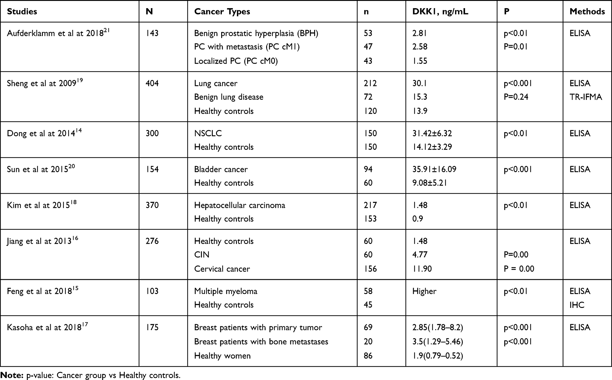 |
Table 1 Different Expression of Serum DKK1 in Different Cancers |
According to different changes in DKK1 expression in various cancers, further studies were conducted to explore the significance of these changes in DKK1. DKK1 is secreted as an antagonist in the Wnt/β-catenin signaling pathway and some studies have revealed the functional role of DKK1 in cancer progression.
The Role in Cancer Progression
DKK1 is considered as a negative regulator of Wnt signaling pathway and plays a vital role in the carcinogenic process. DKK1 is involved in tumorigenesis and invasion of several tumors. Zhou et al have demonstrated that downregulation of DKK1 is responsible for high proliferation capacity of LM-MCF-7 breast cancer cells by flow cytometry and 5-bromo-2-deoxyuridine (BrdU) incorporation assay.25 The specific mechanism involves loss of control of Wnt/catenin signaling pathway. They have found that DKK1 is highly expressed in ovarian somatic (OSC) cells and promoted the invasion of these cells either alone or by activating P-JNK1.26 Takahashi et al utilized a short hairpin RNA (shRNA)-based knockdown system to examine the role of DKK1 in cultured pancreatic carcinoma cells, and the results showed that the knockdown of DKK1 suppressed the migration and invasion of human pancreatic DAC cell line SUIT-2 and its metastatic subline S2-CP8 in vitro.27 In the study regarding the mechanism of miR-1-3p in oral squamous cell carcinoma (OSCC) cells, the levels of miR-1-3p and DKK1 expression in OSCC were detected using reverse-transcription-quantitative PCR and Western blotting. The target relationship between mir-1-3p and DKK1 was determined by dual-luciferase reporter gene analysis. The effects on cell progression were examined by MTT, transwell, colony formation and flow cytometry, and the results confirmed that mir-1-3p inhibited progression of OSCC by inhibiting DKK1 expression.28 All these studies confirmed the role of DKK1 in the invasion and proliferation of some cancers. The proliferation and invasion of cancer cells can be controlled by regulating the levels of DKK1.
DKK1 is involved in tumorigenesis, invasion, and metastases of cancer. Some studies have investigated high levels of DKK1 in 37 human hilar cholangiocarcinoma (HCCA) biopsy samples by immunohistochemistry, due to the correlation of high expression of DKK1 with metastasis to the hilar lymph nodes.29 They also utilized small interference or short hairpin RNA expression vector to knockdown DKK1 for inhibiting cell proliferation, colony formation and migration. Li et al examined cell migration and invasion capability using wound healing assay and boyden chamber assay, and confirmed that the positive expression of DKK1 is related to lymph node metastasis by immunohistochemistry.30 Bone is one of the most common metastatic sites in advanced malignant tumors. Recent studies have revealed that DKK1 is closely related to the occurrence and development of malignant tumors,17 especially to bone metastasis of malignant tumors. Some studies have shown that DKK1 acts as a tumor suppressor in breast cancer cells by inhibiting WNT signals or by mechanisms that do not rely on β-catenin-dependent transcription.31,32 DKK1 protein is overexpressed in the serum of breast cancer patients with bone metastases and those with bone metastases. The use of anti-DKK1 antibody in bone metastasis was developed by studying the mechanism of its action.33 Many in vitro and in vivo preclinical studies have identified the potential therapeutic effect of DKK1 antibody on multiple myeloma (MM) related bone disease.34 Compared with myeloma, DKK1 antibody is considered not effective in treating breast cancer bone metastasis.33 Massive studies have confirmed that DKK1 plays a role in bone metastases of lung cancer.35,36 However, the mechanism by which overexpression of DKK1 predicts poor prognosis in patients with lung bone metastasis is still unclear. Further studies are needed to determine the role of DKK1 in bone metastases in lung cancer. DKK1 is related to invasion and metastasis of various cancers, and its osteolytic effect plays an important role in cancer bone metastasis, and the application of anti-DKK1 antibody in bone metastasis can be developed based on this mechanism.
Through ELISA and a novel time-resolved immunofluorometric assay (TR-IMRA), Sheng et al have investigated the serum concentrations of DKK1 in patients with lung cancer and some other types of cancers, and compared their results with healthy controls.19 The serum DKK1 concentrations were significantly higher in lung cancer patients than others. They also measured CYRFA21-1 in the same set of samples from the NSCLC group and NSE in NSCLC. They found that DKK1 has higher sensitivity and specificity than other tumor markers CYRFA21-1 and NSE. They also confirmed the role of DKK1 in the diagnosis of certain cancers, particularly in lung cancer, and also confirmed that DKK1 can improve the sensitivity and specificity of lung cancer detection by combining the detection of tumor markers CYRFA21-1 and NSE. Kim et al also used in vitro reverse-transcription polymerase chain reaction (RT-PCR), wound healing assays, invasion assays, and ELISAs for detecting DKK1. The results revealed that DKK1 might be a key regulator in HCC progression and also acts as a potential diagnostic indicator.18 Fouad et al have examined and analyzed the serum samples of 50 patients with hepatitis C virus (HCV) related HCC, 20 patients with chronic hepatitis C and 20 patients with HCV-related liver cirrhosis. They found that the serum DKK1 could potentially be used for early diagnosis of HCC.37 A large-scale, multicenter study has also confirmed the clinical significance of DKK1 as a serum protein marker of HCC, especially in the early stage.38 Studies have also shown that the use of DKK1 or in combination with other biomarkers can prove to be clinically helpful to better understand the pathological condition of PDAC and improve its early detection and treatment.39 Some studies have also confirmed it’s the serologic diagnostic value of DKK1 in epithelial ovarian carcinoma40 and pancreatic cancer.41 In general, the serum concentration of DKK1 demonstrated significant changes in certain cancer types and has diagnostic significance. What’s more, some studies have found that DKK1 autoantibodies can act as a diagnostic biomarker in cancer.42 Peng et al have confirmed that DKK1 combined with DKK1 autoantibodies could improve the sensitivity and specificity of esophageal cancer detection. Their results indicated that combining DKK1 with DKK1 autoantibodies may be a valuable tool for early detection of ESCC.43 Also, our team have confirmed the diagnostic value of DKK1 autoantibodies in non-small cell lung cancer.44
In summary, DKK1 has been considered as a novel serologic diagnostic marker of cancer. And in some cancers, DKK1 combined with DKK1 antibodies could improve diagnostic value.
Through biological analysis and identification, They proved that DKK1 might be a candidate for pancreatic ductal adenocarcinoma (PDAC).24 Kaplan-Meier survival analysis then revealed that DKK1 showed a significant association with the overall survival in patients with pancreatic cancer. They also confirmed DKK1 as a therapeutic target and a prognostic marker of PDAC. They also analyzed the expression of DKK1 in PDAC at mRNA and protein levels, and the results showed high expression in pancreatic cancer by real-time PCR. DKK1 expression in PDAC tissue immunohistochemical staining showed close relation to T staging and lymph node metastasis. The study further confirmed that patients with high DKK1 had shorter survival (OS) and relapse-free survival (RFS).45 Sun et al in his study showed that serum DKK1 level was an independent prognostic factor for influencing the OS of bladder cancer patients through multivariate analysis, and a high level of DKK1 was associated with the progression and prognosis of bladder cancer.20 Previous studies have demonstrated that DNA methylation silencing of WNT target gene DKK1 can be used as a prognostic marker of methyl specific PCR in stage II colon cancer (CC) patients.46. In addition, an unbiased random forest analysis of transcriptome data from 247 HCC patients showed that high DKK1 was associated with advanced tumor stage and poor overall survival and disease-free survival.47 In summary, DKK1 has been proved to be related to the prognosis of various cancers and has clinical therapeutic significance.
Progress in Detection Methods
Detection of Serum and Free Fragments
In the early stage, the changes of DKK1 expression in patients were mainly determined by detecting the concentration of DKK1 in the serum. The commonly used methods include enzyme-linked immunosorbent assay and time-immunofluorescence resolution. These methods accurately reflect the changes of DKK1 level. With further research, RT-PCR, reverse transcription PCR, and Western blot methods are also used for quantitative analysis of DKK1. And these changes of DKK1 level in specific sites were detected by immunohistochemistry for metastases. The DKK1 gene in humans is located on 10q11.2 (NM: 012242.2). DKK1 through gene sequencing and polymorphism analysis has become a hot research topic. Koromila et al have amplified and sequenced the entire DKK1 gene sequence, demonstrating that the DKK1 gene polymorphisms showed no significant association with osteoporosis.48 The study showed expression of DKK1 by reconstructing eukaryotic expression vector pcmv-tag-2b (Invitrogen), preparing DKK1 target sequences, and verifying DKK1 expression in non-small cell lung cancer by sequence analysis.30 Bao et al have obtained the total mRNA from the cervical cancer tissues, followed by obtaining human DKK1 gene fragments through RT-PCR and PCR, constructing the recombinant plasmid pcmv-ha2/DKK1 successfully, and identifying the eukaryotic expression products of the gene.49 This laid a foundation for further study on the biological activity of DKK1 protein in tumors. These methods assist in accurately reflecting the expression and distribution differences of DKK1.
Application of DKK1 Autoantibody Detection
Numerous studies have shown that the tumor itself induced the immune response of the body, produced the autoantibodies of anti-tumor related antigens, and detected when the tumor antigen expression remains very low.50,51 Researchers investigated regarding DKK1-related autoantibodies for improving the sensitivity of detection.42,43 At present, the methods used to detect autoantibodies included enzyme linked immunosorbent assay, protein chip, polypeptide chip and antibody chip. During the early stages, our team has confirmed the presence of DKK1 autoantibodies in the serum of patients with non-small cell lung cancer through Western blotting, and detected DKK1 autoantibodies in the serum by indirect ELISA. This indicated that DKK1 autoantibodies play an important role in the diagnosis, treatment and prognosis of non-small cell lung cancer.41 Then Shen et al used polypeptide array technology to detect the epitope of DKK1 autoantibody recognition in the serum of patients with and healthy controls. The results identified 4 high-frequency loci were by DKK1 autoantibodies: aa67-84 (Pep A), aa37-54 (Pep B), aa145-156 (Pep C), and aa247-261 (PepD). The relationship between DKK1 autoantibody subtype and clinicopathological factors was analyzed, as well as the diagnostic value and prognostic value in non-small cell lung cancer. The expression of the 4 DKK1 autoantibody subtypes in NSCLC showed significant up-regulation, and the 3 DKK1 autoantibody subtypes of Pep B, Pep C and Pep D showed association with TNM staging of non-small cell lung cancer and distant metastasis. Also Pep B autoantibodies may play an important role in the occurrence and development of non-small cell lung cancer. Also, their team further clarified the clinical significance of seroautoantibodies and evaluated their feasibility in immunological diagnosis and prognosis.44 Cox regression analysis showed that anti-pepB antibody was considered as an independent prognostic factor of non-small cell lung cancer, suggesting that Pep B antibody subtype was the most valuable biomarker with good prognosis in NSCLC. Whether such unique epitopes can be found in other cancer types, and whether such a method can be used to diagnose cancer, needs further exploration.
Role in Cancer Therapeutics
Based on various roles of DKK1 in diseases, it has lately become a focus of attention in cancer research, both as a biomarker and potential therapeutic target. Numerous studies have shown that the osteolytic effect of DKK1 is vital in the bone metastasis of various cancers. Elevated levels of DKK1 in bone marrow cells and serum inhibited the differentiation of osteoblast precursors. The WNT pathway promotes osteoblast formation and bone formation, and plays a crucial role in the growth, bone balance, or fracture repair.52 Many extracellular Wnt antagonists regulate bone formation by binding directly to Wnt ligands or by competing with Wnt ligands to co-receptors 5 and 6 (LRP5 and LRP6) associated with lipoprotein expression on the surface of bone cells. The possible mechanism is that DKK1 interacts with LRP 5/6 in the WNT pathway, thereby affecting bone formation. They developed a cyclic oligopeptide based on DKK1-LRP 5/6 interaction.53 It inhibits the effect of DKK1 on osteoblast differentiation, and removes the inhibition of Wnt signaling pathway, thus reducing the tumor burden of MM. What’s more, Liu et al have constructed human DKK1 and HSP70 fusion DNA vaccine (hDKK1-hhsp70). Injection into mice can promote the apoptosis of tumor cells, inhibit the proliferation of tumor cells and extend the OS of mice. This might act as an effective immunotherapy in patients with MM.54 Scleroprotein, a secretory factor produced by bone cells, blocked at least a part of the Wnt signaling by binding to LRP5 and LRP6. Deletion of scleroprotein genes and DKK1 leads to increased bone formation in mice and humans, resulting in high bone mass. Antibodies that inhibit sclerosing proteins (scl-ab) or DKK1 (DKK1-ab) are used for treating bone diseases. Combined application of scl-ab and DKK1-ab is used in inhibiting the synergistic effect of bone formation in response to this mechanism. Florio et al have constructed the hetero-ds antibody (hetero-ds) that inhibited these two molecules. The study showed that double inhibition of sclerosing proteins and DKK1 resulted in synergistic bone formation in rodents and non-human primates.55 It also showed better bone repair activity than monotherapy, improving new solutions for the treatment of bone diseases. BHQ880 is a human neutralizing IgG1 anti-DKK1 monoclonal antibody, which has been clinically tested in Phase IB to study its effect on multiple myeloma-related bone diseases, and has been shown to have potential anti-myeloma activity.56 Their research found that anti-DKK1 antibodies have an inhibitory effect on lung cancer cell invasion and growth. In addition, systemic administration of anti-DKK1 antibodies can effectively inhibit the growth of DKK1-positive tumors transplanted into nude mice without any obvious toxicity.57 Further preclinical studies are needed to prove its efficacy and safety in the treatment of individual cancers, the anti-DKK1 antibody can meet the development requirements of human clinical trials.
Conclusion
To sum up, several studies have shown that the expression of DKK1 in various cancers is increased, promoting the development of cancer. It also promotes tumor invasion and metastasis and is associated with the prognosis of cancer. DKK1 in cancer has been used as an effective target for monitoring and treating cancer. MicroRNA is a class of non-coding small RNA molecules (18 to 25 nucleotides) that inhibits transcription, translation of target mRNA, or act as shear target mRNA to promote its degradation. MicroRNA is believed to play an important role in the regulation of development and is associated with various cancers. Recent studies have found that DKK1 acts as the target in various cancers, and miRNA acts directly on DKK1.28,58,59 It also assists in treating cancer, which deserves further research. At present, DKK1 has been used for drug development, vaccine manufacturing and autoantibody application in response to current possible mechanisms of action. Of course, more experiments are needed to further study the specific mechanism of action as well as role of DKK1 as a clinically applicable biomarker and gene therapy target in more diseases.
Acknowledgments
Wentao Yue was supported by The National Natural Science Foundation of China (Grant/Award Numbers:81672838 China), The Beijing Municipal Administration of Hospitals Clinical Medicine Development of Special Funding (No. XMLX201705), and The Beijing Municipal Science & Technology Commission (No. Z181100001718193).
Disclosure
The authors report no conflicts of interest in this work.
References
1. Niehrs C. Function and biological roles of the Dickkopf family of Wnt modulators. Oncogene. 2006;25(57):7469–7481. doi:10.1038/sj.onc.1210054
2. Fedi P, Bafico A, Nieto Soria A, et al. Isolation and biochemical characterization of the human Dkk-1 homologue, a novel inhibitor of mammalian Wnt signaling. J Biol Chem. 1999;274(27):19465–19472. doi:10.1074/jbc.274.27.19465
3. Glinka A, Wu W, Delius H, Monaghan AP, Blumenstock C, Niehrs C. Dickkopf-1 is a member of a new family of secreted proteins and functions in head induction. Nature. 1998;391(6665):357–362. doi:10.1038/34848
4. Cappuccio I, Calderone A, Busceti CL, et al. Induction of Dickkopf-1, a negative modulator of the Wnt pathway, is required for the development of ischemic neuronal death. J Neurosci. 2005;25(10):2647–2657. doi:10.1523/JNEUROSCI.5230-04.2005
5. Caricasole A, Copani A, Caraci F, et al. Induction of Dickkopf-1, a negative modulator of the Wnt pathway, is associated with neuronal degeneration in Alzheimer’s brain. J Neurosci. 2004;24(26):6021–6027. doi:10.1523/JNEUROSCI.1381-04.2004
6. Carter M, Chen X, Slowinska B, et al. Crooked tail (Cd) model of human folate-responsive neural tube defects is mutated in Wnt coreceptor lipoprotein receptor-related protein 6. Proc Natl Acad Sci U S A. 2005;102(36):12843–12848. doi:10.1073/pnas.0501963102
7. Gregory CA, Gunn WG, Reyes E, et al. How Wnt signaling affects bone repair by mesenchymal stem cells from the bone marrow. Ann N Y Acad Sci. 2005;1049(1):97–106. doi:10.1196/annals.1334.010
8. Gunn WG, Conley A, Deininger L, Olson SD, Prockop DJ, Gregory CA. A crosstalk between myeloma cells and marrow stromal cells stimulates production of DKK1 and interleukin-6: a potential role in the development of lytic bone disease and tumor progression in multiple myeloma. Stem Cells. 2006;24(4):986–991. doi:10.1634/stemcells.2005-0220
9. Tian E, Zhan F, Walker R, et al. The role of the Wnt-signaling antagonist DKK1 in the development of osteolytic lesions in multiple myeloma. N Engl J Med. 2003;349(26):2483–2494. doi:10.1056/NEJMoa030847
10. Mazon M, Larouche V, St-Louis M, Schindler D, Carreau M. Elevated blood levels of Dickkopf-1 are associated with acute infections. Immun Inflamm Dis. 2018;6(4):428–434. doi:10.1002/iid3.232
11. Rani S, Chauhan R, Parsad D, Kumar R. Effect of Dickkopf1 on the senescence of melanocytes: in vitro study. Arch Dermatol Res. 2018;310(4):343–350. doi:10.1007/s00403-018-1820-1
12. Ross RD, Shah RC, Leurgans S, Bottiglieri T, Wilson RS, Sumner DR. Circulating Dkk1 and TRAIL are associated with cognitive decline in community-dwelling, older adults with cognitive concerns. J Gerontol a Biol Sci Med Sci. 2018;73(12):1688–1694. doi:10.1093/gerona/glx252
13. Yu C, Seaton M, Letendre S, Heaton R, Al-Harthi L. Plasma dickkopf-related protein 1, an antagonist of the Wnt pathway, is associated with HIV-associated neurocognitive impairment. AIDS. 2017;31(10):1379–1385. doi:10.1097/QAD.0000000000001481
14. Dong LL, Qu LY, Chu LY, Zhang XH, Liu YH. Serum level of DKK-1 and its prognostic potential in non-small cell lung cancer. Diagn Pathol. 2014;9(1):52. doi:10.1186/1746-1596-9-52
15. Feng Y, Zhang Y, Wei X, Zhang Q. Correlations of DKK1 with pathogenesis and prognosis of human multiple myeloma. Cancer Biomarkers Sect a Dis Markers. 2019;24(2):195–201. doi:10.3233/CBM-181909
16. Jiang T, Huang L, Zhang S. DKK-1 in serum as a clinical and prognostic factor in patients with cervical cancer. Int J Biol Markers. 2013;28(2):221–225. doi:10.5301/jbm.5000005
17. Kasoha M, Bohle RM, Seibold A, Gerlinger C, Juhasz-Böss I, Solomayer EF. Dickkopf-1 (Dkk1) protein expression in breast cancer with special reference to bone metastases. Clin Exp Metastasis. 2018;35(8):763–775. doi:10.1007/s10585-018-9937-3
18. Kim SU, Park JH, Kim HS, et al. Serum Dickkopf-1 as a biomarker for the diagnosis of hepatocellular carcinoma. Yonsei Med J. 2015;56(5):1296–1306. doi:10.3349/ymj.2015.56.5.1296
19. Sheng SL, Huang G, Yu B, Qin WX. Clinical significance and prognostic value of serum Dickkopf-1 concentrations in patients with lung cancer. Clin Chem. 2009;55(9):1656–1664. doi:10.1373/clinchem.2009.125641
20. Sun DK, Wang L, Wang JM, Zhang P. Serum Dickkopf-1 levels as a clinical and prognostic factor in patients with bladder cancer. Genet Mol Res GMR. 2015;14(4):18181–18187. doi:10.4238/2015.December.23.5
21. Aufderklamm S, Hennenlotter J, Leidenberger P, et al. Systemic alterations of Wnt inhibitors in patients with prostate cancer and bone metastases. Dis Markers. 2018;2018:1874598. doi:10.1155/2018/1874598
22. González-Sancho JM, Aguilera O, García JM, et al. The Wnt antagonist DICKKOPF-1 gene is a downstream target of beta-catenin/TCF and is downregulated in human colon cancer. Oncogene. 2005;24(6):1098–1103. doi:10.1038/sj.onc.1208303
23. Gao Y, Du X, Zeng J, et al. Prediction and identification of transcriptional regulatory elements at the lung cancer-specific DKK1 locus. Oncol Lett. 2018;16(1):137–144. doi:10.3892/ol.2018.8638
24. Tang Y, Zhang Z, Tang Y, Chen X, Zhou J. Identification of potential target genes in pancreatic ductal adenocarcinoma by bioinformatics analysis. Oncol Lett. 2018;16(2):2453–2461. doi:10.3892/ol.2018.8912
25. Zhou X-L, Qin X-R, Zhang X-D, Ye L-H. Downregulation of Dickkopf-1 is responsible for high proliferation of breast cancer cells via losing control of Wnt/beta-catenin signaling. Acta Pharmacol Sin. 2010;31(2):202–210. doi:10.1038/aps.2009.200
26. Wang S, Zhang S. Dickkopf-1 is frequently overexpressed in ovarian serous carcinoma and involved in tumor invasion. Clin Exp Metastasis. 2011;28(6):581–591. doi:10.1007/s10585-011-9393-9
27. Takahashi N, Fukushima T, Yorita K, Tanaka H, Chijiiwa K, Kataoka H. Dickkopf-1 is overexpressed in human pancreatic ductal adenocarcinoma cells and is involved in invasive growth. Int J Cancer. 2010;126(7):1611–1620. doi:10.1002/ijc.24865
28. Wang Z, Wang J, Chen Z, Wang K, Shi L. MicroRNA-1-3p inhibits the proliferation and migration of oral squamous cell carcinoma cells by targeting DKK1. Biochem Cell Biol. 2018;96(3):355–364. doi:10.1139/bcb-2017-0015
29. Shi X-D, Yu X-H, Wu W-R, et al. Dickkopf-1 expression is associated with tumorigenity and lymphatic metastasis in human hilar cholangiocarcinoma. Oncotarget. 2016;7(43):70378–70387. doi:10.18632/oncotarget.11859
30. Li S, Qin X, Guo X, et al. Dickkopf-1 is oncogenic and involved in invasive growth in non small cell lung cancer. PLoS One. 2013;8(12):e84944–e84944. doi:10.1371/journal.pone.0084944
31. Agur Z, Kirnasovsky OU, Vasserman G, et al. Dickkopf1 regulates fate decision and drives breast cancer stem cells to differentiation: an experimentally supported mathematical model. PLoS One. 2011;6(9):e24225–e24225. doi:10.1371/journal.pone.0024225
32. Mikheev AM, Mikheeva SA, Maxwell J-P, et al. Dickkopf-1 mediated tumor suppression in human breast carcinoma cells. Breast Cancer Res Treat. 2008;112(2):263–273. doi:10.1007/s10549-007-9867-2
33. Mariz K, Ingolf J-B, Daniel H, Teresa NJ, Erich-Franz S. The Wnt inhibitor dickkopf-1: a link between breast cancer and bone metastases. Clin Exp Metastasis. 2015;32(8):857–866. doi:10.1007/s10585-015-9750-1
34. Pinzone JJ, Hall BM, Thudi NK, et al. The role of Dickkopf-1 in bone development, homeostasis, and disease. Blood. 2009;113(3):517–525. doi:10.1182/blood-2008-03-145169
35. Lang J, Zhao Q, He Y, Yu X. Bone turnover markers and novel biomarkers in lung cancer bone metastases. Biomarkers. 2018;23(6):518–526. doi:10.1080/1354750X.2018.1463566
36. Qiao R, Zhong R, Chang Q, et al. Serum dickkopf-1 as a clinical and prognostic factor in non-small cell lung cancer patients with bone metastases. Oncotarget. 2017;8(45):79469–79479. doi:10.18632/oncotarget.18446
37. Fouad YM, Mohamed HI, Kamal EM, Rasek MA. Clinical significance and diagnostic value of serum dickkopf-1 in patients with hepatocellular carcinoma. Scand J Gastroenterol. 2016;51(9):1133–1137. doi:10.3109/00365521.2016.1172337
38. Shen Q, Fan J, Yang XR, et al. Serum DKK1 as a protein biomarker for the diagnosis of hepatocellular carcinoma: a large-scale, multicentre study. Lancet Oncol. 2012;13(8):817–826. doi:10.1016/S1470-2045(12)70233-4
39. Igbinigie E, Guo F, Jiang SW, Kelley C, Li J. Dkk1 involvement and its potential as a biomarker in pancreatic ductal adenocarcinoma. Clin Chim Acta. 2019;488:226–234. doi:10.1016/j.cca.2018.11.023
40. Shizhuo W, Tao J, Shulan Z, Bing Z. The expression and significance of Dickkopf-1 in epithelial ovarian carcinoma. Int J Biol Markers. 2009;24(3):165–170. doi:10.1177/172460080902400306
41. Han S-X, Zhou X, Sui X, et al. Serum dickkopf-1 is a novel serological biomarker for the diagnosis and prognosis of pancreatic cancer. Oncotarget. 2015;6(23):19907–19917. doi:10.18632/oncotarget.4529
42. Yao X, Jiang H, Zhang C, et al. Dickkopf-1 autoantibody is a novel serological biomarker for non-small cell lung cancer. Biomarkers. 2010;15(2):128–134. doi:10.3109/13547500903325662
43. Peng Y-H, Xu Y-W, Guo H, et al. Combined detection of serum Dickkopf-1 and its autoantibodies to diagnose esophageal squamous cell carcinoma. Cancer Med. 2016;5(7):1388–1396. doi:10.1002/cam4.702
44. Shen L, Wu X, Tan J, et al. Combined detection of dickkopf-1 subtype classification autoantibodies as biomarkers for the diagnosis and prognosis of non-small cell lung cancer. Onco Targets Ther. 2017;10:3545–3556. doi:10.2147/OTT.S134162
45. Liu D-J, Xie Y-X, Liu -X-X, et al. The role of Dickkopf-1 as a potential prognostic marker in pancreatic ductal adenocarcinoma. Cell Cycle. 2017;16(17):1622–1629. doi:10.1080/15384101.2017.1356510
46. Kandimalla R, Linnekamp JF, van Hooff S, et al. Methylation of WNT target genes AXIN2 and DKK1 as robust biomarkers for recurrence prediction in stage II colon cancer. Oncogenesis. 2017;6(4):e308–e308. doi:10.1038/oncsis.2017.9
47. Désert R, Mebarki S, Desille M, et al. “Fibrous nests” in human hepatocellular carcinoma express a Wnt-induced gene signature associated with poor clinical outcome. Int J Biochem Cell Biol. 2016;81(Pt A):195–207. doi:10.1016/j.biocel.2016.08.017
48. Koromila T, Georgoulias P, Dailiana Z, et al. CER1 gene variations associated with bone mineral density, bone markers, and early menopause in postmenopausal women. Hum Genomics. 2013;7(1):21. doi:10.1186/1479-7364-7-21
49. Bao GY, Lu KY, Cui SF, Xu L. DKK1 eukaryotic expression plasmid and expression product identification. Genet Mol Res. 2015;14(2):6312–6318. doi:10.4238/2015.June.11.5
50. Reuschenbach M, von Knebel Doeberitz M, Wentzensen N. A systematic review of humoral immune responses against tumor antigens. Cancer Immunol Immunother. 2009;58(10):1535–1544. doi:10.1007/s00262-009-0733-4
51. Lu H, Goodell V, Disis ML. Humoral immunity directed against tumor-associated antigens as potential biomarkers for the early diagnosis of cancer. J Proteome Res. 2008;7(4):1388–1394. doi:10.1021/pr700818f
52. Baron R, Kneissel M. WNT signaling in bone homeostasis and disease: from human mutations to treatments. Nat Med. 2013;19(2):179–192. doi:10.1038/nm.3074
53. Park BM, Kim EJ, Nam HJ, et al. Cyclized oligopeptide targeting LRP5/6-DKK1 interaction reduces the growth of tumor burden in a multiple myeloma mouse model. Yonsei Med J. 2017;58(3):505–513. doi:10.3349/ymj.2017.58.3.505
54. Liu -T-T, Wu Y, Human NT. DKK1 and human HSP70 fusion DNA vaccine induces an effective anti-tumor efficacy in murine multiple myeloma. Oncotarget. 2017;9(1):178–191. doi:10.18632/oncotarget.23352
55. Florio M, Gunasekaran K, Stolina M, et al. A bispecific antibody targeting sclerostin and DKK-1 promotes bone mass accrual and fracture repair. Nat Commun. 2016;7(1):11505. doi:10.1038/ncomms11505
56. Iyer SP, Beck JT, Stewart AK, et al. A Phase IB multicentre dose-determination study of BHQ880 in combination with anti-myeloma therapy and zoledronic acid in patients with relapsed or refractory multiple myeloma and prior skeletal-related events. Br J Haematol. 2014;167(3):366–375. doi:10.1111/bjh.13056
57. Sato N, Yamabuki T, Takano A, et al. Wnt inhibitor Dickkopf-1 as a target for passive cancer immunotherapy. Cancer Res. 2010;70(13):5326–5336. doi:10.1158/0008-5472.CAN-09-3879
58. Wang C, Liao H, Sun H, Zhang Y, Cao Z. MicroRNA-3064-3p regulates the differentiation of cementoblasts through targeting DKK1. J Periodontal Res. 2018;53(5):705–713. doi:10.1111/jre.12554
59. Zhao X, Sun S, Xu J, Luo Y, Xin Y, Wang Y. MicroRNA-152 inhibits cell proliferation of osteosarcoma by directly targeting Wnt/β-catenin signaling pathway in a DKK1-dependent manner. Oncol Rep. 2018;40(2):767–774. doi:10.3892/or.2018.6456
 © 2020 The Author(s). This work is published and licensed by Dove Medical Press Limited. The full terms of this license are available at https://www.dovepress.com/terms.php and incorporate the Creative Commons Attribution - Non Commercial (unported, v3.0) License.
By accessing the work you hereby accept the Terms. Non-commercial uses of the work are permitted without any further permission from Dove Medical Press Limited, provided the work is properly attributed. For permission for commercial use of this work, please see paragraphs 4.2 and 5 of our Terms.
© 2020 The Author(s). This work is published and licensed by Dove Medical Press Limited. The full terms of this license are available at https://www.dovepress.com/terms.php and incorporate the Creative Commons Attribution - Non Commercial (unported, v3.0) License.
By accessing the work you hereby accept the Terms. Non-commercial uses of the work are permitted without any further permission from Dove Medical Press Limited, provided the work is properly attributed. For permission for commercial use of this work, please see paragraphs 4.2 and 5 of our Terms.