")
Back to Journals » Veterinary Medicine: Research and Reports » Volume 6
Surgical treatment of an umbilical hernia in a free-ranging sub-adult African elephant in Samburu National Reserve, Kenya
Authors Mijele D, Njoroge M, Kaitho T
Received 22 September 2014
Accepted for publication 25 March 2015
Published 7 May 2015 Volume 2015:6 Pages 165—170
DOI https://doi.org/10.2147/VMRR.S74756
Checked for plagiarism Yes
Review by Single anonymous peer review
Peer reviewer comments 4
Editor who approved publication: Professor Takashi Agui
Domnic Mijele, Michael Njoroge, Titus Kaitho
Veterinary Services Department, Species Conservation and Management Division, Kenya Wildlife Service, Nairobi, Kenya
Abstract: A 10-year-old male African elephant (Loxodonta africana) at Samburu National Reserve in Northern Kenya, weighing approximately 1,600 kg, presented with an umbilical hernia in October 2013. Umbilical herniorrhaphy was carried out under field conditions. Anesthesia was induced and maintained using etorphine hydrochloride for 3 hours during the surgery. This case report details both the surgical and anesthetic procedure carried out to correct the hernia, and the eventual successful recovery of the elephant from anesthesia. However, the elephant died weeks after the surgery and a postmortem could not reveal the cause of death because predators had scavenged the carcass. The challenges of the surgical procedure and outcome including possible causes of death are highlighted in this report.
Keywords: African elephant, general anesthesia, etorphine hydrochloride, local anesthesia Lignocaine + adrenaline, umbilical herniorrhaphy
Introduction
The African elephant (Loxodonta africana) was listed as Vulnerable (VU A2a) in the 2004 International Union of Conservation of Nature Red List. When a species is listed as “Vulnerable” we are obligated to conserve the species as it is considered to be at a high risk of extinction in the wild.1 Any considered act of intervention is crucial for the survival of the species.
Previously, clinical case reports concerning general anesthesia and surgical intervention in elephants have been associated with high morbidity and mortality.2–7 While an umbilical hernia is a common congenital or acquired condition in domestic animals,8–10 it has been reported to be rare in elephants.2,5 Many smaller hernias appear to resolve spontaneously, but some cases require surgical repair. In the case of this 10-year-old free-ranging African elephant, there was no chance of the hernia resolving unassisted. Thus, since this swelling continued to enlarge, becoming life threatening to the animal, the crucial but bold decision of surgical intervention became inevitable.
Umbilical herniorrhaphy in sub-adult free-ranging African elephants, under field conditions, using etorphine hydrochloride to induce and maintain anesthesia, has not been previously described in literature. Non-surgical repair of uncomplicated, fully reducible umbilical hernias in Asian elephant calves has been successfully carried out in a wildlife Zoo.11
Moreover, surgical correction of an umbilical hernia in a 2-week-old Asian elephant with no complications and a positive outcome has also been performed.12 Additionally, general anesthesia has been induced successfully in captive Asian elephants, using a combination of ketamine, xylazine, and diazepam and maintained with isoflurane in oxygen.12 We maintained anesthesia by occasional top-ups with etorphine hydrochloride and analgesia using local anesthesia (Lignocaine with adrenaline). Etorphine hydrochloride works well in the capture and immobilization of free-ranging elephants.13,14 Etorphine – halothane anesthesia in two 5-year-old captive African elephants, during a 90-minute dental surgery at a hospital setup was successful.15 Moreover, tusk extraction in seven 3.5–21-year-old African elephants using etorphine hydrochloride or carfentanil citrate and halothane in an oxygen mixture has also been reported.16 We continuously monitored anesthesia during the surgery. A similar anesthesia monitoring protocol including cardiothoracic auscultation, palpation of auricular pulse for quality and regularity, checking of rectal temperature, and monitoring of respiratory and heart rates has been described in existing literature.17 Local anesthesia infiltration is rarely attempted in elephants because of the difficulty in administration and the large volumes required, but in this case we were able to achieve analgesia using controlled dosages of Lignocaine, ensuring that the elephant never experienced pain during the surgical procedure.18,19
Case report
Chemical immobilization
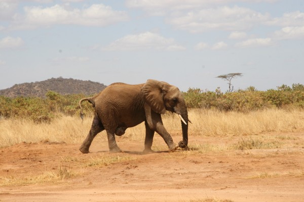
For a detailed examination, the elephant was immobilized using 10 mg of etorphine hydrochloride (0.98%) (M99®; Novartis International AG, Basel, Switzerland) combined with 5,000 iu of hyaluronidase (Kyron Laboratories (Pty) Ltd, Johannesburg, South Africa) in a 3 mL Dan-Inject® dart using a Dan- Inject® rifle (Dan-Inject APS, Sellerup Skovvej, Denmark). Rigorous palpation of the swelling revealed that the hernia contents were soft and could be forced back into the abdominal cavity, we thought that this could be the intestines sitting in the distended region. It was possible to momentarily push the hernia contents back into the abdomen and collapse the swelling (pouch). Aspiration was performed as a confirmatory test to rule out possibilities of abscess or edema in the swelling. Aspiration was aseptically done using a sterile 18 gauge needle attached to a 10 mL syringe, taking care not to puncture the intestines or organs within the bulge. Aspiration revealed that there was no fluid or pus in the swelling, thus, it was described as an umbilical hernia (Figures 1 and 2). This was thought to be a congenital case in which the umbilicus failed to close completely, creating a weak point through which the intestines herniated. In our considered opinion, surgical intervention carried the risks of intense hemorrhage and wound dehiscence soon after surgery due to intense pressure and tension from the abdomen. Additionally, it was a challenge to get the strongest absorbable suture material to close the dense muscles of the elephant abdomen. Hence, surgery was elected for another day. Anesthesia was reversed using 60 mg of diprenorphine hydrochloride (Novartis International AG) administered through the superficial ear veins. Routinely, blood samples had been collected for measurement of packed cell volume, total protein, and complete blood cell count in our laboratory. These blood parameters were confirmed to be within the normal range.
 | Figure 1 A swelling described as an umbilical hernia located at ventral abdomen. |
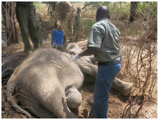
 | Figure 2 Closer view of the swelling/bulge described as an umbilical hernia. |
Later when carrying out the surgery, the elephant was immobilized using the same protocol described above and levels of anesthesia were continuously monitored during this procedure. Vital physiological parameters such as; respiration rate and depth, heart rate, blood pressure (measured using arterial pulse on the ear pinna), color of the mucous membranes, capillary refill time, and temperature were also monitored. This ensured that the elephant did not experience respiratory depression, hypoxia or any other adverse effect due to deep anesthesia. The protocol additionally ensured that the elephant did not wake up in the middle of the operation or feel pain due to waning anesthesia. Temporary shade was set up and placed over the elephant to protect it from direct sunshine. Additionally, a large amount of water was used to prevent possible anesthetic hyperthermia by pouring onto various parts of the body, including the ears.
Surgical process
The elephant was then placed on its dorsal position with one hind leg tied to a vehicle to hold it in dorsal position. A large area of the skin around the hernia pouch was liberally cleaned, scrubbed, and disinfected. The swelling and surrounding abdominal area was cleaned with water using a stiff brush; we used povidone iodine (Betadine® surgical scrub, Mundipharma AG, Basel, Switzerland) alternated with 70% alcohol (surgical spirit) rinse. Finally Betadine® solution was poured on the skin and allowed to dry. Other aseptic techniques included a standard surgical hand scrub, use of sterile surgical gloves, and sterile surgical equipment. A local anesthetic, Lignocaine and adrenaline (Lignocaine hydrochloride 20 mg/mL combined with adrenaline 20 mg/mL) (Norbrook Laboratories (GB) Limited, Northamptonshire, UK), was infiltrated along the intended points of incision. Lignocaine and adrenaline was administered, at a dose rate of 1.25 mg/kg bwt along the incision line using a 21 gauge needle attached to a 20 mL syringe.
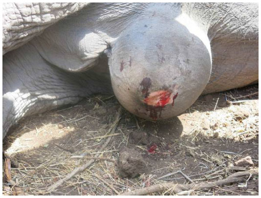
The hernia contents momentarily were pushed by hand into the abdomen (Figure 3) and an anterior-posterior blunt incision was made at the tip of the swelling. The incision was enlarged to about 15 cm wide to allow full exposure of intestines and omentum in the swelling. Hemorrhage was controlled using Kelly curved forceps and suture ligation, depending on the type of sanguineous vessel. Sterile gauze swabs were also used to apply pressure, clean the site, and control bleeding. Hence, any hemorrhaging was minimal throughout the operation. The intestines and the omentum were then pushed back into the abdomen by gloved hands and held in position as the opening into the abdomen was closed using number 3 chromic catgut absorbable suture (Medimax (UK) Ltd, London, UK) in a simple interrupted pattern. The suture was placed using a long curved cutting needle. There was pressure and tension from the protruding abdominal organs but this was managed by using a double strand of suture as well as an assistant surgeon holding the intestines into the abdomen. Once the aperture into the abdomen was securely closed an antibiotic ointment – Opticlox Eye® ointment (cloxacillin 16.7% w/w) (Norbrook Laboratories (GB) Limited) was applied along the suture line. Suturing of abdominal muscles was the next step. Oblique abdominal muscles were sutured using number 3 chromic catgut absorbable suture (Medimax (UK) Ltd) in a simple interrupted pattern, and again, the suture material was folded to form a double strand so that it was strong enough to hold back pressure from the abdominal contents. Another layer of suture was made on the subcutis muscles and abdominal fascia using the same suture material and pattern. Opticlox® eye ointment was applied along every suture line to minimize chances of opportunistic bacterial infection.
 | Figure 3 Intestines had been pushed back into the abdominal cavity to facilitate surgical correction of the umbilical hernia. |
The skin was closed using a threaded nylon suture folded to form two strands attached to a 6 cm long curved cutting needle. Excess skin, fascia, and fatty tissues that formed the hernia pouch were retained and sutured together to help counter the tension from the abdomen. Due to the thickness and texture of the skin, it was difficult to penetrate using a needle. Therefore, a surgical blade attached to a size 4 blade-holder was used to puncture the skin at the point where the needle was to be inserted. The skin was closed by size 4 nylon suture using cruciate suture pattern and in addition, a few simple interrupted suture patterns were placed over the cruciate sutures for reinforcement. A bolus of long-acting oxytetracycline was inserted into the skin suture line to treat any postoperative infection. A large amount of green clay paste was applied over the surgical wound to completely seal it and provide further antibiotic benefit enabling a more rapid recuperation. Other treatments instituted included an injection of 20,000 mg procaine penicillin and 25,000 mg dihydrostreptomycin sulfate (Penstrep®, Procaine penicillin 200 mg dihydrostreptomycin sulfate 250 mg, [Norbrook Laboratories (GB) Limited, UK] and 2,000 mg Flunixin meglumine [Norbrook Laboratories (GB) Limited, UK]) to treat any opportunistic bacterial infections and alleviate pain respectively.
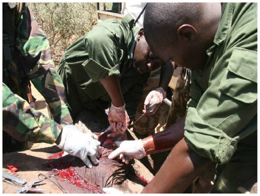
Soon after the surgical process was complete, the ropes tied to the leg were released and the elephant was immediately revived from anesthesia using 60 mg of diprenorphine hydrochloride administered through the superficial ear veins. The elephant regained consciousness and arose steadily but carefully in what appeared to be due to mild pain at the surgical site. Eventually it stood stationary for a while before it moved away (Figure 4) and joined the rest of its family nearby. Attempts to monitor the elephant after the surgical procedure were not successful as the elephant felt disturbed and moved to an unknown location, not to be sighted again for over 3 weeks, where it was eventually found dead and its remains scavenged by predators.
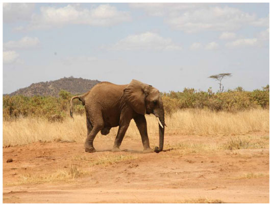 | Figure 4 The elephant walks away after the surgery. |
Discussion
In Samburu National Reserve, Kenya, an umbilical herniorrhaphy in a sub-adult African elephant living in the wild was attempted with a negative outcome. Preparation of the patient for surgery proved a challenge because keeping the elephant off feed was not practical under the circumstances. Typically, in large animal abdominal surgery hay is restricted for 36 hours, grain for 24 hours, and water for 12 hours. On very large hernias, feed can even be restricted for 48 hours.20,21 The elephant underwent a complete physical exam prior to surgery for an umbilical pathology. A complete physical exam included a deep palpation of the umbilical structures, ultrasound evaluation could have been extension of palpation. This is because ultrasonographic examination of affected animal provides information that alters the clinician’s intervention with great enough frequency to recommend its use. Furthermore ultrasound helps to determine which viscera are present in the hernia, further clarifying the extent to which internal structures are distended or infected, amongst other uses.22 Unfortunately, we did not have an ultrasound machine at our disposal.
Neutrophil-lymphocyte reversal/neutrophilia, hyperglobulinemia and hyperfibrinogenemia are all suggestive that umbilical swelling is complicated by infection. The absence of these elevations in these parameters does not however rule out that the swelling may be complicated by infection.22 Laboratory evaluation showed that these parameters in the elephant’s leukogram were within the normal range.
When aseptic surgery is performed, an efficient draping system is mandatory in the opinion of many surgeons.23,24 Draping of the elephant was not done, but the surgeons minimized contact with areas of the animal which were not scrubbed. Abdominal surgical procedures in elephants have challenges, which include difficulties in handling the animal, difficulties associated with the physical mass of abdominal organs, as well as anesthetic risks magnified by the size of the animal.3,4 In horses simple hernias do not necessarily cause any clinical sign and the hernia is usually soft, non-painful, and reducible on palpation.25 In ruminants, umbilical hernias associated with infection have a higher incidence of incisional complications including infections and incisional hernia, as well as peritonitis. Furthermore the most common complication is seroma formation at the incision line.22
During this surgery, intestines, omentum, and fat were found inside the hernia sac. There were no adhesions or fibrinous material. The intestines were neither strangulated nor incarcerated by the hernia ring. In cattle and small ruminants a portion of abomasum and/or greater omentum is commonly found with the hernia sac. The hernia sac may contain omentum, abomasum, or less commonly intestines, although it is difficult to distinguish the specific contents by external palpation. In some cases the contents will be adhered to the hernia sac.22 An incisional hernia is a common complication of colic surgery in horses and, according to reports, occurs in 7%–10% of cases.26,27 Most hernias are preceded by surgical site drainage and infection in the 1st week after surgery.28 Intestinal adhesions are one of the most common complications that limit survival rates after abdominal surgery in horses, and the primary mode of prevention is an atraumatic surgical technique.29 However, exciting advances have been made in certain areas of abdominal surgery in the horse. Recently, methods have been developed to decrease the rate of complications, such as adhesions, incisional infections, and hernias, and have proved effective.30 Use of double strands of number 3 chromic catgut suture material to overcome the inherent challenges of suture strength worked well, however low knot security is a reality. Prior to introduction of synthetic absorbable sutures such as polyglycolic acid and polyglactin 910 (Dexon or Vicryl), Turner and McIlwraith used number 2 or 3 medium chromic catgut in many umbilical hernias with satisfactory results.23
Umbilical herniorrhaphy in sub-adult free living African elephants can be attempted using etorphine hydrochloride as general anesthetic and a Lignocaine and adrenaline combination as local anesthetic. However, any surgical procedures performed in adult or sub-adult free-ranging wild elephants are bound to have multiple post-operative risks and challenges. Risks such as wound or suture dehiscence, wound gaping, wound infection, along with the difficulties of consistent, necessary post-operative monitoring of the elephant in the wild are expected, posing a real challenge. Confinement of such elephants is recommended to enhance post-surgical monitoring, however, confinement might not be practical due to aggression of wild elephants and difficulties in constructing a strong enough confinement facility to restrain the animal during post-surgical monitoring. In this case, the cause of death could not be accurately confirmed since the carcass was already scavenged, making a postmortem examination ineffectual. There were many possible post-operative complications including surgical wound dehiscence, wound infection, intestinal adhesions, peritonitis amongst others. These are conditions which can be well managed in confinement or in captive situations rather than in free-ranging wild elephants. Furthermore, general anesthesia and surgical interventions in elephants have been reported to be associated with high morbidity and mortality rates.2–6 Thus, while umbilical hernias have been corrected in juvenile elephants (2 weeks to 5 months old),11,12 the Samburu African elephant in question was a 10-year-old sub-adult male, complicating this matter further.
Conclusion
The surgical process lasted 3 hours and anesthesia was topped-up twice with 2 mg of etorphine hydrochloride; after 1- and half-hour intervals. Prognosis of the case was guarded after the surgical intervention, and despite efforts to closely monitor the elephant, reports state that the animal succumbed to death 3 weeks post-surgery, due to multiple post-operative complications.
Acknowledgments
We acknowledge support of the veterinary surgeons team by the David Sheldrick Wildlife Trust (DSWT) who airlifted the team to Samburu National Reserve through the Sky Vet Initiative. Thank you DSWT for collaborating with Kenya Wildlife Service to aid our conservation work. Last but not least, we sincerely thank Lina Sideras (DSWT), Kanyiha Mbogori (Boston, MA, USA Contributor), and Kagendo Mbogori – linguist (Melbourne, Australia Contributor) who were English copy editors.
Disclosure
The authors report no conflicts of interest in this work.
References
International Union for Conservation of Nature [homepage on the Internet]. Blanc J, 2008. Loxodonta africana. The IUCN Red List of Threatened Species. Version 2014.2. Available from: http://www.iucnredlist.org. Accessed September 10, 2014. | |
Singh BS. Umbilical hernia in an elephant calf. Indian Vet J. 1971;48(5):533–536. | |
Fowler ME. Problems with immobilizing and anesthetizing elephants. Proc Am Ass Zoo Vet. 1981:87–91. | |
Oosterhuis JE. The performance of caesarian section on an Asian elephant (Elephas maximus indicus). Proc Am Zoo Vet. 1990:157–158. | |
Pathak S, Saikia CJ, Lahon DK, et al. Attempted ventral herniorrhaphy in an Asian elephant (Elephas maximus) using xylaxine sedation. J Zoo Wildl Med. 1990;21:234–235. | |
Honeyman VL, Pettifer GR, Dyson DH. Arterial blood pressure and blood gas values in normal standing and laterally recumbent African (Loxodonta Africana) and Asian (Elephas maximus) elephants. J Zoo Wildl Med. 1992;23:205–210. | |
Wiedner EB, Peddie J, Peddie LR, et al. Strangulating intestinal obstructions in four captive elephants (Elephas maximus and Loxodonta Africana). J Zoo Wildl Med. 2012;43(1):125–130. | |
Fretz PB, Hamilton GF, Barber SM, Ferguson JG. Management of umbilical hernias in cattle and horses. J Am Vet Med Assoc. 1983;183(5):550–552. | |
Smeak D. Abdominal hernias. In: Slatter D, editor. Textbook of Small Animal Surgery. 2nd ed. Philadelphia, Pennsylvania: W.B. Saunders Co; 1993:883–923. | |
Saperstein G. Congenital defects and hereditary disorders in ruminants. In: Smith BP, editor. Large Animal Internal Medicine. 3rd ed. St Louis, Missouri: Mosby Inc.; 2002:883–923. | |
Wiedner EB, Gray C, Rich P, et al. Non surgical repair of an umbilical hernia in two Asian elephant calves (Elephas maximus). J Zoo Wildl Med. 2008;39:248–251. | |
Abou-Madi N, Kollias GV, Hackett RP, Durcharme NG, Gleed RD, Moakler JP. Umbilical herniorrhaphy in a juvenile Asian elephant (Elephas maximus). J Zoo Wildl Med. 2004;35(2):221–225. | |
Kock MD, Martin RB, Kock N. Chemical Immobilization of Free-Ranging African Elephants (Loxodonta africana) in Zimbabwe, Using Etorphine (M99) Mixed with Hyaluronidase, and Evaluation of Biological Data Collected Soon after Immobilization. J Zoo Wildl Med. 1993;24(1):1–10. | |
Burroughs R, Hofmeyer M, Morkel P, Kock MD, Kock R, Meltzer D. Chemical immobilization – individual species requirements. Mega herbivores – Elephant. In: Kock MD, Burroughs R, editors. Chemical and Physical Restraint of Wild Animals. 2nd ed. Greyton, South Africa: International Wildlife Veterinary Services (Africa); 2012:212–218. | |
Stegmann GF. Etorphine-halothane anaesthesia in two five-year-old African elephants (Loxodonta africana). J S Afr Vet Assoc. 1999;70(4):164–166. | |
Welsch B, Jacobson ER, Kollias GV, Kramer L, Gardner H, Page CD. Tusk extraction in the African elephant (Loxodonta africana). J Zoo Wildl Med. 1989;20(4):446–453. | |
Osofsky SA. A practical anesthesia monitoring protocol for free-ranging adult African elephants (Loxodonta africana). J Wildl Dis. 1997;33(1):72–77. | |
Schaftenaar W. Vaginal vestibulotomy in an Asian elephant (Elephas maximus). Proc Am Ass Zoo Vet. 1996:434–439. | |
Nayar KN, Chandrasekharan K, Radhakrishnan K. Management of surgical affections in captive elephants. J Indian Vet Ass Kerala. 2002;7(3):55–59. | |
Iowa State University Veterinarian. Wheat JD. Surgical Repair of Umbilical and Inguinal Hernias in the Bovine. Iowa State University Veterinarian: 1952 Vol 14: Iss 1, Article 3. Available from: http://lib.dr.iastate.edu/cgi/viewcontent.cgi?article=1746&context=iowastate_veterinarian. Accessed April 7, 2015. | |
Duke University and Duke University Medical Center [homepage on the Internet]. Duke University Animal Care and Use Program. Available from: http://vetmed.duhs.duke.edu/guidelines_for_general_surgery_in_animals.htm. Accessed April 7, 2015. | |
Scott RRH. Umbilical hernia. In: Scott RR Haskell, editor. Blackwell’s Five-Minute Veterinary Consult: Ruminant. 1st ed. Ames Iowa USA: Wiley-Blackwell. 2008:902–905. | |
Turner AS, McIlwraith CW. Techniques in Large Animal Surgery. 2nd ed. Philadelphia: Lippincott Williams & Wilkins; 1989:1–8 and 254–259. | |
Baines S, Lipscomb V, Hutchinson T, editors. BSAVA Manual of Canine and Feline Surgical procedures. A foundation Manual. British Small Animal Veterinary Association (BSAVA); 2012:198–209. | |
Lavoie JP, Hinchcliff KW. Hernias (Umbilical and inguinal). In: Lavoie JP, Hinchcliff KW, editors. Blackwell’s Five-Minute Veterinary Consult: Equine. 2nd ed. Ames Iowa USA: Wiley-Blackwell; 2008:373. | |
Mair TS, Smith LJ. Survival and complication rates in 300 horses undergoing surgical treatment of colic. Part 3: long-term complications and survival. Equine Vet J. 2005;37(4):310–314. | |
French NP, Smith J, Edwards GB, Proudman CJ. Equine surgical colic: risk factors for postoperative complications. Equine Vet J. 2002;34(5):444–449. | |
Wilson DA, Baker GJ, Boero MJ. Complications of celiotomy incisions in horses. Vet Surg. 1995;24(6):506–514. | |
Gorvy DA, Barrie Edwards G, Proudman CJ. Intra-abdominal adhesions in horses: a retrospective evaluation of repeat laparotomy in 99 horses with acute gastrointestinal disease. Vet J. 2008;175(2):194–201. | |
Kelmer G. Update on Recent Advances in Equine Abdominal Surgery. Vet Clin North Am Equine Pract. 2009;25(2):271–82. |
 © 2015 The Author(s). This work is published and licensed by Dove Medical Press Limited. The full terms of this license are available at https://www.dovepress.com/terms.php and incorporate the Creative Commons Attribution - Non Commercial (unported, v3.0) License.
By accessing the work you hereby accept the Terms. Non-commercial uses of the work are permitted without any further permission from Dove Medical Press Limited, provided the work is properly attributed. For permission for commercial use of this work, please see paragraphs 4.2 and 5 of our Terms.
© 2015 The Author(s). This work is published and licensed by Dove Medical Press Limited. The full terms of this license are available at https://www.dovepress.com/terms.php and incorporate the Creative Commons Attribution - Non Commercial (unported, v3.0) License.
By accessing the work you hereby accept the Terms. Non-commercial uses of the work are permitted without any further permission from Dove Medical Press Limited, provided the work is properly attributed. For permission for commercial use of this work, please see paragraphs 4.2 and 5 of our Terms.