")
Back to Archived Journals » Research and Reports in Forensic Medical Science » Volume 4
Role of physical properties of dental restorative biomaterials in criminalistics
Authors Pereira C , Costa J, Santos J, de Mendonça M
Received 27 March 2014
Accepted for publication 22 April 2014
Published 20 June 2014 Volume 2014:4 Pages 19—24
DOI https://doi.org/10.2147/RRFMS.S51279
Checked for plagiarism Yes
Review by Single anonymous peer review
Peer reviewer comments 2
Cristiana Palmela Pereira,1–4 João Franco Costa,5 Jorge Costa Santos,6,7 Maria Cristina de Mendonça6,7
1Legal Medicine and Forensic Sciences, Faculty of Medicine, University of Lisbon, Lisbon, 2Departments of Pharmacology and Therapeutic and Dental Morphology, Faculty of Dental Medicine, University of Lisbon, Lisbon, 3Integrate Research from the Research Centre of Statistics and Applications of the University of Lisbon, Lisbon, 4South Branch of the Portuguese National Institute of Legal Medicine and Forensic Sciences, Lisbon, 5Judicial Police, Lisbon, 6Medical Legal Doctors, South Branch of the Portuguese National Institute of Legal Medicine and Forensic Sciences, Lisbon, 7Faculty of Medicine, University of Lisbon, Lisbon, Portugal
Abstract: The body of an 89-year-old woman was discovered at home by a member of her family. Only part of the body, the legs, were found intact without burn damage. The rest of the body was burned to ashes without the possibility to identify the cadaver. Forensic Dentistry had two main goals to resolve in this forensic pathology casework, and they were: 1) establish the identification of the cadaver; and 2) estimate the temperature and the direction of the fire by the analysis and interpretation of the skeletal intraoral dental materials altered by thermal processes. This case study manuscript focuses only on the second objective. The effects of fire on teeth are influenced by the temperature applied and by its duration. Additionally, adjacent tissues as well as temperature alterations caused by substances used to quench the fire have been shown to affect the thermal impact on teeth and their restoration. Bodies may be subjected to various temperatures, depending on the origin of the fire and the conditions that promote continuation of the blaze. Fire effects on certain dental materials can indicate the direction and the temperature of a fire. This is based on the physical, mechanical, and chemical changes of different dental materials resulting from high temperatures. The distinct properties of various dental materials, well defined, can influence and predict their performance under different degrees of fire. In this case study, the cadaver had two main groups of dental materials: metal alloys and polymers. The importance of the detailed degree of predictability of fire performance data from thermal decomposition data should not be underestimated; polymers cannot burn if they do not break down and metal alloys cannot burn if the melting point is not reached, independent of the variable time of the fire.
Keywords: fire investigation, forensic dentistry, dental materials properties
Introduction
Fire death investigations involve collaborative effort among law enforcement, arson investigators, forensic pathologists, anthropologists, and forensic odontologists to reconstruct the circumstances of the scene, the manner of death, and victim identity. The primary purpose of a body fire investigation is to establish the origin of the fire, the temperature of the fire, the body’s position, and determine the likely cause; and thus, conclude whether the death was accidental, natural, or deliberate.12
The field of forensic dentistry, or the more professional term, forensic odontology, is the application of dentistry to the law. Forensic dentistry is now an integral part of criminal investigations.
The purpose of this case study is to review and present the aim and the application of forensic dentistry in a new field. Dental science plays a vital role in the detection and solution of crime scenes, including resolving the circumstances of deaths.3
Case report
General findings at the scene
The body of an 89-year-old woman was discovered at home by a member of her family. Only part of the body, the legs, were found intact, without burn damage from the fire. The rest of the body was burned to ashes (Figure 1).
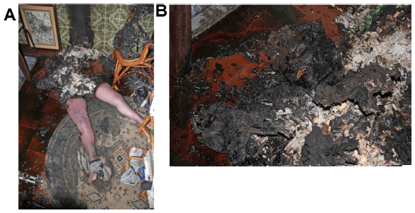 | Figure 1 General findings at the scene. |
This is what forensic investigators call an alleged spontaneously combusted corpse. The woman may have ignited herself accidentally. The point of ignition was not near the place where the cadaver was found. On arrival, the firefighters observed that the gas cooker was turned on. Her clothes went up in flames before she fell to the floor and they functioned as an external source of fire. She started to take off some of her clothes, which were found on the tile floor of the kitchen, burned, before she fell to the floor. There was little damage in the kitchen, apart from the charred ceiling above the body and the burned floor beneath. No reasonable explanation has ever been found for this kind of fire situation. The best explanation is that the fat rendered from a burning body can act in the same manner as the fuel in an oil lamp or candle. If the body is positioned in such a way that the oils rendered from it are accessible to the flames, they will continue to fuel the fire.
Objectives
Forensic dentistry had two main goals to resolve in this forensic pathology casework, and they were: 1) establish the identification of the cadaver; and 2) estimate the temperature and the direction of the fire by the analysis and interpretation of the skeletal intraoral dental materials altered by thermal processes. This case study manuscript focuses only on the second objective. Fire effects on certain dental materials, based on the physical, mechanical, and chemical properties, can indicate the fire direction and the fire temperature, and may help to provide an accurate assessment of the circumstances of the death.
Examination of the skull
After removing the soft tissues from the skull, the hard tissues showed several degrees of burn damage. The skull had more extensive burn destruction in the lower third than in the upper third (Figure 2). The right side of the skull showed more burn damage than the left side, with more bone fragmentations, mainly in the lower jaw.
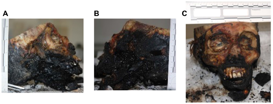 | Figure 2 The (A and B) lateral and (C) frontal views of the skull. |
Examination of the teeth
Analysis of the intraoral cavity showed two acrylic resin removable prosthodontic rehabilitations: both upper and lower. In the lower one, we can observe that the fire direction was from right to left and the fire intensity was higher on the right side than on the left side. The lower prosthodontic rehabilitation was carbonized on the right side but not on the left side. The morphology of the acrylic teeth was conserved on the left side of the lower jaw. When we compared the upper and the lower rehabilitations, we observed that the fire intensity was higher in the lower one (Figure 3).
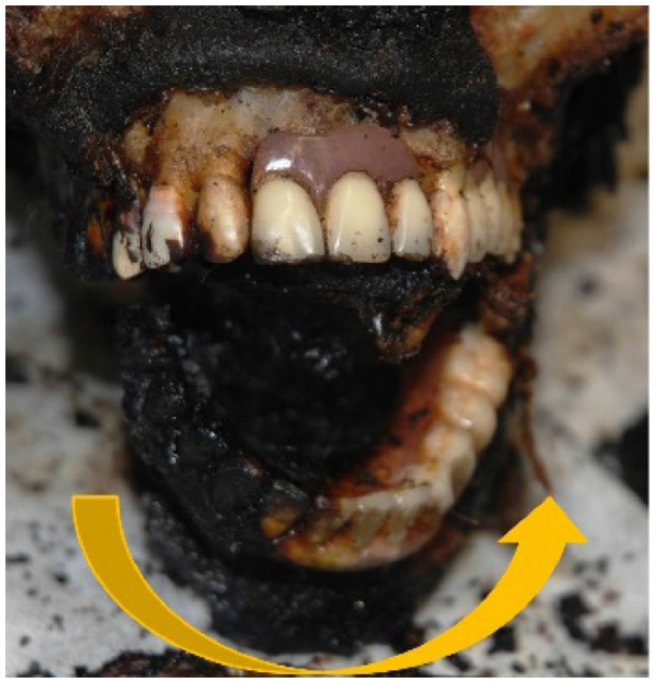 | Figure 3 Frontal view of the oral cavity. |
The alveolar bone from the upper jaw was less carbonized than that from the lower jaw. The left side of the oral cavity was lightly burned (Figure 4). The lower rehabilitation had a stainless steel hook for the last tooth and was not burned or molten. This structure is different from the clasp unit of the direct retainer. The rehabilitation was manufactured with an alloy of cobalt–chromium–molybdenum, for which the melting point is higher than stainless steel.4 On the right side of the oral cavity, we observed that the lower jaw was fully carbonized, with specific areas of the jaw calcined and missing (Figure 4). The lower acrylic resin removable prosthodontic rehabilitation was partially carbonized and was missing part of the base structure and some of the teeth.
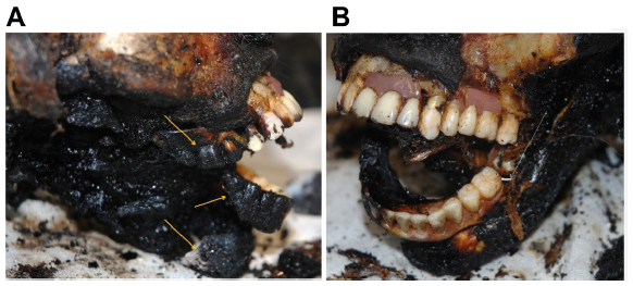 | Figure 4 The (A) right and (B) left lateral views of the oral cavity. |
On the upper jaw, the structure of the denture base and the morphology of the teeth were intact and the burn damage was less severe than in the rehabilitation from the lower jaw. The fire direction of the upper jaw was from the right side to the left side. In summary, the intensity of the fire was higher on the right side and in the lower jaw.
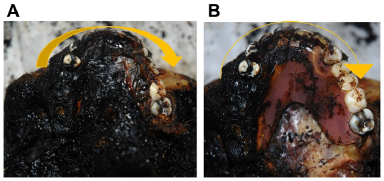
When we removed the lower denture, the fire intensity and the fire direction were the same in the vestibular and lingual surfaces of the rehabilitation. The right side of the rehabilitation was carbonized, and was missing the acrylic resin structure and likely a stainless steel hook at the distal end of the rehabilitation as well (Figure 5).
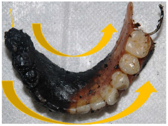 | Figure 5 Lower denture where the direction of the fire was from the right to the left side (large arrows), on the lingual and vestibular surfaces. |
There were two types of acrylic according to the polymer, polymethyl methacrylate, which can be thermoplastic or thermosetting.5 The one present in this cadaver was a thermosetting polymer. The differences between the two types of this polymer are a concern in terms of the glass transition temperature, melting crystallization temperature, and carbonization temperature.6,7 Thermosetting polymers that form irreversible chemical bonds during the curing process do not melt, but carbonize, and the glass transition temperature is very high. The carbonization temperature is 900°C–1,000°C for thermosetting polymers.1
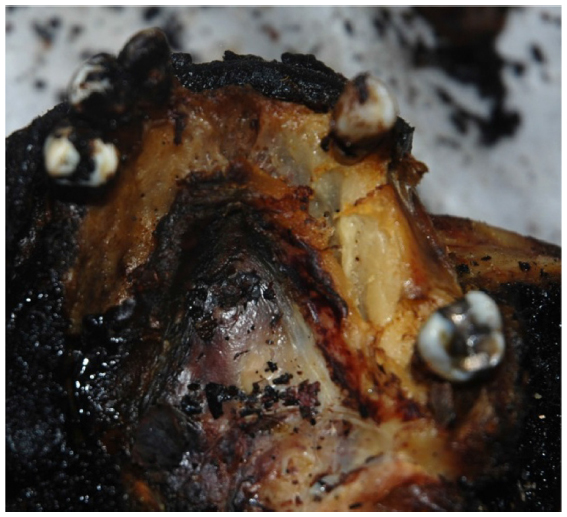
Inside the base of the lower denture, on the right side, the temperature was near 900°C, where was missing the thermosetting polymer structure of the rehabilitation (Figure 6). The left side of the rehabilitation structure was intact; it was not carbonized because the fire intensity was less than the thermosetting polymer carbonization temperature.
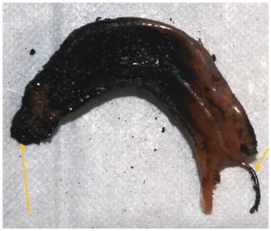 | Figure 6 Inside the base of the lower denture. |
Another way of estimating the temperature is the presence of the stainless steel hook on the left side. This is a metal alloy of iron and carbon, which we call austenitic stainless steel, for which the melting point is 700°C.8 On the left side, the temperature was less than 700°C, but if a hook was also present on the right side, the temperature would have been higher than this melting point temperature.
The lower jaw mainly presented two different colors of the alveolar bone (Figure 7).9,10 The alveolar bone was white on the right side and dark in front and on the left side of the mandible. These burn injury patterns again confirm the fire direction, from the right side to the left side. The bone was calcined on the right side of the mandible and was charred on the anterior, and slightly burned on the left side.
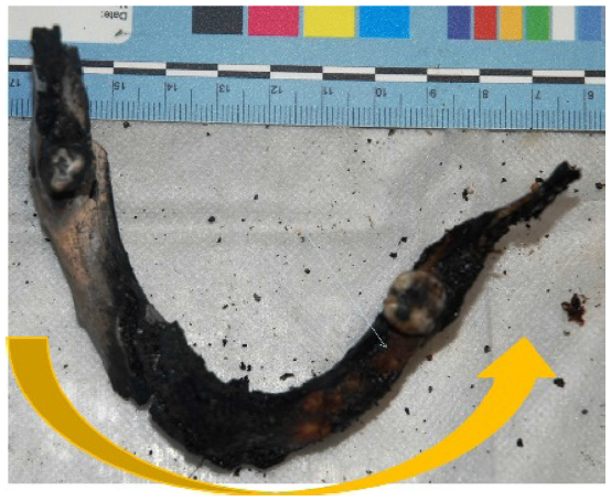 | Figure 7 Upper view of the lower jaw with color alteration of bone caused by the fire. |
The lower border of the mandible showed the same pattern of the fire damage (Figure 8) in that it was white in the right side (calcined), black on the anterior and left sides (charred), and slightly burned on the distal end of the left side of the lingual surface of the mandible.6,7,11 The lower jaw had two teeth: tooth 37 and tooth 47 (Figure 9) and both had antemortem dental restorations. The second molar from the left side on the oclusal surface had a restoration of amalgam (Figure 9). The filling was present but was dark. The second molar of the right side had a cavity compatible with an amalgam filling (Figure 9). The alloy was molten and there were traces of residual alloy on the floor of the cavity.
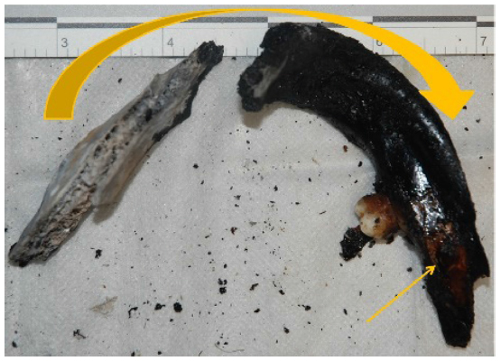 | Figure 8 The inferior border of the lower jaw with different patterns of burn damage. |
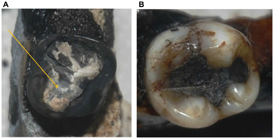 | Figure 9 The teeth (A) 47 and (B) 37. |
The amalgam filling consisted of a eutectic alloy. The advantage of these alloys is that the low melting point allows work in the mouth. Conventional amalgam restorations darkened and pulverized at 300°C and roasting began at 800°C.8 At 1,000°C in situ in the filling, we only saw the shine of the silver in the second molar from the right side.12 From the color of the filling from the second molar of the left side, we estimate that the fire temperature was near 300°C.
The visual appearance of the upper jaw was also compatible with a fire direction from the right to the left (Figure 10). The temperature intensity was higher on the right side than on the left. After removing the soft tissues and cleaning, the pattern injuries were the same in the lower jaw (Figures 10 and 11). The acrylic resin from the upper denture was carbonized on the distal end of the right side (Figure 12), and this pattern was the same on the inner surface (Figure 12).
 | Figure 10 (A) The upper jaw with the denture before removing the soft tissues and (B) after removing the soft tissues. |
 | Figure 11 The burn damage on the upper jaw bone. |
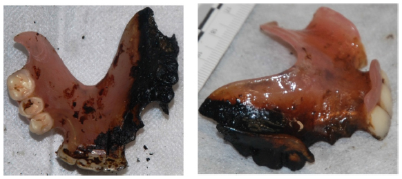 | Figure 12 The burn damage on the denture on the external and internal surfaces. |
Conclusion
Forensic dentists have become a very important part in the investigation of death circumstances together with criminalistics. The forensic dentist in this case carries considerable responsibility since their scientific opinion is frequently asked when all other paths of resolution have been exhausted. In this case study, the circumstances of the death were the variables to be resolved, by estimating the temperature exposure by examining the fire effects on the dental restoration biomaterials. The conclusion resolved by forensic dentistry was that the temperature between the right and the left sides of the oral cavity was significantly different based on the physical properties of the different dental biomaterials. On the right side, the temperature was near 1,000°C and on the left side, approximately 300°C. This explanation was given because of the position of the head when the woman fell on the tile floor. The dissipation of the temperature was less in the right side because the floor acted as an insulating material. The cadaver fell in the same position as she was discovered.
Acknowledgment
This research was partially sponsored by national funds through the Fundação Nacional para a Ciência e Tecnologia, Portugal – FCT (PEst-OE/MAT/UI0006/2014).
Disclosure
The authors report no conflicts of interest in this work.
References
Meng LY, Park SJ. Investigation of narrow pore size distribution on carbon dioxide capture. Bull Korean Chem Soc. 2012;33(11):3749–3754. | |
Pereira CP, Santos JC. How to do identify single cases according to the quality assurance from IOFOS. The positive identification of an unidentified body by dental parameters: a case of homicide. J Forensic Leg Med. 2013;20(3):169–173. | |
Pereira CP, Costa JF, Santos JC, de Mendonça MC. The role of forensic dentistry in fire scene investigation: determine the direction and the temperature of fire by dental biomaterials evidence in a body by not-so-spontaneous human combustion. J Forensic Odontostomatol. 2013; 31 Suppl 1:42–43. | |
Balogun SA, Esezobor DE, Agunsoye JO. Effect of melting temperature on the wear characteristics of austenitic manganese steel. J Miner Mater Charact Eng. 2008;7(3):277–289. | |
Jagger RG. Effect of the curing cycle on some properties of a polymethylmethacrylate denture base material. J Oral Rehabil. 1978;5(2):151–157. | |
Pereira C. Fotopolimerização. Aplicação em Medicina Dentária [Photopolymerization. Applications in Dental Medicine]. Acta Fotobiológica. 2001;16:23–26. | |
Portugal J, Bernardo MF, Pereira C, Ortet J, Leitão J. Effect of light curing time on the effectiveness of composite polymerization. Conf Proc Dent Mater. 2002;18(8):A17, abstr 44. | |
Marshall SJ, Marshall GW Jr. Dental amalgam: the materials. Adv Dent Res. 1992;6:94–99. | |
Pereira C. Commingled Assemblage from Earthquake 1755 of Lisbon: Forensic Anthropology Study in Forensic Science. Hauppauge, NY: Nova Science Publishers; 2012:149–168. | |
Pereira CP, Antunes MT. Antropologia Dentária Forense: identificação humana através de elementos anatómicos orais – maxilares e dentes in Medicina Dentária Forense [Forensic Dental Anthropology: Human Identification by oral anatomic elements - jaws and teeth, in Forensic Dentistry]. Lidel; 2012. | |
Pereira C, Mendonça MC, Antunes MT, Santos JS. Identificação forense de lesões traumáticas em esqueletos cranianos alterados pela acção do fogo: Amostra populacional de Vítimas do Terramoto de 1755, em Lisboa. Conference Proceedings of 8º Congresso Nacional de Medicina Legal [Forensic Identification of traumatic lesions observed in skulls changed by fire: Population Sample from the victims from the earthquake 1755 in Lisbon. Conference Proceedings of the 8th National Meeting of Legal Medicine]; 2009. | |
Sakaguchi RL, Powers JM. Restorative materials – metals. In: Craig’s Restorative Dental Materials. 13th ed. St Louis, MO: Mosby; 2012:199–251. |
 © 2014 The Author(s). This work is published and licensed by Dove Medical Press Limited. The full terms of this license are available at https://www.dovepress.com/terms.php and incorporate the Creative Commons Attribution - Non Commercial (unported, v3.0) License.
By accessing the work you hereby accept the Terms. Non-commercial uses of the work are permitted without any further permission from Dove Medical Press Limited, provided the work is properly attributed. For permission for commercial use of this work, please see paragraphs 4.2 and 5 of our Terms.
© 2014 The Author(s). This work is published and licensed by Dove Medical Press Limited. The full terms of this license are available at https://www.dovepress.com/terms.php and incorporate the Creative Commons Attribution - Non Commercial (unported, v3.0) License.
By accessing the work you hereby accept the Terms. Non-commercial uses of the work are permitted without any further permission from Dove Medical Press Limited, provided the work is properly attributed. For permission for commercial use of this work, please see paragraphs 4.2 and 5 of our Terms.