")
Back to Journals » Cancer Management and Research » Volume 12
Prognostic Impact of DHRS9 Overexpression in Pancreatic Cancer
Authors Li H , Zhou J, Zhao F, Yu J, Xu L
Received 29 February 2020
Accepted for publication 24 June 2020
Published 20 July 2020 Volume 2020:12 Pages 5997—6006
DOI https://doi.org/10.2147/CMAR.S251897
Checked for plagiarism Yes
Review by Single anonymous peer review
Peer reviewer comments 3
Editor who approved publication: Professor Seema Singh
Huang-bao Li,1 Jun Zhou,1 Fengqing Zhao,1 Jiayin Yu,2 Longsheng Xu3
1Department of Hepatobiliary and Pancreatic Surgery, First Hospital of Jiaxing, First Affiliated Hospital of Jiaxing University, Jiaxing, Zhejiang, People’s Republic of China; 2Department of Pathology, First Hospital of Jiaxing, First Affiliated Hospital of Jiaxing University, Jiaxing, Zhejiang, People’s Republic of China; 3Department of Anesthesiology and Pain Research Center, First Hospital of Jiaxing. The First Affiliated Hospital of Jiaxing University, Jiaxing, Zhejiang, People’s Republic of China
Correspondence: Huang-bao Li Tel +86 15868300272
Email [email protected]
Purpose: Pancreatic cancer (PC) has poor prognosis despite systemic treatment. Dehydrogenase/reductase member 9 (DHRS9) has been reported to be involved in many events of tumorigenesis, but its prognostic impact in PC remains undetermined. Thus, this study aimed to explore the association between DHRS9 expression and the prognosis of PC and investigate the possible mechanism by which DHRS9 is involved in PC progression.
Patients and Methods: The study used data: from Gene Expression Omnibus (GEO) and our institution to compare the DHRS9 expression between PC and adjacent normal tissues; from The Cancer Genome Atlas (TCGA) and our institution to assess the clinicopathological characteristics and prognosis of PC patients in high and low DHRS9 expression groups; and from TCGA to predict the potential mechanism of DHRS9 in PC. Western blot assay was used to identify DHRS9 expression in specimens collected from five patients who underwent surgery in our institute. Furthermore, immunohistochemistry (IHC) was then used to identify DHRS9 expression in the specimens of 109 patients who underwent surgery at our institute. Kaplan–Meier and Cox regression analyses were used to assess the prognostic significance of DHRS9 expression among PC patients.
Results: All the IHC, Western blot, and GEO datasets indicated that compared to normal tissues, DHRS9 was significantly overexpressed in PC tissues. IHC results demonstrated that the strong intensity of DHRS9 expression was significantly correlated with vascular infiltration (P < 0.05). Further, high DHRS9 expression was identified as a prognostic risk factor for overall survival. Functional analysis of DHRS9 co-expressed genes indicated that DHRS9 was involved in mitogen-activated protein kinases/extracellular signal-regulated kinase (MAPK/ERK) signaling pathway.
Conclusion: DHRS9 is upregulated in PC tissue, and high DHRS9 expression is correlated with poor prognosis in PC. DHRS9 may affect the oncological process of PC through MAPK/ERK pathway.
Keywords: pancreatic cancer progression, prognosis, MAPK/ERK pathway, TCGA, GEO
Introduction
Pancreatic cancer (PC), the most lethal solid malignancy, has a poor prognosis worldwide,1 and its health burden is increasing, including in both the United States and China.2,3 Due to the special anatomical site and non-specific symptoms in the early stages of the disease, only 15–20% of patients have the opportunity to undergo radical surgery, and the remaining 80–85% of patients are diagnosed with locally advanced or distant metastases.4 Although systemic therapies such as surgery, chemotherapy, radiotherapy, biotherapy, and immunotherapy can improve the prognosis of patients with PC, the 5-year survival rate is still less than 5%.5
Dehydrogenase/reductase member 9 (DHRS9), a member of the short-chain dehydrogenases/reductases family, is originally considered to be involved in the metabolism of retinol.6 However, further research indicates that DHRS9 is involved in the biosynthesis of all-trans retinoic acid (ATRA).7 Because ATRA is involved in many events of tumorigenesis,8 DHRS9 is also speculated to be involved in the development of tumors. Previous studies have confirmed that the low expression of DHRS9 in oral squamous cell carcinoma8 and colon cancer9 predicts poor prognosis. In contrast, from the Kaplan–Meier plotter database, high expression of DHRS9 mRNA in stomach adenocarcinoma and ovarian cancer is associated with poor prognosis,10 thus, the prognostic impact of DHRS9 may differ between cancer types.
The poor prognosis of PC despite systemic treatment highlights the need for further investigations on the molecular mechanisms of PC growth and metastasis and predictive biomarkers with prognostic value to guide treatment. In this regard, it is reasonable to investigate the importance of differentially expressed DHRS9 in PC and its prognostic value. However, unlike in other cancers, the correlation between DHRS9 expression and PC prognosis is unknown. Thus, this study aimed to explore the association between DHRS9 expression and the prognosis of PC. Towards this goal, the differential expression of DHRS9 mRNA or protein in PC and normal tissues were compared using the Gene Expression Omnibus (GEO) database, The Cancer Genome Atlas (TCGA) database, and our dataset. In addition, we explored the prognostic value of DHRS9 expression in PC and investigated the possible mechanism by which DHRS9 is involved in PC progression, using bioinformatics methods.
Patients and Methods
Patients and Study Design
This study was approved by the Ethics Committee of First Hospital of Jiaxing (approval number: LS2018-176). The need for informed consent was waived owing to the retrospective nature of the study design. However, we obtained the authorization of patients or family members to use the follow-up information.
We evaluated patients with PC who underwent radical surgery between November 2011 and June 2019 in the First Hospital of Jiaxing, China. In total, 193 samples from 109 patients were used for immunohistochemical (IHC) staining. Five pairs of fresh samples were used for Western blot assay. Data on demographic characteristics and clinicodemographic variables including sex, age, tumor site, tumor maximum diameter, lymph node metastasis, grade, vascular infiltration, nerve infiltration, and the American Joint Committee on Cancer TNM staging (8th edition) were collected from medical records. However, of these patients, 10 were lost to follow-up; the clinicodemographic characteristics of the 99 patients with follow-up are shown in Table 1. The follow-up period was defined as the time interval from the date of surgery to death or the last follow-up (August 17, 2019). In addition, the clinicopathological information of 178 patients with DHRS9 mRNA expression was obtained from the TCGA database as a reference study. However, this dataset has no records of vascular infiltration and nerve infiltration. Details of the Jiaxing dataset and the TCGA dataset are shown in Table 1.
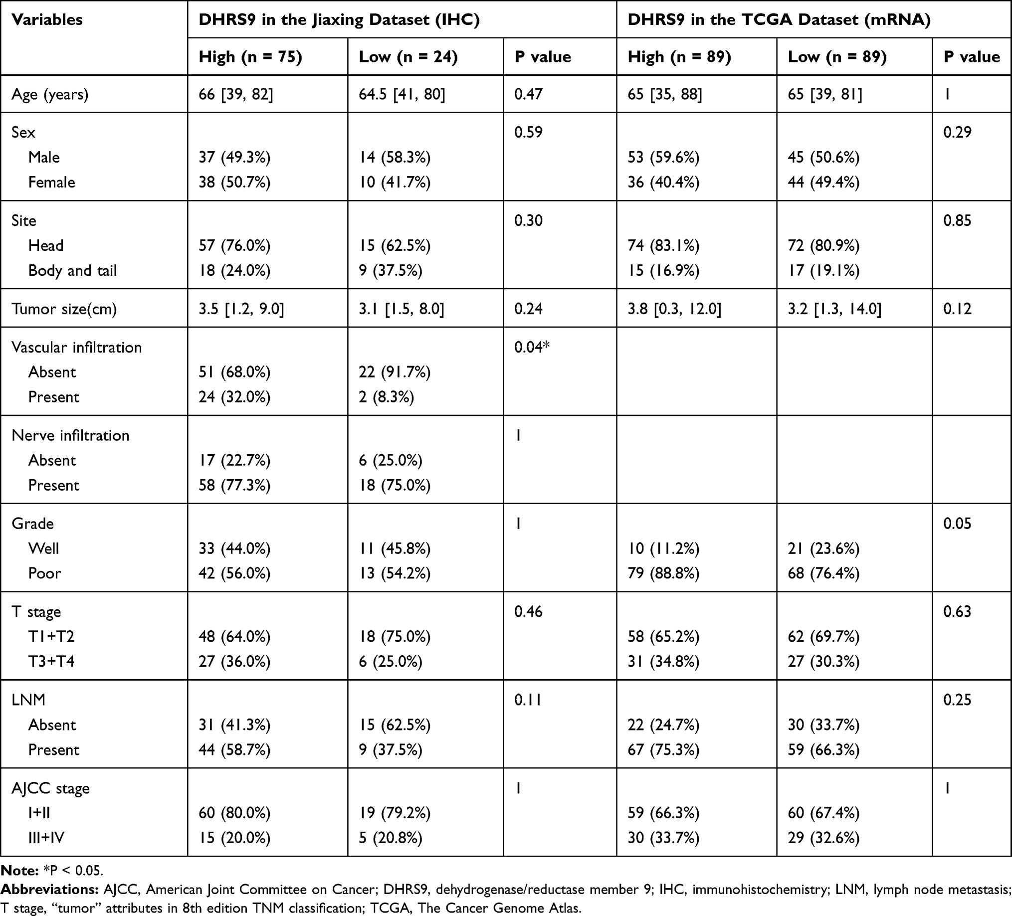 |
Table 1 Association Between Clinicopathologic Variables and DHRS9 Expression in the Two Datasets |
Bioinformatics Analysis
The expression of DHRS9 mRNA in cancerous and adjacent normal tissues or normal pancreatic tissue (222 cases and 173 cases) were obtained from the GEO (GSE16515, GSE15471, GSE62452, GSE63158, GSE102238) and TCGA databases, and the expression of DHRS9 mRNA in PC cell lines and pancreatic duct epithelium cell lines (20 cases and 1 case) was obtained from the GSE40098. The detailed data can be viewed in the database according to the GSE number (https://www.ncbi.nlm.nih.gov/geo/) and is also presented in Supplementary Table 1.
Western Blot Analysis
Total protein was extracted from the five pairs of PC and adjacent normal tissues and quantified these using a BCA Protein Assay (Servicebio, Wuhan, China). The protein samples were separated by sodium dodecyl sulfate polyacrylamide gel electrophoresis (SDS-PAGE) and transferred onto polyvinylidene fluoride (PVDF) membranes for immunoblotting. Immunoblotting was performed with antibodies against DHRS9 (LifeSpan BioSciences, American, catalog no.LS-C145077/57,544, 1:200). And GAPDH (Servicebio, Wuhan, China). The results were visualized with a chemiluminescent detection system and autoradiography film.
Immunohistochemistry
To verify the reliability of the bioinformatics analysis, the expression of DHRS9 in 102 tumor tissues and 91 adjacent normal tissues from 109 surgically treated PC patients at our institute was determined via IHC staining. Formalin-fixed, paraffin-embedded sections of all 193 samples were subjected to the EnVision method using DHRS9 polyclonal antibodies (LifeSpan BioSciences, American, catalog no.LS-C145077/57,544, 1:250). Phosphate-buffered saline was used as a negative control instead of the primary antibody. Sections were counterstained with hematoxylin and eosin.
Two independent experienced investigators examined all slides. The intensity of the cellular staining was scored as 0 for no staining; 1, weak; 2, moderate; and 3, strong. The proportion of the stained tumor cells was scored as 0 for 0–5% stained cells; 1, 6–25%; 2, 26–0%; 3, 51–75%; and 4, >75%. These intensity and proportion scores were then multiplied and calculated as the final score. A score ≥ 6 was defined as DHRS9 overexpression, whereas a score < 6 was regarded as low expression. The cases in the Jiaxing dataset were then accordingly grouped into the low and the high expression groups. The cases in the TCGA dataset were also grouped into the low and the high expression groups, but according to the median value of DHRS9 expression, which reflected the expression of DHRS9 at the mRNA level.
Statistical Analysis
All statistical analyses were performed using R (https://www.r-project.org/R version 3.6.1). Data cleaning was based on the “dplyr” package, survival analysis was based on the “survminer” and “survival” packages, the three-line table was based on the “Table 1” package, and other data processing was based on the basic functions of R. Continuous variables were presented as the median (range) and compared using the Mann–Whitney U-test. Meanwhile, categorical variables were expressed as numbers (%) and compared using the chi-square test. The Kaplan–Meier method was used for survival analysis, and univariate and multivariate Cox regression analyses were used to assess the prognostic factors associated with overall survival (OS). P-values < 0.05 were considered significant.
Result
Patient Characteristics
In total, there were 24 cases with low expression and 75 cases with high expression in the Jiaxing dataset. Meanwhile, in the TCGA dataset, there were 178 cases categorized into the low and high expression groups by the median DHRS9 mRNA expression. There were no significant differences in patient characteristics between the two datasets. The median age was 65 (39–82) years, the proportion of male patients was higher, and most of the tumors (72.7% in Jiaxing set and 82.0% in TCGA set) were located in the head of the pancreas. Sex, age, tumor site, tumor diameter, tumor grade, T stage, lymph node metastasis, and TNM stage (P > 0.05) were also not significantly different between the high and the low expression groups in the two datasets. In the Jiaxing dataset, high DHRS9 expression was associated with vascular infiltration (P < 0.05) (Table 1).
mRNA and Protein Levels of DHRS9 is Upregulated in Pancreatic Cancer
The five datasets from the GEO database (GSE16515, GSE15471, GSE62452, GSE63158, and GSE102238) showed that compared with the adjacent normal tissues or normal pancreatic tissue, DHRS9 was overexpressed in PC tissues (Figure 1A–D). The GSE16515 and GSE15471 datasets used the same platform, and thus these two datasets were processed together (Figure 1A). In addition, compared with immortalized non-malignant human pancreatic duct epithelial (HPDE) cell line, DHRS9 mRNA was generally overexpressed in PC cell lines (GSE40098). The relative expression of DHRS9 mRNA in the nine leading cancer cell lines and the HPDE cell line is shown in Figure 1E. The results of IHC staining to confirm the results of the bioinformatics analysis are shown in Figure 1F. In addition, Western blot assay was performed to determine the protein expression levels of DHRS9 on the five pairs of samples in Figure 1G. DHRS9 protein expression was higher in the tumor tissues than in the adjacent normal tissues in cases 1–4, but in the fifth case, the result was reversed. The low and high expressions of DHRS9 are shown in Figure 2. The DHRS9 expression in PC tissues was significantly higher than that in the adjacent normal tissues (P < 0.001).
 |
Figure 1 Comparison of DHRS9 mRNA or protein expression between (A) PC and normal tissues (GSE16515 and GSE15471); (B) PC and adjacent normal tissues (GSE62452); (C) PC and adjacent normal tissues (GSE63158); (D) PC and adjacent normal tissues (GSE102238); (E) nine leading PC cell lines and the HPDE cell line (GSE40098); (F) Intensity of IHC in PC and adjacent normal tissues (Jiaxing set); (G) Western blot of five couple cases in PC and adjacent normal tissues (Jiaxing set). Abbreviations: ANT, adjacent normal tissues; DHRS9, Dehydrogenase/reductase member 9; GEO, Gene Expression Omnibus; HPDE, immortalized non-malignant human pancreatic duct epithelial cell line; IHC, immunohistochemistry; PC, pancreatic cancer; T, tumor; TCGA, The Cancer Genome Atlas. |
 |
Figure 2 Immunostaining of DHRS9 in pancreatic cancer. (A) Negative; (B) Low; (C) Median; (D) And high expression. Abbreviation: DHRS9, Dehydrogenase/reductase member 9. |
High DHRS9 Expression is Associated with Poor Prognosis
As shown in Figure 3, high DHRS9 expression in the GEO dataset (Figure 3A, P < 0.05, survival data integrated from GSE62452 and GSE102238), TCGA dataset (Figure 3B, P < 0.001), and the study dataset (Figure 3C, P < 0.001) indicated poor OS. The results of univariate and multivariate Cox regression analyses to validate the impact of DHRS9 expression on clinical outcomes are shown in Tables 2 and 3. In univariate analysis, high DHRS9 expression was a prognostic risk factor for OS in both the Jiaxing dataset (Table 2, hazard ratio [HR] = 3.15, P < 0.001) and TCGA dataset (Table 3, HR = 2.04, P < 0.001). In multivariate analysis, high DHRS9 expression remained an independent risk factor for poor prognosis in both the Jiaxing dataset (Table 2, HR = 2.27, P = 0.031) and the TCGA dataset (Table 3, HR = 1.75, P = 0.010). Lymph node metastasis was also an independent risk factor in the multivariate Cox analysis in the two datasets. Other risk factors in each dataset are also shown in Tables 2 and 3.
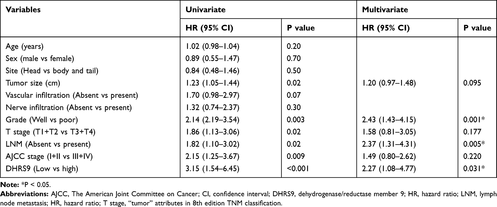 |
Table 2 Univariate and Multivariate Cox Regression Analyses of Prognostic Factors in the Jiaxing Set |
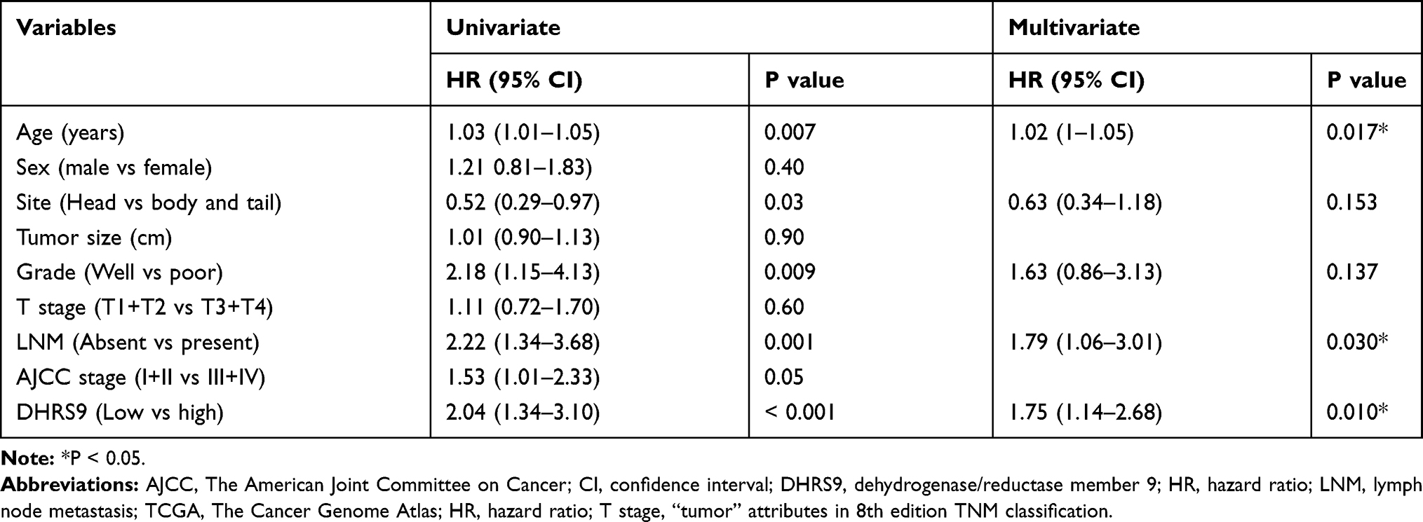 |
Table 3 Univariate and Multivariate Cox Regression Analyses of Prognostic Factors in the TCGA Set |
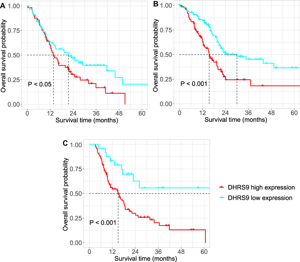 |
Figure 3 Kaplan-Meier survival analysis of overall survival according to DHRS9 expression in pancreatic cancer according to the (A) GEO dataset (GSE62452 and GSE102238); (B) TCGA dataset; and (C) The Jiaxing set. Abbreviations: DHRS9, Dehydrogenase/reductase member 9; GEO, Gene Expression Omnibus; TCGA, The Cancer Genome Atlas. |
Potential Mechanisms of DHRS9 in Pancreatic Cancer
To explore the potential mechanisms by which DHRS9 impacted the prognosis of PC, the study mined the gene set co-expressed with DHRS9 from the TCGA database. According to the Pearson correlation between genes, 642 related genes with P value < 0.001 were retained for the next function analyses. The gene set was introduced into Metascape (http://metascape.org/gp/index.html#/main/step1),11 and it was found that these genes may play a role in tumor-related pathways such as the Wnt signaling pathway, serine/threonine kinase signaling pathway, MAPK signaling pathway, response to extracellular stimulus and ubiquitin-dependent protein catabolic process (Figure 4A). Since the three biological processes, serine/threonine kinase signaling pathway, MAPK signaling pathway, and response to extracellular stimulus were all related to MAPK signaling pathway,12 we speculated that DHRS9 may be mainly involved in MAPK signaling pathway. By searching the literature, this study collected key genes in the MAPK signaling pathway.12,13 Through correlation analysis based on TCGA dataset, it was found that the key genes that were highly related to DHRS9 were mainly concentrated in the ERK1/2 pathway (Figure 4B).
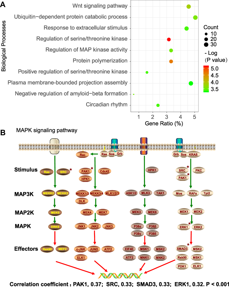 |
Figure 4 Potential mechanism by which DHRS9 impacts the prognosis of pancreatic cancer. (A) Biological processes in which DHRS9 is potentially involved; (B) Key genes of MAPK signaling pathway and genes highly co-expressed with DHRS9. The mark * in the figure indicates that the correlation coefficient is greater than 0.3 and p <0.001. Abbreviations: DHRS9, Dehydrogenase/reductase member 9; MAPK, mitogen-activated protein kinase; MAP2K, mitogen-activated protein kinase kinase; MAP3K, mitogen-activated protein kinase kinase kinase. |
Discussion
This study explored the prognostic impact of mRNA level of DHRS9 in PC using the GEO and TCGA databases and verified the results through Western blot assay and IHC analysis of samples from our institution. In addition, the possible mechanism by which DHRS9 was involved in PC progression was also investigated through bioinformatics. The results indicated that compared with normal pancreatic tissue, DHRS9 was highly expressed in PC tissues, and high DHRS9 expression was associated with poor prognosis. Further, DHRS9 was involved in tumor-related pathways such as the MAPK signaling pathway. In both the Jiaxing dataset and the TCGA dataset, DHRS9 expression showed no significant correlation with parameters such as sex, age, tumor diameter, tumor grade, lymph node metastasis, T stage, and TNM stage. However, it was associated with vascular infiltration in the Jiaxing dataset.
The most common cause of death in patients with PC after surgery is local recurrence or distant metastasis.14 Our findings indicated that tumor tissues may promote vascular invasion through the overexpression of DHRS9, which accelerates the recurrence and metastasis of PC. All-trans retinoic acid is not only a target for antitumor therapy, but also interacts with other substances to play a synergistic antitumor effect.15 Earlier studies suggested that DHRS9 exerts its anti-tumor activity as a rate-limiting enzyme in the all-trans retinoic acid biosynthesis process.16 The relationship between DHRS9 expression and prognosis in oral squamous cell carcinoma and colon cancer appears to confirm this view.8,9 However, existing research suggests that DHRS9 not only plays a role in suppressing cancer, but also in promoting cancer in some tumors such as gastric cancer and ovarian cancer.10 The results of this study also demonstrated that, compared with normal pancreatic tissue, DHRS9 was highly expressed in PC tissue. In addition, patients with high expression had a worse prognosis than those with low expression. The results of the above researches suggest that DHRS9 may play contrasting roles through distinct mechanisms. However, to date, there are no studies conducted to identify the mechanism by which DHRS9 promotes tumorigenesis. One study reported that DHRS9 can act as a marker of regulatory macrophages with immunosuppressive activity.17 Indicating that DHRS9 may promote tumor progression by affecting tumor immunity.
Co-expression of genes usually suggests that they perform similar functions in biology.18 This study explored the gene set co-expressed with DHRS9 in PC through bioinformatics and then further explored the function of this gene set. The results showed that DHRS9 may play a role in multiple tumor-related pathways (Figure 4A). Since the three biological processes, serine/threonine kinase signaling pathway, MAPK signaling pathway, and response to extracellular stimulus were all related to MAPK signaling pathway,12 the study speculated that DHRS9 may be mainly involved in MAPK signaling pathway. By searching the literature, the study collected key genes in the MAPK signaling pathway.12,13 Through correlation analysis based on TCGA dataset, it was found that key genes that were highly related to DHRS9 were mainly concentrated in the ERK1/2 pathway (Figure 4B). Based on these findings, we speculate that DHRS9 may exert cancer-promoting activity in PC by participating in the biological processes of the MAPK/ERK1/2 signaling pathway.
The study explored the relationship between DHRS9 overexpression and the prognosis of PC, and the potential mechanism. However, several limitations to this study need to be considered when interpreting the findings. First, the precise molecular mechanism by which DHRS9 acts as a tumor promotor in PC and the roles of DHRS9 in PC cell lines and animal models need to be verified in vitro. Despite these limitations, our results clearly indicate that DHRS9 might be a useful diagnostic and therapeutic target in PC patients.
In conclusion, the results of this study showed that DHRS9 overexpression is associated with a poor outcome in PC patients. Functional analysis of DHRS9 co-expressed genes suggests that DHRS9 is involved in multiple tumor-related processes.
Data Sharing Statement
The data for statistical processing are available from the first author and the corresponding author, Huangbao Li, at [email protected].
Ethical Approval
This study was conducted in line with the tenets of the 1964 Declaration of Helsinki and its later amendments or comparable ethical standards and was approved by the Institutional Review Board of First Hospital of Jiaxing.
Informed Consent
Because the study was retrospective and the data used were obtained from a public database, informed consent could not be obtained from the patients.
Author Contributions
All authors made substantial contributions to conception and design, acquisition of data, or analysis and interpretation of data; took part in drafting the article or revising it critically for important intellectual content; gave final approval of the version to be published; and agree to be accountable for all aspects of the work.
Disclosure
The authors report no conflicts of interest in this work.
References
1. Tiriac H, Belleau P, Engle DD, et al. Organoid profiling identifies common responders to chemotherapy in pancreatic cancer. Cancer Discov. 2018;8(9):1112–1129. doi:10.1158/2159-8290.CD-18-0349
2. Siegel RL, Miller KD, Jemal A. Cancer statistics, 2020. CA Cancer J Clin. 2020;70(1):7–30. doi:10.3322/caac.21590
3. Lin QJ, Yang F, Jin C, et al. Current status and progress of pancreatic cancer in China. World J Gastroenterol. 2015;21(26):7988–8003. doi:10.3748/wjg.v21.i26.7988
4. Khorana AA, Mangu PB, Berlin J, et al. Potentially curable pancreatic cancer: American society of clinical oncology clinical practice guideline update. J Clin Oncol. 2017;35(20):2324–2328. doi:10.1200/JCO.2017.72.4948
5. Ducreux M, Seufferlein T, Van Laethem JL, et al. Systemic treatment of pancreatic cancer revisited. Semin Oncol. 2019;46(1):28–38. doi:10.1053/j.seminoncol.2018.12.003
6. Soref CM, Di Y-P, Hayden L, et al. Characterization of a novel airway epithelial cell-specific short chain alcohol dehydrogenase/reductase gene whose expression is up-regulated by retinoids and is involved in the metabolism of retinol. J Biol Chem. 2001;276(26):24194–24202. doi:10.1074/jbc.M100332200
7. Kropotova ES, Zinovieva OL, Zyryanova AF, et al. Altered expression of multiple genes involved in retinoic acid biosynthesis in human colorectal cancer. Pathol Oncol Res. 2014;20(3):707–717. doi:10.1007/s12253-014-9751-4
8. Shimomura H, Sasahira T, Nakashima C, et al. Downregulation of DHRS9 is associated with poor prognosis in oral squamous cell carcinoma. Pathology. 2018;50(6):642–647. doi:10.1016/j.pathol.2018.06.002
9. Hu L, Chen HY, Han T, et al. Downregulation of DHRS9 expression in colorectal cancer tissues and its prognostic significance. Tumour Biol. 2016;37(1):837–845. doi:10.1007/s13277-015-3880-6
10. Nagy Á, Lanczky A, Menyhart O, et al. Validation of miRNA prognostic power in hepatocellular carcinoma using expression data of independent datasets. Sci Rep. 2018;8(1):9227. doi:10.1038/s41598-018-27521-y
11. Zhou Y, Zhou B, Pache L, et al. Metascape provides a biologist-oriented resource for the analysis of systems-level datasets. Nat Commun. 2019;10(1):1523. doi:10.1038/s41467-019-09234-6
12. Kyriakis JM, Avruch J. Mammalian MAPK signal transduction pathways activated by stress and inflammation: a 10-year update. Physiol Rev. 2012;92(2):689–737. doi:10.1152/physrev.00028.2011
13. Hoang VT, Yan TJ, Cavanaugh JE, et al. Oncogenic signaling of MEK5-ERK5. Cancer Lett. 2017;392:51–59. doi:10.1016/j.canlet.2017.01.034
14. Ansari D, Gustafsson A, Andersson R. Update on the management of pancreatic cancer: surgery is not enough. World J Gastroenterol. 2015;21(11):3157–3165. doi:10.3748/wjg.v21.i11.3157
15. Schultze E, Collares T, Lucas CG, et al. Synergistic and additive effects of ATRA in combination with different anti-tumor compounds. Chem Biol Interact. 2018;285:69–75. doi:10.1016/j.cbi.2018.02.021
16. Napoli JL. Physiological insights into all-trans-retinoic acid biosynthesis. Biochim Biophys Acta. 2012;1821(1):152–167. doi:10.1016/j.bbalip.2011.05.004
17. Riquelme P, Amodio G, Macedo C, et al. DHRS9 is a stable marker of human regulatory macrophages. Transplantation. 2017;101(11):2731–2738. doi:10.1097/TP.0000000000001814
18. van Dam S, Vosa U, van der Graaf A, et al. Gene co-expression analysis for functional classification and gene-disease predictions. Brief Bioinform. 2018;19(4):575–592. doi:10.1093/bib/bbw139
 © 2020 The Author(s). This work is published and licensed by Dove Medical Press Limited. The full terms of this license are available at https://www.dovepress.com/terms.php and incorporate the Creative Commons Attribution - Non Commercial (unported, v3.0) License.
By accessing the work you hereby accept the Terms. Non-commercial uses of the work are permitted without any further permission from Dove Medical Press Limited, provided the work is properly attributed. For permission for commercial use of this work, please see paragraphs 4.2 and 5 of our Terms.
© 2020 The Author(s). This work is published and licensed by Dove Medical Press Limited. The full terms of this license are available at https://www.dovepress.com/terms.php and incorporate the Creative Commons Attribution - Non Commercial (unported, v3.0) License.
By accessing the work you hereby accept the Terms. Non-commercial uses of the work are permitted without any further permission from Dove Medical Press Limited, provided the work is properly attributed. For permission for commercial use of this work, please see paragraphs 4.2 and 5 of our Terms.