")
Back to Journals » International Journal of Nanomedicine » Volume 13 » T-NANO 2014 Abstracts
Preferential binding of fullerene and fullerenol with the N-terminal and middle regions of amyloid beta peptide: an in silico investigation
Authors Pandya V, Baweja L, Dhawan A
Received 18 October 2016
Accepted for publication 21 November 2016
Published 15 March 2018 Volume 2018:13(T-NANO 2014 Abstracts) Pages 71—73
DOI https://doi.org/10.2147/IJN.S125011
Checked for plagiarism Yes
Review by Single anonymous peer review
Peer reviewer comments 2
Editor who approved publication: Professor Lei Yang
Vishal Pandya,1 Lokesh Baweja,1,2 Alok Dhawan1,2
1Division of Biological & Life Sciences, School of Arts & Sciences, (Formerly, Institute of Life Sciences), Ahmedabad University, Ahmedabad, Gujarat, 2Nanotherapeutics & Nanomaterial Toxicology Group, CSIR-Indian Institute of Toxicology Research, Mahatma Gandhi Marg, Lucknow, Uttar Pradesh, India
Abstract: Amyloid beta (Aβ) deposits are implicated in the pathogenesis of debilitating neurodegenerative disorders such as Alzheimer’s disease. In the present study, the interactions of carbon-based nanoparticles (NPs) such as fullerene and fullerenol having different surface chemistry with Aβ were investigated using molecular dynamics simulations and docking studies. A detailed analysis of docking results showed that in 68% of the Aβ conformations, fullerene and fullerenol showed interactions with the N-terminal region of the peptide. However, the high-affinity binding site (E=−48.31 kJ/mol) of fullerene resides in the hydrophobic middle region of the peptide, whereas fullerenol interacts favorably with the charged N-terminal region with a binding energy of −50.42 kJ/mol. The above differences in binding could be attributed to the surface chemistry of fullerene and fullerenol. Moreover, the N-terminal and middle regions of Aβ play an important role in Aβ aggregation. Therefore, the binding of fullerene and fullerenol could inhibit amyloid aggregation. This information will be helpful in designing NPs for targeting amyloid-related disorders.
Keywords: fullerene, fullerenol
Introduction
The fast developing field of nanotechnology has made a significant impact on numerous areas of science and technology. Understanding the interaction between nanoparticles (NPs) and biomolecules1,2 is essential for NP-based biotechnology and biomedical applications such as gene delivery, inhibiting protein amyloidosis, tumor therapy, and cellular imaging. Carbon-based NPs such as fullerene and fullerenol have been proposed for inhibiting amyloid aggregation. However, the mechanism of interaction of fullerene and fullerenol with amyloid beta (Aβ) peptide is not well understood. Therefore, an attempt has been made to investigate the interaction of fullerene and fullerenol with Aβ using computational studies. This study will provide detailed insight into the interaction of fullerene and fullerenol with Aβ, which, in turn, would be useful for designing NPs for targeting amyloid-related disorders.
Materials and methods
The structure of Aβ (1–40) was obtained from the Protein Data Bank (1BA4). The Aβ was simulated further for 50 ns using GROMACS (Version 4.5.5) to generate an ensemble of Aβ conformations using g_cluster algorithm. Nineteen dominant conformations of Aβ were docked with fullerene and fullerenol using PatchDock server3 to predict the most probable binding site of fullerene and fullerenol in Aβ. Fullerene and fullerenol were generated using GaussView. The docking results were analyzed using PyMOL and graphs were plotted using Σ(Sigma) plot.
Results and discussion
The interaction energy of Aβ–NPs complexes is represented in Figure 1. It can be observed from the figure that fullerene showed the highest affinity, with conformation number 17, whereas fullerenol showed high affinity with conformation number 8 of Aβ.
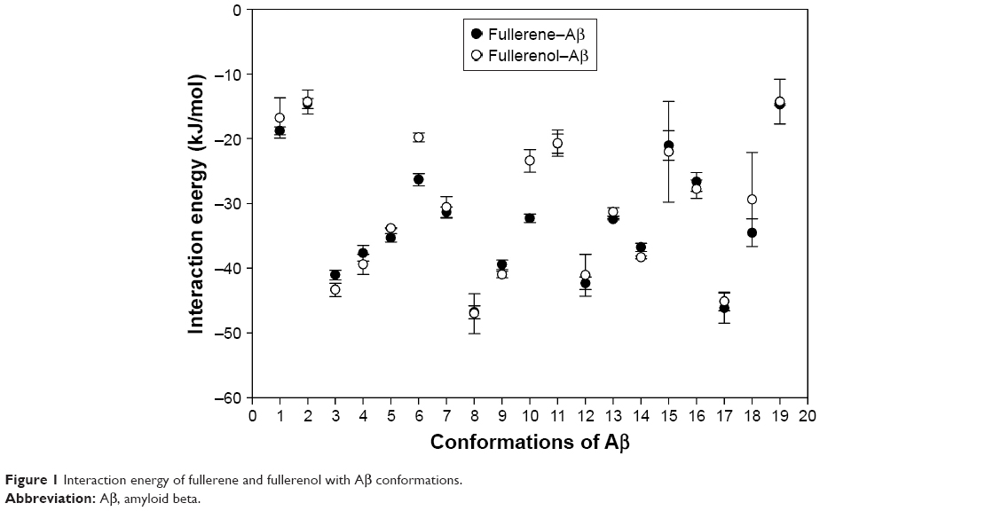 | Figure 1 Interaction energy of fullerene and fullerenol with Aβ conformations. |
The snapshots of Aβ–fullerene and Aβ–fullerenol are represented in Figure 2. The high-affinity binding site (E=−48.31 kJ/mol) of fullerene resides in the hydrophobic middle region of the peptide, whereas fullerenol interacts favorably with the charged N-terminal region with a binding energy of −50.42 kJ/mol. Aβ has three different regions, which are N-terminal, middle region, and C-terminal. Docking studies have shown that these NPs preferentially bind to the N-terminal and middle regions of the peptide (Figure 2). To confirm which portion of the peptide is actively involved in the interaction with fullerene and fullerenol, we calculated the occupancy of binding sites by NPs in Aβ. The results showed that fullerene and fullerenol occupied the N-terminal in ~68% of Aβ conformations and fullerene occupied the middle region in ~47% of Aβ conformations, whereas the C-terminal showed least occupancy by fullerene and fullerenol (Figure 3).
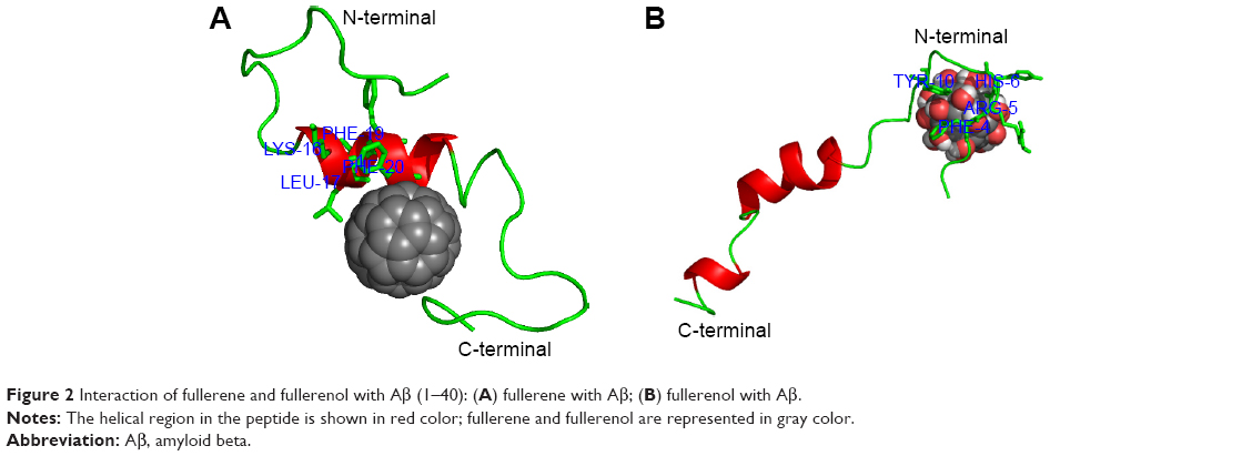 | Figure 2 Interaction of fullerene and fullerenol with Aβ (1–40): (A) fullerene with Aβ; (B) fullerenol with Aβ. |
 | Figure 3 Most probable binding sites of peptide with fullerene and fullerenol. |
We could infer that the C-terminal is not actively involved in the interaction as the other two regions of Aβ. The middle region of the peptide and specifically the residues 16–20 (KLVFF) are involved in Aβ polymerization/aggregation.4 The aromatic residues and the charged residues in these regions may form π–π, hydrogen bonding, and van der Waals interactions with fullerene and fullerenol.
Taking together, the above analysis showed that the binding of the NPs to Aβ may modulate their aggregation.
Conclusion
We investigated the effect of fullerene and fullerenol NPs on the Aβ (1–40) peptides by performing docking and simulation studies. Our docking studies demonstrated high-affinity binding of NPs with the peptide at the N-terminal and middle regions, which is in agreement with the previous experimental study results. These results provide novel insight into the inhibition mechanism5 of fullerene and fullerenol on the aggregation of Aβ (1–40). Further molecular dynamics simulations will be performed to validate our docking results.
Acknowledgments
Funding from the vLife Sciences, Centre of Excellence, In Silico Technology, Institute of Life Sciences, Ahmedabad University, and the financial assistance for the Centre for Nanotechnology Research and Application (CENTRA) by the Gujarat Institute for Chemical Technology (Grant no ILS/GICT/2013/003) are gratefully acknowledged. The European Union Seventh Framework Program (FP7/2007–2013) under the Grant agreement No 263147 (Nano Valid, Development of reference methods for hazard identification, risk assessment and life cycle analysis of engineered nanomaterials) is also acknowledged. The Division of Biological & Life Sciences, Ahmedabad University manuscript communication number is 041.
Disclosure
The authors report no conflicts of interest in this work.
References
Lokesh B, Balamurugan K, Subramanian V, Dhawan A. Hydration patterns of graphene-based nanomaterials (GBNMs) play a major role in the stability of a helical protein: a molecular dynamics simulation study. Langmuir. 2013;29(46):14230–14238. | ||
Matteo C, Francesco Z. Baiting proteins with C60. ACS Nano. 2010;4(4):2283–2299. | ||
Klein J. Probing the interactions of proteins and nanoparticles. Proc Natl Acad Sci U S A. 2007;104(7):2029–2030. | ||
Ito M, Johansson J, Stomberg M, Nilsson L. Effects of ligands on unfolding of amyloid β-peptide central helix: mechanistic insights from molecular dynamics simulations. PLoS One. 2012;7(1):e30510. | ||
Jeong E, Minyung L. Fullerene inhibits β-amyloid peptide aggregation. Biochem Biophys Res Commun. 2003;303(2):576–579. |
 © 2018 The Author(s). This work is published and licensed by Dove Medical Press Limited. The full terms of this license are available at https://www.dovepress.com/terms.php and incorporate the Creative Commons Attribution - Non Commercial (unported, v3.0) License.
By accessing the work you hereby accept the Terms. Non-commercial uses of the work are permitted without any further permission from Dove Medical Press Limited, provided the work is properly attributed. For permission for commercial use of this work, please see paragraphs 4.2 and 5 of our Terms.
© 2018 The Author(s). This work is published and licensed by Dove Medical Press Limited. The full terms of this license are available at https://www.dovepress.com/terms.php and incorporate the Creative Commons Attribution - Non Commercial (unported, v3.0) License.
By accessing the work you hereby accept the Terms. Non-commercial uses of the work are permitted without any further permission from Dove Medical Press Limited, provided the work is properly attributed. For permission for commercial use of this work, please see paragraphs 4.2 and 5 of our Terms.