")
Back to Journals » Clinical Ophthalmology » Volume 11
Normal flora of conjunctiva and lid margin, as well as its antibiotic sensitivity, in patients undergoing cataract surgery at Phramongkutklao Hospital
Authors Ratnumnoi R, Keorochana N , Sontisombat C
Received 25 March 2016
Accepted for publication 21 September 2016
Published 27 January 2017 Volume 2017:11 Pages 237—241
DOI https://doi.org/10.2147/OPTH.S109247
Checked for plagiarism Yes
Review by Single anonymous peer review
Peer reviewer comments 2
Editor who approved publication: Dr Scott Fraser
Ravee Ratnumnoi, Narumon Keorochana, Chavalit Sontisombat
Department of Ophthalmology, Phramongkutklao Hospital, Bangkok, Thailand
Objective: This study aimed to evaluate the normal flora of conjunctiva and lid margin, as well as its antibiotic sensitivity.
Design: This was a prospective cross-sectional study.
Patients and methods: A prospective study was conducted on 120 patients who underwent cataract surgery at the Phramongkutklao Hospital from September 2014 to October 2014. Conjunctival and lid margin swabs were obtained from patients before they underwent cataract surgery. These swabs were used to inoculate blood agar and chocolate agar plates for culturing. After growth of the normal flora, the antibiotic sensitivity method using tobramycin, moxifloxacin, levofloxacin, and cefazolin was applied.
Main outcome measures: Normal flora of conjunctiva and lid margin, along with its antibiotic sensitivity, from patients who underwent cataract surgery was assessed.
Results: A total of 120 eyes were included in this study, and bacterial isolation rates were identified. Five bacteria from the lid margin were cultured, namely, coagulase-negative staphylococcus (58.33%), Streptococcus spp. (2.5%), Corynebacterium (1.67%), Micrococcus spp. (1.67%), and Staphylococcus aureus (0.83%). Two bacteria from the conjunctiva were cultured, namely, coagulase-negative staphylococcus (30%) and Streptococcus spp. (0.83%). Results of antibiotic sensitivity test showed that all isolated bacteria are sensitive to cefazolin 100%, tobramycin 98.67%, levofloxacin 100%, and moxifloxacin 100%.
Conclusion: Coagulase-negative staphylococci are the most common bacteria isolated from conjunctiva and lid margin.
Keywords: conjunctival flora, lid margin flora, cataract surgery, sensitivity, antibiotics
Introduction
Cataract surgery, an intraocular surgery, is considered one of the most frequent procedures performed in the ophthalmology unit. In addition, the number of patients who undergo this type of surgery has increased continuously. With the significant number of operations being performed, postoperative endophthalmitis represents a serious complication. Its prevalence rate has been estimated to be between 0.07% and 0.12%.1–3 The appearance of this complication mostly leads to worsening of patients’ vision and may result in permanent loss of vision if not treated properly. Moreover, the prognosis in this case is poor.
Coagulase-negative staphylococcus, Staphylococcus aureus, and Streptococcus spp. are the common causes of postoperative endophthalmitis.4 In most cases, intraocular infection occurs as a result of bacteria colonizing the area around the eye, eyelid, lid margin, and conjunctiva.5 In addition, Thoms et al6 found that there was a greater risk of endophthalmitis in those who did not receive topical antibiotics until the day after surgery. Assessment of compliance in a prospective study indicated that there was a reduction in periocular flora with the administration of preoperative topical antibiotics.7 Currently, because the preoperative usage of antibiotic eye drops is frequent, bacteria are prone to be drug resistant.
According to a report from the Phramongkutklao Hospital, from 2011 to 2013, there was an increase in the incidence of postoperative endophthalmitis. As a result, several types of antibiotics have been adopted pre- and postoperatively. A guideline was prepared for both pre- and postoperative use of antibiotics during cataract surgery. Moxifloxacin (eye drops) is applied 4 times, every 15 minutes prior to surgery in the preoperative period, whereas cefazolin intracameral, vigamox (eye drops and intracameral), tobrex (eye drops), and levofloxacin (eye drops) are antibiotics of choice for postoperative administration. Even though there are guidelines for pre- and postoperative procedures for cataract surgeries, with usage of antibiotics (topical eye drops, subconjunctival injection, intracameral injection, and systemic antibiotics) being prevalent among ophthalmologists, there are still cases of postoperative endophthalmitis. Root cause analysis was conducted after higher incidence of postoperative endophthalmitis; the methods included cultures of all flora from the surface of all surgical instruments as well as the operating room air. Finally, bacteria cultured from the air needle were found to be the cause of the outbreak.
Taking this serious postoperative complication into account, researchers then perceived the importance of conducting research on bacteria cultured from the lid margin and conjunctiva, together with their response to antibiotics and their drug resistance.
Patients and methods
This research was conducted with patients who had plans to undergo any type of cataract surgery, such as phacoemulsification with intraocular lens, extracapsular cataract extraction (ECCE), intracapsular cataract extraction, and so on, between September and October 2014. This sample included 120 patients scheduled for cataract surgery at Phramongkutklao Hospital.
This research was approved by the Phramongkutklao Institutional Review Board.
Patients
A total of 120 patients (120 eyes) who underwent cataract surgery since September 2014 were enrolled in this research. The additional criteria included consensual patients, aged between 30 and 80 years, and no record of topical and systemic antibiotic use within 7 days prior to sample collection. Patients with blepharitis and periorbital infection were excluded from this research.
Methods
A total of 120 patients (120 eyes) who underwent cataract surgery at Phramongkutklao Hospital and met the inclusion criteria were included in the study. All volunteers had to sign informed consent forms. Data regarding the patients’ history of treatment and medications had to be approved by the hospital director.
Sample specimens were obtained prior to the operation day by using sterile cotton swabs; the samples were then placed for bacterial culture in blood agar and chocolate agar plates. Bacteria found on the plates were then tested for susceptibility to the drugs tobramycin, moxifloxacin, levofloxcin, and cefazolin. Statistical analysis was performed using IBM SPSS statistic (version 23), chi-square test, and Fisher’s exact test, considering the prevalence rate of positive culture of coagulase-negative staphylococcus as 71%. The data were recorded on a predesigned sheet and maintained on an Excel spreadsheet. The baseline characteristics of these research subjects were recorded.
Results
In this study, the baseline characteristics of the 120 participants were as follows: females, 59.17%; males, 40.83%; age group according to the inclusion criteria, 30–80 years. Hypertension was the most common underlying disease in the patients (Table 1).
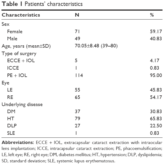 | Table 1 Patients’ characteristics |
Coagulase-negative staphylococci were the most common bacteria found on the lid margin (58.33%) and conjunctiva (30%) (Table 2).
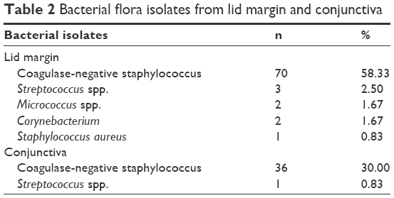 | Table 2 Bacterial flora isolates from lid margin and conjunctiva |
The results showed that coagulase-negative staphylococci found on lid margin area were associated with hypertension (P=0.006). In the same way, coagulase-negative staphylococci found on conjunctiva area were associated with hypertension (P=0.001), and also associated with diabetes mellitus (P=0.028) (Table 3 and 4).
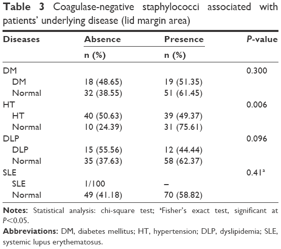 | Table 3 Coagulase-negative staphylococci associated with patients’ underlying disease (lid margin area) |
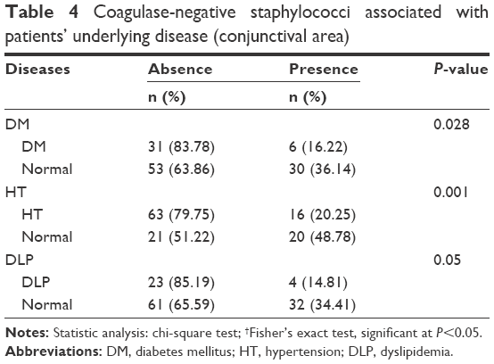 | Table 4 Coagulase-negative staphylococci associated with patients’ underlying disease (conjunctival area) |
In this study, an increase of age was also related to higher amount of bacterial colonization (Tables 5 and 6). Furthermore, there was a significant difference in the number of bacteria found in the conjunctival area, with patients with diabetes mellitus having more bacteria than those without diabetes mellitus (P=0.028).
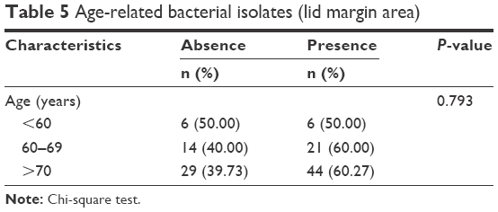 | Table 5 Age-related bacterial isolates (lid margin area) |
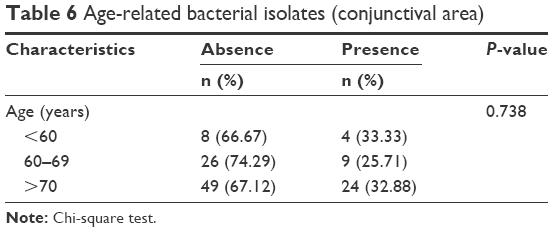 | Table 6 Age-related bacterial isolates (conjunctival area) |
The results of the antibiotic sensitivity test showed that all isolated bacteria were sensitive to all antibiotics used in this study. Only 1.33% of isolated bacteria were resistant to tobramycin (Table 7).
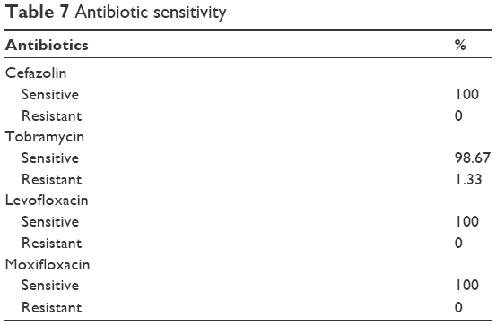 | Table 7 Antibiotic sensitivity |
Discussion
Postoperative endophthalmitis is a serious complication of cataract surgeries, with the potential for developing permanent visual loss. Even though there are guidelines for pre- and postoperative procedures for cataract surgeries, with usage of antibiotics (topical eye drops, subconjunctival injection, intracameral injection, and systemic antibiotics) being prevalent among ophthalmologists, there are still cases of postoperative endophthalmitis.
According to the study conducted by Taban et al,8 the incidence rate of postoperative endophthalmitis following cataract surgery has surprisingly increased. Of the 3,140,650 patients who underwent cataract surgeries in the period 1963–2003, the incidence of an infection in the period 1963–2003 was 0.128%, while it accounted for 0.265% in the period from 2000 to 2003. The same trend was also noted at Phramongkutklao Hospital.
There are a number of risk factors related to postoperative infection, including age, underlying disease, history of use of antibiotics and steroids, types of cataract surgery, intraoperative complications, postoperative antibiotic use, and patients’ personal hygiene.
Antibiotic use is now prevalent, and sometimes, it is overused. Overuse of antibiotics can result in an increase of drug resistance or, even worse, alteration of normal flora. Therefore, this study aimed to study the normal flora of the eye and its sensitivity to the commonly used antibiotics, especially those that individuals in Phramongkutklao Hospital used.
Trinavarat and Atchaneeyasakul9 studied postoperative endophthalmitis after cataract surgery; among the 5 eyes with postoperative endophthalmitis after ECCE surgery, 2 eyes were infected with coagulase-negative staphylococci. When implementing phacoemulsification surgery with 31 eyes, 7 eyes were found to contain coagulase-negative staphylococci.
This study indicated that more bacteria were found in the lid margin area than in the conjunctival area. Coagulase-negative staphylococci were the most common bacteria found in both lid margin in 70 (58.33%) and conjunctiva in 36 eyes (30%), whereas the second and third most common bacteria were Streptococcus spp. and Corynebacterium.
Based on the guidelines of the European Society of Cataract and Refractive Surgeons on prevention, investigation, and management of postoperative endophthalmitis, August 2007, the most common organisms found in patients with postoperative intraocular infection were coagulase-negative staphylococci (33%–77%) and S. aureus (10%–21%); coagulase-negative staphylococci, the most common bacteria found in the conjunctival culture, were found to be drug resistant, with 14% resistant to levofloxacin and 17.9% to tobramycin.10 The study of normal conjunctival flora by Keshav and Basu11 found that coagulase-negative staphylococci (81.5%) and S. aureus (3.7%) are the normal flora inhabiting the lid margin and conjunctiva of normal populations and are the main causes of acute postoperative endophthalmitis.
In addition, this study showed that an increase in age is related to higher amount of bacteria. Hoshi et al5 also found that both patients with lacrimal duct obstruction and immunosuppressed patients are more likely to be colonized by pathogens that cause postoperative endophthalmitis. The isolation rate at the lid margin area was not significant between diabetics and nondiabetics, similar to the results of the study by Suto et al.12
Antibiotic sensitivity in this study indicated that almost all bacteria are sensitive to all types of antibiotics used in this study. Similar to the study of Keshav and Basu,11 the study of normal conjunctival flora and its antibiotic sensitivity found maximum sensitivity to vancomycin, gentamycin, chloramphenicol, and ciprofloxacin.11 However, this does not match with the results of some studies. For example, the study of Suto et al12 demonstrated that coagulase-negative staphylococci, the most common bacteria, are resistant to tobramycin (17.9%) and levofloxacin (14%).12
Conclusion
Coagulase-negative staphylococci are the most common bacteria isolated from the conjunctival and lid margin areas. Bacterial isolation from the lid margin is greater than that from the conjunctival area. Almost all the bacterial florae are found to be highly sensitive to all the antibiotics tested in this study.
Acknowledgments
The authors thank Dr Raveewan Choontanom, MD, Ophthalmologist, Department of Ophthalmology, Phramongkutklao Hospital, who provided both helpful comments and editorial support in the preparation of this manuscript. Additionally, the authors are also grateful to Dr Sudarat Thunyaharn, MSc, Microbiologist, Department of Microbiology, Faculty of Medicine, Phramongkutklao Hospital, for his valuable contribution to the collection of study information, sample collection and incubation, and the interpretations of antibiotic sensitivity in this study.
Disclosure
The authors report no conflicts of interest in this work.
References
Kattan HM, Flynn HW Jr, Pflugfelder SC, et al. Nasocomial endophthalmitis survey. Ophthalmology. 1991;91:3–7. | ||
Results of endophthalmitis Vitrectomy Study. A randomized trial of immediate vitrectomy and of intravenous antibiotics for the treatment of postoperative bacterial endophthalmitis. Arch Ophthalmol. 1995;113(12):1479–1496. | ||
Javitt JC, Vitale S, Canner JK, et al. National outcome of cataract extraction. Arch Ophthalmol. 1991;109(8):1085–1089. | ||
Han DP, Wisniewski SR, Kelsey SF, Doft BH, Barza M, Pavan PR. Microbiologic yields and complication rates of vitreous needle aspiration versus mechanized vitreous biopsy in the endophthalmitis Vitrectomy study. Retina. 1999;19(2):98–102. | ||
Hoshi S, Hashida M, Urabe K. Risk factors for aerobic bacterial conjunctival flora in preoperative cataract patients. Eye (Lond). Epub 2016 Jul 15. | ||
Thoms SS, Musch DC, Soong HK. Postoperative endophthalmitis associated with sutured versus unsutured clear cornea cataract extraction. Br J Ophthalmol. 2007;91(6):728–730. | ||
Miño de Kaspar H, Kreutzer TC, Aguirre-Romo I, et al. A prospective randomized study to determine the efficacy of preoperative topical levofloxacin in reducing conjunctival bacterial flora. Am J Ophthalmol. 2008;145(1):136–142. | ||
Taban M, Behrens A, Newcomb RL, et al. Acute endophthalmitis following cataract surgery, a systematic review of the literature. Arch Ophthalmol. 2005;123(5):613–620. | ||
Trinavarat A, Atchaneeyasakul LO. Surgical techniques of cataract surgery and subsequent postoperative endophthalmitis. J Med Assoc Thai. 2005;88(suppl 9):S1–S5. | ||
Barry P, Behrens-Baumann W, Pleyer U, Seal D. ESCRS Guidelines on prevention, investigation and management of post-operative endophthalmitis. Version 2. 2007. Available from: http://www.escrs.org/vienna2011/programme/handouts/ic-100/ic-100_barry_handout.pdf. Accessed June 11, 2014. | ||
Keshav BR, Basu S. Normal conjunctival flora and their antibiotic sensitivity in Omanis undergoing cataract surgery. Oman J Ophthalmol. 2012;5(1):16–18. | ||
Suto C, Morinaga M, Yagi T, Tsuji C, Toshida H. Conjunctival sac bacterial flora isolated prior to cataract surgery. Infect Drug Resist. 2012;5:37–41. |
 © 2017 The Author(s). This work is published and licensed by Dove Medical Press Limited. The full terms of this license are available at https://www.dovepress.com/terms.php and incorporate the Creative Commons Attribution - Non Commercial (unported, v3.0) License.
By accessing the work you hereby accept the Terms. Non-commercial uses of the work are permitted without any further permission from Dove Medical Press Limited, provided the work is properly attributed. For permission for commercial use of this work, please see paragraphs 4.2 and 5 of our Terms.
© 2017 The Author(s). This work is published and licensed by Dove Medical Press Limited. The full terms of this license are available at https://www.dovepress.com/terms.php and incorporate the Creative Commons Attribution - Non Commercial (unported, v3.0) License.
By accessing the work you hereby accept the Terms. Non-commercial uses of the work are permitted without any further permission from Dove Medical Press Limited, provided the work is properly attributed. For permission for commercial use of this work, please see paragraphs 4.2 and 5 of our Terms.