Back to Journals » International Journal of Nanomedicine » Volume 21
Nature-Inspired Alternatives to PEG for Next-Generation Long-Circulating Nanocarriers
Received 20 April 2026
Accepted for publication 30 May 2026
Published 8 June 2026 Volume 2026:21 618454
DOI https://doi.org/10.2147/IJN.S618454
Checked for plagiarism Yes
Review by Single anonymous peer review
Peer reviewer comments 3
Editor who approved publication: Professor Eng San Thian
Nasrullah Jan,1 Mokhtar Rejili2
1Department of Rheumatology and Clinical Immunology, The First Affiliated Hospital of Xiamen University, School of Medicine, Xiamen University, Xiamen, 361000, People’s Republic of China; 2Department of Biology, College of Sciences, Imam Mohammad Ibn Saud Islamic University (IMSIU), Riyadh, 11623, Saudi Arabia
Correspondence: Nasrullah Jan, Department of Rheumatology and Clinical Immunology, The First Affiliated Hospital of Xiamen University, School of Medicine, Xiamen University, Xiamen, 361000, People’s Republic of China, Email [email protected]
Abstract: Polyethylene glycol (PEG) has long been considered as the gold standard for imparting stealth properties to nanocarriers. However, the recent emergence of anti-PEG antibodies has spurred innovation toward nature-inspired alternatives. By mimicking the structural and functional architectures of biological systems such as cell membranes and protein corona, nanocarriers can be engineered to achieve prolonged circulation, superior biocompatibility, and active targeting capabilities. This perspective presents nature-inspired alternatives to PEG, including cell membranes and the protein corona, for extending the circulation time of nanocarriers. It also highlights current challenges associated with these nature-inspired alternatives and outlines future research directions to overcome these hurdles in designing next-generation long-circulating nanocarriers. The infographic explores nature-inspired alternatives to PEG for long-circulating nanocarriers. It includes: 1. Classic PEGylated Nanocarriers: Highlights PEG as the standard stealth coating for decades that prevents immune detection and extends circulation time. 2. Problem: Anti-PEG Antibodies: Demonstrates how these antibodies accelerate clearance and reduce therapeutic efficacy. 3. Bio-Inspired Alternatives: i. Cell Membrane-Coated Nanocarriers: Employ cell membranes to achieve biomimetic stealth, enabling immune evasion, prolonged circulation, improved biocompatibility, and enhanced targeting. ii. Dysopsonin Corona-Coated Nanocarriers: Utilize a dysopsonin corona (e.g., albumin, clusterin) to provide natural stealth by reducing opsonization and extending circulation time. These nature-inspired coatings effectively address the challenges posed by anti-PEG immunity.Nature-inspired alternatives to PEG, tackling anti-PEG antibodies with bio-inspired stealth coatings.
Keywords: long circulation, ABC phenomenon, cell membranes, protein corona
Introduction
The PEG Paradigm and Its Pitfalls
PEGylation, the covalent attachment of polyethylene glycol (PEG) to therapeutic agents, is a well-established and clinically approved drug delivery method that has been used for decades to extend drug circulation time and reduce immunogenicity, with minimal side effects.1 The use of PEG has been approved by the U.S. Food and Drug Administration (FDA) as a base or vehicle in cosmetics, foods, and pharmaceuticals, including oral, topical, injectable, nasal, and rectal formulations. It is slightly toxic and is easily eliminated from the body via feces or kidneys.2 PEGylation technology has emerged as a widely recognized approach for prolonging the blood residence time of various therapeutics, including small molecules and proteins, leading to significant advancements in the development of drug-loaded nanoparticles and biologics. The hydrophilic PEG chains form a hydration layer around the nanocarriers, which prevents the recognition and binding of nanocarriers by opsonins through spatial repulsion. Consequently, PEGylation is considered to decrease the phagocytosis and clearance of nanocarriers from the systemic circulation by the mononuclear phagocyte system (MPS).3 The impact of PEGylation on the pharmaceutical industry is evident from the multibillion-dollar market value of PEGylated drugs, as over 30 PEGylated products are currently used in clinics, with many more undergoing clinical trials. For further information, please refer to work published by Gao et al4 Table 1 summarizes some FDA-approved PEGylated nanocarriers.
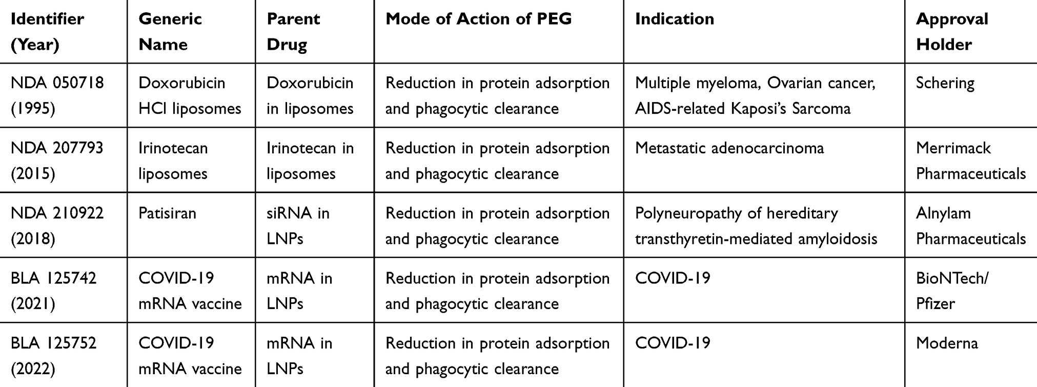 |
Table 1 FDA Approved PEGylated Nanocarriers |
Despite the success of PEGylated therapeutics, recent studies have revealed that PEG is not as biologically inert as previously believed. Repeated administration can induce the production of anti-PEG antibodies, resulting in the accelerated blood clearance (ABC) phenomenon and reduced therapeutic efficacy.5,6 In this phenomenon, the second dose of PEGylated nanocarriers is rapidly cleared from the bloodstream when injected within a certain time interval from the first dose, due to their accelerated accumulation in the liver. This phenomenon alters the biodistribution of the encapsulated drug, leading to decreased therapeutic efficacy and potentially causing adverse effects.7 In preclinical animal models, the initial administration of PEGylated nanocarriers induces the production of anti-PEG antibodies. These antibodies specifically recognize and bind to the surface of subsequently administered PEGylated nanocarriers, triggering activation of the complement system. This immune response significantly accelerates the in vivo clearance of nanocarriers.8 Similarly, clinical studies in humans have frequently reported elevated titers of anti-PEG IgM and IgG antibodies in patients treated with PEGylated biotherapeutics. The presence of these antibodies is associated with reduced therapeutic efficacy, increased toxicity, and a higher incidence of adverse effects. Collectively, these findings challenge the widely held assumption that PEG is nonimmunogenic. Instead, PEGylated nanocarriers commonly provoke an immune response, wherein the induced anti-PEG antibodies contribute to diminished therapeutic outcomes by mediating the ABC phenomenon.9 Therefore, drug delivery scientists are actively seeking for alternative solutions to PEG to enhance the circulation time and efficacy of therapeutics.
Recently, biologically inspired materials, such as cell membrane-coated nanoparticles and endogenous dysopsonin coronas, have been employed in drug delivery applications. Unlike synthetic polymer-coated stealth nanoparticles, which primarily rely on hydration coronas for their function, these alternative nanoparticles incorporate active biological components to achieve anti-phagocytic effects in vivo.10 Specifically, cell membrane constituents are extracted from healthy cells and subsequently coated onto the surfaces of nanocarriers, effectively masking them as native cells. This “cell membrane camouflage” strategy reduces immune system recognition and prolongs circulation time. Consequently, it facilitates immune evasion and promotes targeted interactions with specific cell types, thereby enhancing the specificity and efficacy of drug delivery systems.11 The protein corona contains dysopsonins, such as albumin, histidine-rich glycoprotein, clusterin, Apo A4, and Apo C3, which extend the blood circulation time of nanoparticles.12–14 Albumin, the most abundant serum protein, is particularly associated with dysopsonin-mediated immune evasion, thereby prolonging nanoparticles circulation time.15
This perspective introduces nature-inspired alternatives to PEG, such as cell membranes and protein coronas, to overcome the ABC phenomenon and prolong the circulation time of nanocarriers.
Natural Architectures for Next-Generation Long-Circulating Nanocarriers
Cell Membranes
Cells, the fundamental units of the body, perform a wide range of functions, including the ability to interface and interact with the surrounding environment. Rather than synthetically replicating these functions, researchers are increasingly leveraging naturally isolated cell membranes to enhance the biointerfacing capabilities of nanoparticles.16 Cell membrane coating involves isolating cell membranes from various source cells and applying these membranes onto nanoparticles. The cell membrane is isolated from source cells using various techniques, including hypotonic lysis, dounce homogenization, ultrasonication, and freeze-thaw cycles. Careful attention must be paid to preserve the integrity and functionality of the cell membrane. The isolated cell membranes are then coated onto nanoparticles cores. Both organic (eg., liposomes, PLGA) and inorganic (eg. metal-based) nanoparticle cores are used, each offering distinct advantages in terms of therapeutic efficacy, drug loading, and stability.17 The coating of membrane onto nanoparticles is typically achieved through techniques such as sonication, extrusion, and electroporation. Sonication uses ultrasonic waves to coat membrane vesicles onto nanoparticles. Ultrasonic parameters should be optimized to enhance fusion efficiency while minimizing protein denaturation and drug leakage. In the extrusion process, nanoparticle cores and membrane vesicles are passed through polycarbonate membranes with varying pore sizes to fuse the membrane with the nanoparticles. Although this process is effective and yields stable results, it is not suitable for large-scale production. In electroporation, an electric pulse creates transient pores in the cell membrane, allowing nanoparticles to enter the cell membrane. This process significantly preserves the membrane proteins and integrity.18 The resulting cell membrane-coated nanoparticles have inherent properties, such as prolonged circulation, immune evasion, biocompatibility, and homotypic targeting19 (Figure 1). Zhang’s lab was the pioneer in reporting cell membrane-coated nanotechnology as a long-circulating biomimetic nanosystem in 2011. The researchers isolated cell membranes from red blood cells (RBCs) and coated them onto PLGA nanoparticles. The resulting RBC membrane-coated nanoparticles conferred stealth properties to polymeric nanoparticles. These RBC membrane-coated nanoparticles exhibited a circulation half-life significantly longer than that of their PEGylated counterparts.20 Subsequent research has explored coating nanoparticles with membranes derived from platelets,21 cancer cells,22 leukocytes,23 and stem cell.24
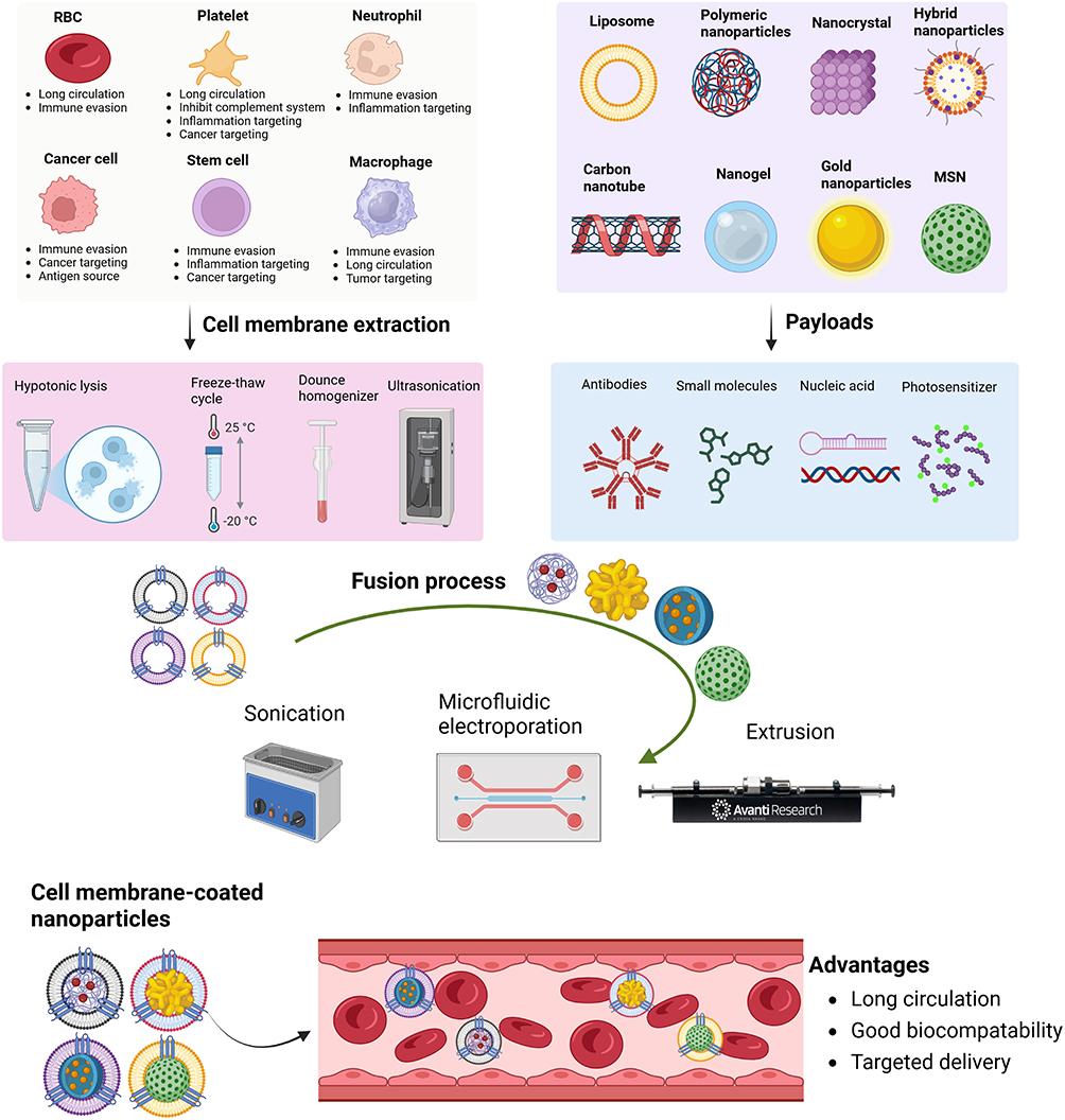 |
Figure 1 Schematic illustration of method of preparation and inherent properties of cell membrane-coated nanoparticles. (https://www.BioRender.com). |
RBC Membrane
RBCs, commonly known as erythrocytes, are abundant in the human body and play crucial roles in oxygen transport. The lipid bilayer of RBC exhibits distinct asymmetry, with phosphatidylethanolamine and phosphatidylserine predominantly located on the inner leaflet, while sphingomyelin and phosphatidylcholine are primarily found on the outer leaflet. This arrangement helps maintain membrane fluidity and prevents unwanted cell aggregation. Additionally, negatively charged sialylated glycoproteins on the surface reduce nonspecific protein binding and inhibit activation of the complement system. Key surface proteins such as CD47, CD59, and CD55 protect RBCs from phagocytosis and complement-mediated destruction. Furthermore, the absence of mitochondria and nuclei decreases the cells’ metabolic activity and reduces their potential to elicit immune responses.25 The high density of CD47 proteins expressed on their surfaces signals immune cells to inhibit immune attacks, allowing RBCs to circulate without being targeted by the host immune system. This immune-evasive property of RBC membranes makes them particularly advantageous as a coating material for nanoparticles, as they enhance circulation longevity and improve biocompatibility.26,27 Building on this concept, Rao et al functionalized Fe3O4 nanoparticles with RBC membranes. In vitro macrophage uptake and in vivo pharmacokinetic studies demonstrated that due to the presence of CD47, Fe3O4 nanoparticles evaded immune clearance via the CD47/SIRP-α signaling pathway. Upon repeated administration of the RBC membrane-coated Fe3O4 nanoparticles, no ABC phenomenon was observed. Therefore, the researchers concluded that RBC membranes are a superior alternative to PEG for prolonging the systemic circulation time of nanoparticles.28 Similarly, Piao et al found that RBC membranes serve as alternative coating materials to PEG to extend the blood residence time of gold nanocages used in photothermal therapy.29
Platelet Membrane
Platelet membrane coating endows nanoparticles with properties such as prolonged circulation time, immune evasion, and enhanced targeting of inflammation, tumors, and other pathological sites. The prolonged circulation is attributed partly to their “marker-of-self” receptor, CD47, and sialylated glycans, which prevents macrophage-mediated phagocytosis of platelet membrane-coated nanoparticles. In addition to CD47, platelet membranes also contain P-selectins, glycoprotein (GP) Ib, and integrins such as αIIbβ3 and α5β1.30 P-selectins mediate interactions with inflamed or damaged tissues, improving targeting efficiency in cardiovascular diseases.31 They can also bind to overexpressed CD44 on tumors, facilitating dynamics and complex tumor-platelet cross-talk critical for tumor growth and metastasis. GPIb binds to exposed collagen in damaged vasculature to initiate tissue repair. With the help of GPIb, platelets can also bind directly to pathogenic bacteria, aiding in their removal from the body. Integrins such as αIIbβ3 and α5β1 binds to the extracellular matrix in the tumor milieu, optimizing therapeutic cargo delivery and enhancing treatment outcomes.32 Immunomodulatory proteins like CD40L and CD62P are also expressed on platelets membranes, making platelet membrane-coated nanoparticles highly applicable in immunotherapy.31 With such broad and dynamic biointerfacing capabilities, platelet membrane-coated nanoparticles have become attractive drug carriers for long-circulation and targeted delivery applications.
WBC Membrane
Leukocytes, or white blood cells (WBCs), are immune cells responsible for protecting the body against infections, repairing tissue injuries, engulfing foreign invaders, and resisting pathogens or diseases. Leukocytes, such as neutrophils, macrophages, NK cells, and T cells, play essential roles in many important diseases, including infections, cancers, and inflammatory disorders. Their functional diversity have inspired the development of WBC membrane-coated nanoparticles that inherit entire source cell antigens. WBC membrane-coated nanoparticles act as source cell decoys and simulate their broad biointerfacing properties, with intriguing therapeutic potentials.33 Proteins expressed on the cell membranes of immune cells confer a diverse array of functionalities to nanoparticles, including extended circulation time in the bloodstream, enhanced antigen recognition for improved targeting, optimized cellular interactions, controlled drug release profiles, and reduced in vivo toxicity.34
Cancer Cell Membrane
Cancer cells utilize various complex immune evasion mechanisms that enable them to grow uncontrollably. The ability of cancer cells to escape immune surveillance involves a complex interplay between tumor cells, the tumor microenvironment, and immune cells. Cancer cells employ several strategies to evade the immune system, including creating an immunosuppressive microenvironment, altering antigen presentation, and inhibiting immune cell functions. These strategies enable cancer cells to persist and proliferate despite immune surveillance.35 For homotypic targeting, galectin-3, T antigen (Thomsen-Friedenreich glycoantigen), and epithelial cell adhesion molecule (EpCAM) on cancer cell membranes facilitate homotypic adhesion among cancer cells. Moreover, antigens present on cancer cell membranes can be effectively utilized in cancer immunotherapy. Therefore, coating nanoparticles with cancer cell membranes retains tumor-specific antigens and homotypic targeting capabilities, enriching drug delivery approaches for cancer immunotherapy.36
Stem Cell Membrane
The unique properties of stem cells primarily arise from the diverse array of receptors on their membranes, including chemokine, cytokine, cell-matrix, growth factor, and cell-cell communication receptors. Chemokines play a crucial role in stem cell migration, adhesion, and homing to tumors or injured sites. Growth factor receptors are the second most abundant type of stem cell membrane receptor and are associated with stem cell differentiation and migration. Cytokine receptors guide stem cells toward injured areas. Cell-matrix receptors such as CD44 are involved in cell homing, adhesion, and migration. Notch, a transmembrane receptor, mediates signaling between the cells and the nucleus. Therefore, coating nanoparticles with mesenchymal stem cell membranes is an effective method for selective targeting, enabling entry into specific cells.37
Protein Corona
After systemic administration, nanoparticles are exposed to several thousands of proteins in the blood. These proteins are adsorbed on the surface of nanoparticles and form a layer around the nanoparticles called a protein corona. The protein corona changes the physicochemical characteristics of nanoparticles, such as size, surface charge, functionality, and surface composition, thus providing a new biological identity to nanoparticles. This nanoparticle–protein complex determines various biological responses, such as circulation time, cellular uptake, bioavailability, and even toxicity.38 Historically, the formation of a protein corona has been associated with adverse delivery outcomes due to opsonization. Opsonins are proteins that bind to the surface of exogenous particles, including synthetic nanomaterials in the bloodstream, thereby marking them for recognition, sequestration, and subsequent degradation by MPS.39 However, there is also a silver lining. The protein corona contains dysopsonins, such as albumin, histidine-rich glycoprotein, clusterin, Apo A4, and Apo C3, which prolong the blood circulation time of nanoparticles.12–14 Albumin is the most abundant serum protein and is associated with dysopsonin-mediated immune evasion, prolonging the circulation time of nanoparticles.15 Albumin is the predominant protein in blood plasma, constituting approximately 60% of the total blood protein content. As a highly water-soluble globular protein, albumin exhibits biodegradability, biocompatibility, non-immunogenicity, and well-established clinical safety. Its distinctive pocket-like structural features, defined by specific chemical configurations and conformations, enable interactions with a wide range of pharmaceutical agents, potentially protecting these compounds from metabolic degradation and elimination in vivo. With a half-life of approximately 19 days and the ability to evade renal clearance, albumin effectively prolongs the systemic circulation time of nanomedicines. The internalization of albumin by tumor cells primarily occurs through receptor-mediated transcytosis involving gp60 receptors and the secreted protein acidic and rich in cysteine (SPARC) pathway, facilitating active tumor targeting without the need for external ligand modification.40 Due to these advantages, the preformed albumin corona provides a protective coating to the polymeric nanoparticles. The albumin-nanoparticle nexus inhibits plasma protein adsorption and decreases complement activation, which ultimately prolongs the circulation time and reduces the toxicity of polymeric nanoparticles (Figure 2).41 Similarly, in situ albumin corona protected maleimide-decorated nanoparticles from phagocytosis in the systemic circulation, prevented the ABC phenomenon after subsequent dose administration, and facilitated nanoparticle accumulation at tumor sites.42 Albumin corona has enabled the targeted delivery of drug nanocarriers to the liver,43 kidney,44 and heart,45 thus regulating their biodistribution. Clusterin (also known as apolipoprotein-J) is a major protein of the corona that is attached to the surface of polymer-modified nanoparticles. The incorporation of polymer-modified nanoparticles with clusterin reduced macrophage uptake, providing a dyspopsonin-mediated immune evasion function of clusterin.12 Aoyama et al also reported that clusterin provides a stealth effect to non-PEGylated silver and silica nanoparticles. Binding of clusterin to both nanoparticles suppressed the cellular uptake of nanoparticles in human macrophage-like cells (THP-1 cells).46 Histidine-rich glycoprotein is the most abundant protein in the hard corona of SiO2 nanoparticles. Histidine-rich glycoproteins compete and bind with high affinity to the nanoparticle surface, forming a stable and homogenous corona that inhibits the binding and uptake of nanoparticles by macrophages.14 CD47 is a “marker of the self.” Building on this understanding, multiple research groups have engineered vectors, including nanoparticles and lentiviruses, functionalized with CD47 to evade immune cell detection.47–49 In 2013, the laboratory of D. Discher introduced simplified variants of CD47, termed “minimal self-peptides,”50 which have demonstrated significant efficacy in preventing macrophage-mediated clearance of various nanomedicines such as albumin-based nanocarriers,51 silica nanoparticles,52 and lipid nanoparticles.53 Recently, Ponton et al reported the cloaking of albumin and CD47 peptides on mesoporous silica nanoparticles for enhanced immune evasion and synergistic combination therapy. The results showed that albumin and CD47 peptide provided long circulation times to mesoporous silica nanoparticles and mitigated macrophage phagocytosis by up to 3.5-folds.54 These findings suggest that dysopsonins in the protein corona can be utilized as substitutes for PEG to provide stealth ability to the nanocarriers.
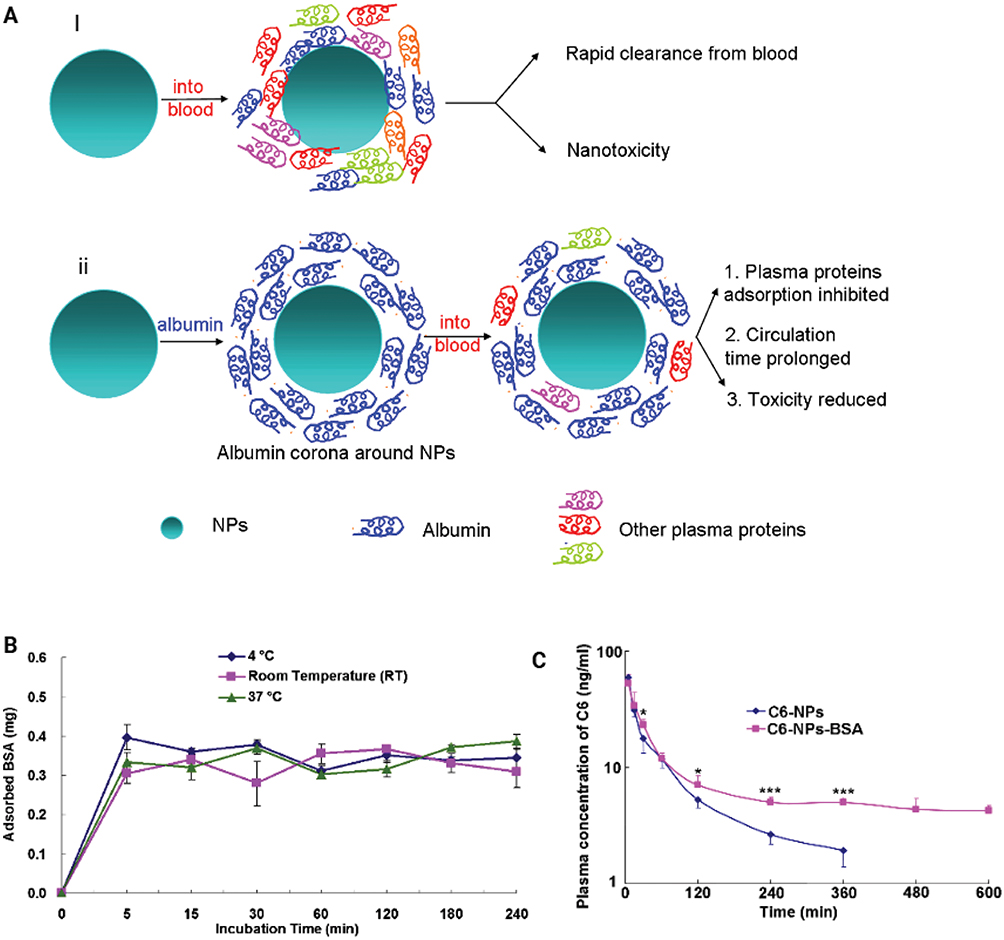 |
Figure 2 (A) Schematic illustration of the biological responses to nanoparticles in the absence and presence of albumin Corona. (I) Upon entry into the bloodstream, various plasma proteins adsorbed onto the bare nanoparticles, leading to accelerated clearance and toxicity. (ii) The presence of preformed albumin corona on the surface of NPs effectively inhibits the adsorption of plasma proteins, thereby extending circulation time and mitigating the associated toxicity of the nanoparticles. (B) Adsorption kinetics of both soft and hard BSA corona. (C) Pharmacokinetic profiles of coumarin-6 in nanoparticles and nanoparticles albumin corona after I.V. administration into the healthy male SD rats. Adopted from41 with permission. Copyright Elsevier, 2013. |
Table 2 summarizes several representative studies of biomimetic nanosystems as nature-inspired alternatives to PEG, highlighting their surface proteins, improvements in circulation half-life, and key features.
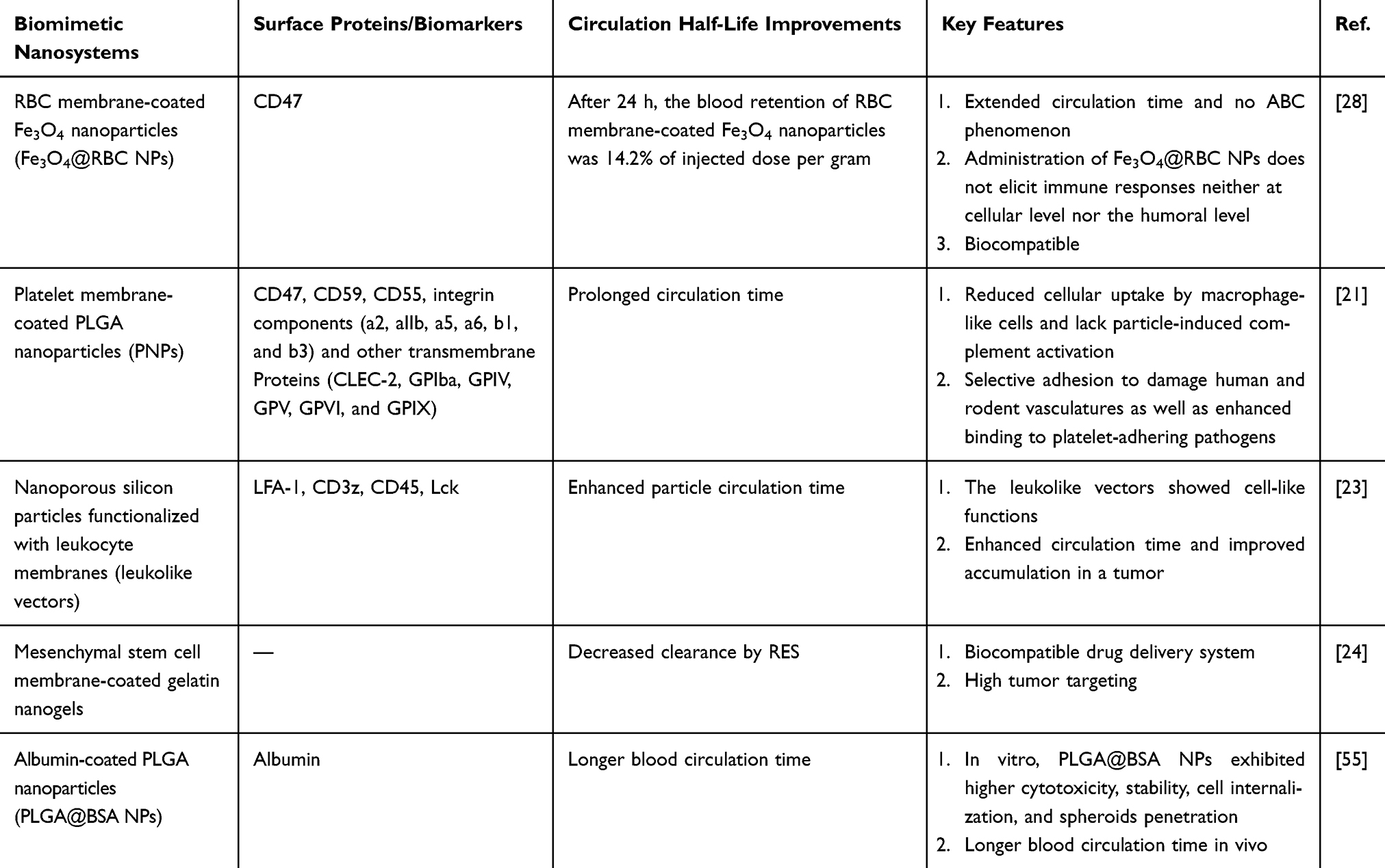 |
Table 2 Summary of Biomimetic Nanosystems as Nature-Inspired Alternatives to PEG, Highlighting Their Surface Proteins, Improvements in Circulation Half-Life, and Key Features |
Current Challenges and Future Prospects
Despite the considerable advantages of cell membrane-coated nanoparticles —such as prolonged blood circulation, biocompatibility, immune evasion, and targeted efficiency—this technology is still in its infancy. From bench to bedside, several theoretical challenges and technical barriers need to be overcome. Currently, only a few defined membrane proteins are responsible for immune evasion and targeting properties; however, numerous other proteins may target different organs and potentially trigger immune responses. Therefore, a three-dimensional microenvironment should be recreated to facilitate cell-cell and cell-extracellular matrix interactions, enabling differentiation between functional proteins and those that may provoke immune responses.56
The large-scale production of cell membrane-coated nanoparticles remains a significant challenge to their widespread application. Current cell membrane coating techniques primarily rely on membrane extrusion and sonication, each with distinct advantages and disadvantages. Specifically, sonication provides high production efficiency but results in low uniformity, whereas membrane extrusion achieves high uniformity at the cost of production efficiency. These limitations could be overcome by employing advanced fabrication methods such as microfluidic technologies, three-dimensional printing, and particle replication in non-wetting templates.26
Preclinical data on cell membrane-coated nanoparticles face challenges in the reproducibility of experimental results and safety assessments. Significant heterogeneity exists in the sources of cell membranes. Variations in cell lines, culture conditions, passage numbers, and extraction methods lead to differences in membrane structure, composition, and protein expression. Similarly, preparation techniques, particle size ranges, extrusion cycles, and coating ratios, and other parameters of nanoparticles lacks standardized protocols. Therefore, future research should shift from individual laboratory practices toward standardized, verifiable, reproducible, and scalable protocols to establish a robust data foundation for subsequent clinical translation. Regarding safety assessments, the ultrasonic disruption and membrane isolation steps are prone to contamination if sterile conditions are not strictly maintained. Moreover, traditional sterilization methods can damage membrane structure and function due to the sensitivity of cell membranes to high temperatures and disinfectants. Consequently, a comprehensive safety evaluation system should be established to conduct systemic toxicological studies involving multiple doses, large-animal models, and long-term assessments to clarify the safety of cell membrane-coated nanoparticles.57
Conclusion
In the post-PEG era, this perspective highlights the paradigm shift in the design of long-circulating nanocarriers by adopting nature-inspired architectures. While PEG has served as an indispensable benchmark for decades, the challenges posed by the ABC phenomenon and anti-PEG antibodies have catalyzed the exploration of more sophisticated biomimetic strategies. As detailed in this perspective, the direct utilization of cell membranes, from erythrocytes to immune cells, offers a remarkable degree of biological authenticity, effectively translating the innate “self-recognition” mechanisms of parental cells to synthetic cores. Concurrently, rational exploitation of the protein corona, either through pre-coating or by engineering surfaces that recruit specific dysopsonins, represents a complementary approach that works in harmony with the biological processes of the body. Collectively, these nature-inspired platforms not only address the immunological shortcomings of PEG, but also introduce multifunctionality, such as enhanced biocompatibility and the potential for active targeting without complex chemical modifications. However, transitioning these biomimetic systems from the laboratory to the clinic requires addressing significant challenges, including standardization of manufacturing, scale-up reproducibility, and comprehensive immunogenicity profiling. Future research will likely focus on advanced membrane coatings or precisely engineered corona-forming moieties to create next-generation nanocarriers that are not merely invisible to the immune system, but also active participants in the biological milieu. Ultimately, by learning from and harnessing nature’s own architecture, we are poised to develop more intelligent, safer, and more effective therapeutic vehicles for a new era of nanomedicine.
Acknowledgment
The authors acknowledge the support and funding provided by the Deanship of Scientific Research at Imam Mohammad Ibn Saud Islamic University (IMSIU) (grant number IMSIU-DDRSP-RP26).
Funding
This work was supported and funded by the Deanship of Scientific Research at Imam Mohammad Ibn Saud Islamic University (IMSIU) (grant number IMSIU-DDRSP-RP26).
Disclosure
The authors declare no conflict of interest.
References
1. Swierczewska M, Lee KC, Lee S. What is the future of PEGylated therapies?. Expert Opin. Emerg. Drugs. 2015;20(4):531–11. doi:10.1517/14728214.2015.1113254
2. Harris JM, Chess RB. Effect of pegylation on pharmaceuticals. Nat Rev Drug Discov. 2003;2(3):214–221. doi:10.1038/nrd1033
3. Pan J, Wang Y, Chen Y, et al. Emerging strategies against accelerated blood clearance phenomenon of nanocarrier drug delivery systems. J Nanobiotechnol. 2025;23(1):138. doi:10.1186/s12951-025-03209-0
4. Gao Y, Joshi M, Zhao Z, Mitragotri S. PEGylated therapeutics in the clinic. Bioeng. Transl. Med. 2024;9(1):e10600. doi:10.1002/btm2.10600
5. Dams ET, Laverman P, Oyen WJ, et al. Accelerated blood clearance and altered biodistribution of repeated injections of sterically stabilized liposomes. J Pharmacol Exp Ther. 2000;292(3):1071–1079. doi:10.1016/S0022-3565(24)35391-1
6. Ishida T, Kiwada H. Accelerated blood clearance (ABC) phenomenon upon repeated injection of PEGylated liposomes. Int J Pharm. 2008;354(1–2):56–62. doi:10.1016/j.ijpharm.2007.11.005
7. Ishihara T, Takeda M, Sakamoto H, et al. Accelerated blood clearance phenomenon upon repeated injection of PEG-modified PLA-nanoparticles. Pharm Res. 2009;26(10):2270–2279. doi:10.1007/s11095-009-9943-x
8. Jan N, Shah H, Shi G. Biomimetic approaches against accelerated blood clearance (ABC) phenomenon of nanoparticulate drug delivery systems. Int J Pharm. 2025;680:125753. doi:10.1016/j.ijpharm.2025.125753
9. Miao G, He Y, Lai K, et al. Accelerated blood clearance of PEGylated nanoparticles induced by PEG-based pharmaceutical excipients. J Control Release. 2023;363:12–26. doi:10.1016/j.jconrel.2023.09.003
10. Fam SY, Chee CF, Yong CY, Ho KL, Mariatulqabtiah AR, Tan WS. Stealth coating of nanoparticles in drug-delivery systems. Nanomaterials. 2020;10(4):787. doi:10.3390/nano10040787
11. Saadh MJ, Mustafa MA, Kumar A, et al. Stealth nanocarriers in cancer therapy: a comprehensive review of design, functionality, and clinical applications. AAPS Pharm Sci Tech. 2024;25(6):140. doi:10.1208/s12249-024-02843-5
12. Schöttler S, Becker G, Winzen S, et al. Protein adsorption is required for stealth effect of poly (ethylene glycol)-and poly (phosphoester)-coated nanocarriers. Nat Nanotechnol. 2016;11(4):372–377. doi:10.1038/nnano.2015.330
13. Ritz S, Schöttler S, Kotman N, et al. Protein Corona of nanoparticles: distinct proteins regulate the cellular uptake. Biomacromolecules. 2015;16(4):1311–1321. doi:10.1021/acs.biomac.5b00108
14. Fedeli C, Segat D, Tavano R, et al. The functional dissection of the plasma Corona of SiO 2-NPs spots histidine rich glycoprotein as a major player able to hamper nanoparticle capture by macrophages. Nanoscale. 2015;7(42):17710–17728. doi:10.1039/C5NR05290D
15. Shaw J, Pearson RM. Nanoparticle personalized biomolecular Corona: implications of pre-existing conditions for immunomodulation and cancer. Biomater. Sci. 2022;10(10):2540–2549. doi:10.1039/D2BM00315E
16. Fang RH, Kroll AV, Gao W, Zhang L. Cell membrane coating nanotechnology. Adv. Mater. 2018;30(23):1706759. doi:10.1002/adma.201706759
17. Zhao X, Liu Q, Xu Z, et al. Cell membrane-coated nanoparticles: advanced drug delivery systems for chronic wound healing therapy. Int J Nanomed;2026. 563555. doi:10.2147/IJN.S563555
18. Jan N, Shah H, Khan S, et al. T cell-inspired therapeutic delivery platforms: from nanomedicines to cell therapy. Mater Today Bio. 2026;37:102909. doi:10.1016/j.mtbio.2026.102909
19. Sultana P, Kim YK, Cho SJ, Asadujjaman M, Jee J-P. Advances in cell membrane-coated nanoparticles: multifunctional platforms for targeted drug delivery, precision phototherapy, and enhanced immunotherapy. Biomater. Sci. 2025;13(19):5232–5259. doi:10.1039/D5BM00660K
20. C-MJ H, Zhang L, Aryal S, Cheung C, Fang RH, Zhang L. Erythrocyte membrane-camouflaged polymeric nanoparticles as a biomimetic delivery platform. Proc Natl Acad Sci. 2011;108(27):10980–10985. doi:10.1073/pnas.1106634108
21. C-MJ H, Fang RH, Wang K-C, et al. Nanoparticle biointerfacing by platelet membrane cloaking. Nature. 2015;526(7571):118–121. doi:10.1038/nature15373
22. Fang RH, C-MJ H, Luk BT, et al. Cancer cell membrane-coated nanoparticles for anticancer vaccination and drug delivery. Nano Lett. 2014;14(4):2181–2188. doi:10.1021/nl500618u
23. Parodi A, Quattrocchi N, Van De Ven AL, et al. Synthetic nanoparticles functionalized with biomimetic leukocyte membranes possess cell-like functions. Nat Nanotechnol. 2013;8(1):61–68. doi:10.1038/nnano.2012.212
24. Gao C, Lin Z, Jurado-Sánchez B, Lin X, Wu Z, He Q. Stem cell membrane-coated nanogels for highly efficient in vivo tumor targeted drug delivery. Small. 2016;12(30):4056–4062. doi:10.1002/smll.201600624
25. Kim Y-A, Lee MH, Sohn HS, Kim HY. From circulation to regeneration: blood cell membrane-coated nanoparticles as drug delivery platform for immune-regenerative therapy. Pharmaceutics. 2026;18(1):66. doi:10.3390/pharmaceutics18010066
26. Jan N, Yehia RM, Mohammed M, et al. Cell membrane-coated nanoparticles: a new frontier in atherosclerosis therapy. Chem Eng J. 2025;524:169183. doi:10.1016/j.cej.2025.169183
27. Nguyen PHD, Jayasinghe MK, Le AH, Peng B, Le MT. Advances in drug delivery systems based on red blood cells and their membrane-derived nanoparticles. ACS nano. 2023;17(6):5187–5210. doi:10.1021/acsnano.2c11965
28. Rao L, Bu LL, Xu JH, et al. Red blood cell membrane as a biomimetic nanocoating for prolonged circulation time and reduced accelerated blood clearance. Small. 2015;11(46):6225–6236. doi:10.1002/smll.201502388
29. Piao J-G, Wang L, Gao F, You Y-Z, Xiong Y, Yang L. Erythrocyte membrane is an alternative coating to polyethylene glycol for prolonging the circulation lifetime of gold nanocages for photothermal therapy. ACS nano. 2014;8(10):10414–10425. doi:10.1021/nn503779d
30. Han H, Bartolo R, Li J, Shahbazi M-A, Santos HA. Biomimetic platelet membrane-coated nanoparticles for targeted therapy. Eur. J. Pharm. Biopharm. 2022;172:1–15. doi:10.1016/j.ejpb.2022.01.004
31. Syed Altaf RR, Simon K, Mohan A, et al. Fusing biomimetic engineering with nanotechnology: the rise of platelet membrane-coated nanoparticles as a versatile platform for biomedical innovation. Adv Ther. 2025;8(11):e00262. doi:10.1002/adtp.202500262
32. Wang S, Duan Y, Zhang Q, et al. Drug targeting via platelet membrane–coated nanoparticles. Small Struct. 2020;1(1):2000018. doi:10.1002/sstr.202000018
33. Wang D, Wang S, Zhou Z, et al. White blood cell membrane-coated nanoparticles: recent development and medical applications. Adv. Healthcare Mater. 2022;11(7):2101349. doi:10.1002/adhm.202101349
34. Oroojalian F, Beygi M, Baradaran B, Mokhtarzadeh A, Shahbazi MA. Immune cell membrane-coated biomimetic nanoparticles for targeted cancer therapy. Small. 2021;17(12):2006484. doi:10.1002/smll.202006484
35. Tufail M, Jiang C-H, Li N. Immune evasion in cancer: mechanisms and cutting-edge therapeutic approaches. Signal Transduct Target Ther. 2025;10(1):227. doi:10.1038/s41392-025-02280-1
36. Zhong Z, Deng W, Wu J, et al. Cell membrane coated nanoparticles as a biomimetic drug delivery platform for enhancing cancer immunotherapy. Nanoscale. 2024;16(18):8708–8738. doi:10.1039/D4NR00284A
37. Wang M, Xin Y, Cao H, et al. Recent advances in mesenchymal stem cell membrane-coated nanoparticles for enhanced drug delivery. Biomater. Sci. 2021;9(4):1088–1103. doi:10.1039/D0BM01164A
38. Nguyen VH, Lee B-J. Protein Corona: a new approach for nanomedicine design. Int J Nanomed. 2017;Volume 12:3137–3151. doi:10.2147/IJN.S129300
39. Dilliard SA, Siegwart DJ. Passive, active and endogenous organ-targeted lipid and polymer nanoparticles for delivery of genetic drugs. Nature Rev Mater. 2023;8(4):282–300. doi:10.1038/s41578-022-00529-7
40. Zhou Y, Wang X, Zhang D, et al. Precision-guided stealth missiles in biomedicine: biological carrier-mediated nanomedicine hitchhiking strategy. Adv. Sci. 2025;12(21):2504672. doi:10.1002/advs.202504672
41. Peng Q, Zhang S, Yang Q, et al. Preformed albumin Corona, a protective coating for nanoparticles based drug delivery system. Biomaterials. 2013;34(33):8521–8530. doi:10.1016/j.biomaterials.2013.07.102
42. Li Z, Li D, Li Q, et al. In situ low-immunogenic albumin-conjugating-Corona guiding nanoparticles for tumor-targeting chemotherapy. Biomater. Sci. 2018;6(10):2681–2693. doi:10.1039/C8BM00692J
43. Cai H, Ma Y, Wu Z, et al. Protein Corona influences liver accumulation and hepatotoxicity of gold nanorods. NanoImpact. 2016;3:40–46. doi:10.1016/j.impact.2016.09.005
44. Qin S, Wu B, Gong T, Zhang Z-R, Fu Y. Targeted delivery via albumin Corona nanocomplex to renal tubules to alleviate acute kidney injury. J Control Release. 2022;349:401–412. doi:10.1016/j.jconrel.2022.07.013
45. Lomis N, Sarfaraz ZK, Alruwaih A, Westfall S, Shum-Tim D, Prakash S. Albumin nanoparticle formulation for heart-targeted drug delivery: in vivo assessment of congestive heart failure. Pharmaceuticals. 2021;14(7):697. doi:10.3390/ph14070697
46. Aoyama M, Hata K, Higashisaka K, Nagano K, Yoshioka Y, Tsutsumi Y. Clusterin in the protein Corona plays a key role in the stealth effect of nanoparticles against phagocytes. Biochem. Biophys. Res. Commun. 2016;480(4):690–695. doi:10.1016/j.bbrc.2016.10.121
47. Wang F, Yuan Z, McMullen P, et al. Near-infrared-light-responsive lipid nanoparticles as an intelligent drug release system for cancer therapy. Chem. Mater. 2019;31(11):3948–3956. doi:10.1021/acs.chemmater.9b00150
48. Sosale NG, Ivanovska T II, Tsai RK, et al. “Marker of Self” CD47 on lentiviral vectors decreases macrophage-mediated clearance and increases delivery to SIRPA-expressing lung carcinoma tumors. Mol Ther Meth Clin Develop. 2016;3:16080. doi:10.1038/mtm.2016.80
49. Alyami EM, Tarar A, Peng C-A. Less phagocytosis of viral vectors by tethering with CD47 ectodomain. J Mat Chem B. 2022;10(1):64–77. doi:10.1039/D1TB01815A
50. Rodriguez PL, Harada T, Christian DA, Pantano DA, Tsai RK, Discher DE. Minimal” Self” peptides that inhibit phagocytic clearance and enhance delivery of nanoparticles. Science. 2013;339(6122):971–975. doi:10.1126/science.1229568
51. Dou W-T, Guo C, Zhu L, et al. Targeted near-infrared fluorescence imaging of liver cancer using dual-peptide-functionalized albumin particles. Chem. Biomed. Imaging. 2023;2(1):47–55. doi:10.1021/cbmi.3c00078
52. Liu C, Yu D, Ge F, Yang L, Wang Q. Fluorescent and mass spectrometric evaluation of the phagocytic internalization of a CD47-peptide modified drug-nanocarrier. Anal. Bioanal.Chem. 2019;411(18):4193–4202. doi:10.1007/s00216-019-01825-y
53. Papp TE, Zeng J, Shahnawaz H, et al. CD47 peptide-cloaked lipid nanoparticles promote cell-specific mRNA delivery. Mol Ther. 2025;33(7):3195–3208. doi:10.1016/j.ymthe.2025.03.018
54. Pontón I, Núñez S, Díaz-Perlas C, Oller-Salvia B, Semino C, Sánchez-García D. CD47 peptide-albumin cloaked mesoporous silica nanoparticles for enhanced macrophage evasion and synergistic combination therapy. J Drug Delivery Sci Technol. 2025;107851.
55. Li Z, Li D, Zhang W, Zhang P, Kan Q, Sun J. Insight into the preformed albumin Corona on in vitro and in vivo performances of albumin-selective nanoparticles. Asian J. Pharm. Sci. 2019;14(1):52–62. doi:10.1016/j.ajps.2018.07.002
56. Li Y, Sun H, Cao D, et al. Overcoming biological barriers in cancer therapy: cell membrane-based nanocarrier strategies for precision delivery. Int J Nanomed. 2025;Volume 20:3113–3145. doi:10.2147/IJN.S497510
57. Sui X, Li X, Zhang P, Sun X, Zhao Y, Yuan X. Application and advances of cell membrane-coated nanoparticles in diabetic wound healing. Int J Nanomed. 2026;21:592135. doi:10.2147/IJN.S592135
 © 2026 The Author(s). This work is published and licensed by Dove Medical Press Limited. The
full terms of this license are available at https://www.dovepress.com/terms
and incorporate the Creative Commons Attribution
- Non Commercial (unported, 4.0) License.
By accessing the work you hereby accept the Terms. Non-commercial uses of the work are permitted
without any further permission from Dove Medical Press Limited, provided the work is properly
attributed. For permission for commercial use of this work, please see paragraphs 4.2 and 5 of our Terms.
© 2026 The Author(s). This work is published and licensed by Dove Medical Press Limited. The
full terms of this license are available at https://www.dovepress.com/terms
and incorporate the Creative Commons Attribution
- Non Commercial (unported, 4.0) License.
By accessing the work you hereby accept the Terms. Non-commercial uses of the work are permitted
without any further permission from Dove Medical Press Limited, provided the work is properly
attributed. For permission for commercial use of this work, please see paragraphs 4.2 and 5 of our Terms.