")
Back to Journals » Therapeutics and Clinical Risk Management » Volume 10
Mutation p.R156H of KRT10 responsible for severe phenotype of epidermolytic ichthyosis in a Chinese family
Zhiliang Li,1,* Qiao Liu,2,* Aimin Wang,2 Hongsheng Wang,1 Chengrang Li1
1Department of Dermatology, Institute of Dermatology, Chinese Academy of Medical Sciences and Peking Union Medical College, Nanjing, People's Republic of China; 2Hainan Provincial Hospital of Skin Disease, Hainan, People's Republic of China
*These authors contributed equally to this work
Epidermolytic ichthyosis is a rare genetic disorder characterized by diffuse erythroderma from the time of birth with subsequent appearance of thick, brown scales and occasional blister formation. Mutation has been found in keratin 1 (K1) and keratin 10 (K10) genes.1 Epidermolytic hyperkeratosis (EHK) is mostly inherited in a dominant mode. We report a Chinese family of EHK sufferers and their mutation findings.
Dear editor
Epidermolytic ichthyosis is a rare genetic disorder characterized by diffuse erythroderma from the time of birth with subsequent appearance of thick, brown scales and occasional blister formation. Mutation has been found in keratin 1 (K1) and keratin 10 (K10) genes.1 Epidermolytic hyperkeratosis (EHK) is mostly inherited in a dominant mode. We report a Chinese family of EHK sufferers and their mutation findings.
The pedigree of the EHK family showed an autosomal dominant inheritance pattern (Figure 1). The proband, a 41 year-old Chinese woman, had erythroderma on the head, trunk, and extremities. She exhibited skin blistering, erosions, erythrodermic, and hyperkeratosis on the skin since infancy and the blistering and erosion gradually diminished with age. Physical examination revealed hyperkeratosis of the entire body (Figure 2A) and desquamation of the palm (Figure 2B). Her son, a 13 year-old boy, has a similar history and clinical manifestation. He was born with typical extensive erythroderma and bullae, and his skin was covered with hyperkeratotic patches of scales. Hyperkeratotic plaques can be seen on the back of the hand (Figure 2C), this lesion healed quickly after retinoid acid treatment (Figure 2D). Pathological examination of the upper portion of his left arm showed granular degeneration in the suprabasal layers (Figure 2E).
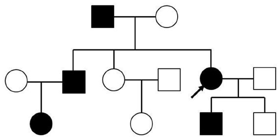 | Figure 1 Pedigree figure. |
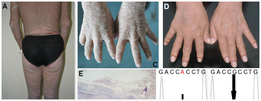 | Figure 2 Clinical, histopathological, and gene sequencing of the patients. |
Direct sequencing of the whole coding regions of KRT1 and KRT10 was performed. A heterozygous KRT10 missense mutation c.467G>A(p.Arg156His) was identified in the proband and her son, while not in normal controls (Figure 2F and G). The mutation hotspots of EHK are located at the conserved sequences in helix initiative and helix terminal motifs of K1 and K10.2 The mutation of the reported family, c.467G>A, just occurred at the hotspots. A previous study has revealed that the relationship between the genotype and phenotype in these patients was complex, not only dependent on the position of the mutation but also on the actual amino acid substitution.3,4 The p.R156 of KRT10 can be substituted by many amino acids such as serine, glycine, cysteine, histidine, and leucine, of which histidine is the most similar to arginine. Theoretically, the p.Arg156His mutation should be accompanied by a mild clinical manifestation,3 but the reported patients experienced severe conditions. So we cannot conclude that the more similar between two amino acid substitution in the same codon, the milder consequence will be brought. The p.Arg156His mutation is rather common in previous reports,2,4 but the clinical manifestations vary greatly. Other genetic or epigenetic factors that were not identified may influence disease severity.
In conclusion, we confirm the diagnosis of epidermolytic ichthyosis in the two patients according to the genetic testing and this is beneficial for their genetic counseling. The results reveal that the mutation p.R156H of KRT10 is responsible for severe clinical manifestations in a Chinese family with EHK, and also confirm the complexity of the genotype–phenotype correlation in EHK.
Acknowledgment
We are indebted to the Foundation of PUMC innovation team, Teaching and Research Fund Project of PUMC (Grant No 2011zlgc0114, 303-05-8050).
Disclosure
No potential conflict of interest relevant to this work was reported.
References
DiGiovanna JJ, Bale SJ. Clinical heterogeneity in epidermolytic hyperkeratosis. Arch Dermatol. 1994;130(8):1026–1035. | |
Yang JM, Nam K, Kim SW, et al. Arginine in the beginning of the 1A rod domain of the keratin 10 gene is the hot spot for the mutation in epidermolytic hyperkeratosis. J Dermatol Sci. 1999;19(2):126–133. | |
Sun XK, Ma LL, Xie YQ, Zhu XJ. Keratin 1 and keratin 10 mutations causing epidermolytic hyperkeratosis in Chinese patients. J Dermatol Sci. 2002;29(3):195–200. | |
Arin MJ, Oji V, Emmert S, et al. Expanding the keratin mutation database: novel and recurrent mutations and genotype-phenotype correlations in 28 patients with epidermolytic ichthyosis. Br J Dermatol. 2011;164(2):442–447. |
 © 2014 The Author(s). This work is published and licensed by Dove Medical Press Limited. The full terms of this license are available at https://www.dovepress.com/terms.php and incorporate the Creative Commons Attribution - Non Commercial (unported, v3.0) License.
By accessing the work you hereby accept the Terms. Non-commercial uses of the work are permitted without any further permission from Dove Medical Press Limited, provided the work is properly attributed. For permission for commercial use of this work, please see paragraphs 4.2 and 5 of our Terms.
© 2014 The Author(s). This work is published and licensed by Dove Medical Press Limited. The full terms of this license are available at https://www.dovepress.com/terms.php and incorporate the Creative Commons Attribution - Non Commercial (unported, v3.0) License.
By accessing the work you hereby accept the Terms. Non-commercial uses of the work are permitted without any further permission from Dove Medical Press Limited, provided the work is properly attributed. For permission for commercial use of this work, please see paragraphs 4.2 and 5 of our Terms.