")
Back to Journals » Research and Reports in Urology » Volume 11
Lumbrokinase effects on pro- and anti-apoptotic gene expression in Wistar rats with testicular torsion
Authors Danarto R, Heriyanto DS, Risan M, Yuri P
Received 16 April 2019
Accepted for publication 13 August 2019
Published 19 September 2019 Volume 2019:11 Pages 249—254
DOI https://doi.org/10.2147/RRU.S212431
Checked for plagiarism Yes
Review by Single anonymous peer review
Peer reviewer comments 2
Editor who approved publication: Dr Jan Colli
Raden Danarto,1 Didik Setyo Heriyanto,2 Muhammad Risan,1 Prahara Yuri1
1Division of Urology, Department of Surgery, Faculty of Medicine, Universitas Gadjah Mada/Dr. Sardjito Hospital, Yogyakarta 55281, Indonesia; 2Department of Anatomical Pathology, Faculty of Medicine, Universitas Gadjah Mada/Dr. Sardjito Hospital, Yogyakarta 55281, Indonesia
Correspondence: Prahara Yuri
Division of Urology, Department of Surgery, Faculty of Medicine, Public Health and Nursing, Universitas Gadjah Mada/Dr. Sardjito Hospital, Jl. Kesehatan No. 1, Yogyakarta 55281, Indonesia
Email [email protected]
Background: Testicular reperfusion is believed to be the mechanism by which testicular injury occurs in the ischemic testis. This study was performed to determine the therapeutic efficacy of lumbrokinase for treating ischemia-reperfusion (IR) injury-induced bilateral testicular torsion.
Methods: Twenty-four male rats were equally divided into the following groups: torsion only (T), torsion plus lumbrokinase (TL), torsion-detorsion only (TD) and torsion-detorsion plus lumbrokinase (TDL) groups. The right testicle in each groups sample was rotated 720° for 4 h, followed by orchiectomy. The rats in the TD (TD and TDL) groups additionally underwent detorsion for 1 h after the initial rotation. Testicular tissues were collected for measuring anti-apoptotic B-cell lymphoma-2 (BCL-2) and pro-apoptotic BCL-2-associated X protein (BAX) gene expression levels using real-time polymerase chain reaction.
Results: Pro- and anti-apoptotic gene expression levels were increased in the TD groups. Lumbrokinase was significantly effective in lowering BAX expression levels, particularly those in the TDL group compared with those in the TD group (P<0.05). Lumbrokinase did not significant change BCL-2 expression levels.
Conclusion: The administration of lumbrokinase before orchiectomy can protect against IR-induced testicular damage by reducing pro-apoptotic gene expression levels.
Keywords: testicular torsion, pro-apoptotic genes, anti-apoptotic genes, lumbrokinase
Background
Testicular torsion is the most common cause of acute scrotum and is commonly attributed to excess mobility of the testis with a “bell-clapper deformity”, wherein the tunica vaginalis abnormally fixes proximally on the cord.1 The annual prevalence of torsion is 8.6 per 100,000 males aged 10–19 years in the United States. Testicular torsion is more common on the left side than on the right, with a 1.2:1 ratio, possibly caused by slightly longer spermatic cords on the left.2 Testicular torsion can occur at any age, with the peak occurrence at 14 years and a second peak at 1 year of age.3 The occurrence of testicular torsion at 1 year of age is the most common cause of acute scrotum (83%). Between 3 and 13 years of age, the most frequent form of testicular torsion is the testicular appendix. After 17 years of age, epididymitis is the most frequent diagnosis (75%).4 Despite improvements in early diagnosis and changes in management (ie earlier surgical intervention), infertility remains one of the main sequelae of testicular torsion.5
Testicular reperfusion is believed to be the main cause of testicular injury in ischemic testes. Restoring blood flow after a period of testicular ischemia is necessary for maintaining cell function and testicular viability; however, reintroduction of oxygen can initiate a cascade of events that exacerbates testicular tissue injury via the formation of reactive oxygen species (ROS).6,7
To prevent testicular ischemia-reperfusion (IR) injury in animal models, numerous pharmacological agents and treatments have been evaluated, including the use of free radical scavengers, antioxidant drugs, neutrophil elastase inhibitors, NO donors, anti-inflammatory drugs, PDE5 inhibitors, K-ATP channel blockers and anti-coagulants. These treatments have different mechanisms of action in protecting testicular tissue from damage.
Lumbrokinase, derived from Lumbricus rubellus extracts,8 was recently identified as a group of bioactive proteolytic enzymes. Previous studies demonstrated its many beneficial properties such as anti-inflammatory, anti-oxidative, anti-fibrotic, anti-microbial and anti-cancer effects.9,10 Lumbrokinase can be easily absorbed by the intestinal tract, without any effects on its activity. Previous studies of IR injury have demonstrated that the anti-oxidative and anti-inflammatory effects of lumbrokinase can reduce testicular germ cell apoptosis.6,7,11
This study aimed to evaluate the therapeutic efficacy of lumbrokinase for treating IR-induced unilateral testicular torsion.
Methods
For this experimental, post-test, control group study, all animal laboratory study protocols were approved by the Ethical Review Board of Universitas Gadjah Mada. We followed American Veterinary Medical Association (AVMA) for the welfare of the animals. All rats used in this study were obtained from the Experiment Centre Department of Universitas Gadjah Mada. All experimental procedures were performed in the same facility. The study was conducted with 24 rats (Rattus novergicus), each weighing 250–350 g. This sample size was obtained by random sampling using formula (t-1)(r-1) ≥15) which minimal sample 6 for each group. Standard food was provided to all rats without limitation. The rats were divided into the following four groups: two testicular torsion (T) groups and two testicular torsion-detorsion (TD) groups. In the T group, the rats underwent either testicular torsion only (T) or testicular torsion with lumbrokinase administration (TL). In the TD group, the rats underwent testicular torsion-detorsion only (TD) or testicular torsion-detorsion with lumbrokinase administration (TDL).
Before all surgical procedures, ketamine (2 mg/kg) was administered as anesthesia to all rats under sterile conditions. A midline incision was made in the scrotum to open the tunica vaginalis and shift the left testicle into the surgical field. In the T group, a clockwise 720° rotation of the testicle was made, and the tissue was tied within the hemiscrotal using a 5–0 silk suture for 3 h. The same material was used to close the incision. After 4 h, a bilateral orchiectomy was performed, and an ambient temperature of 37 °C was maintained. In the TL group, the same procedure as in the T group was performed, except that 1 h before bilateral orchiectomy, 80 mg/kg lumbrokinase was administered. In the TD group, after 4 h of torsion, the testicle underwent detorsion for 1 h, followed by bilateral orchiectomy. In the TDL group, the same procedure as in the TD group was performed, except that 1 h before bilateral orchiectomy, 80 mg/kg lumbrokinase was administered. All rats that had undergoing orchiectomy was terminated using cervical dislocation under anesthetic situation.
Tissue processing
Collected samples were fixed with 10% liquid formalin. All excised testicles were analyzed for B-cell lymphoma-2 (BCL-2, anti-apoptotic) and BCL-2-associated X protein (BAX, pro-apoptotic) gene expression levels using real-time polymerase chain reaction (Table 1). All analysis procedures were performed in the pathology-anatomy laboratory of Gadjah Mada University. Histological evaluation was completed in blinded manner.
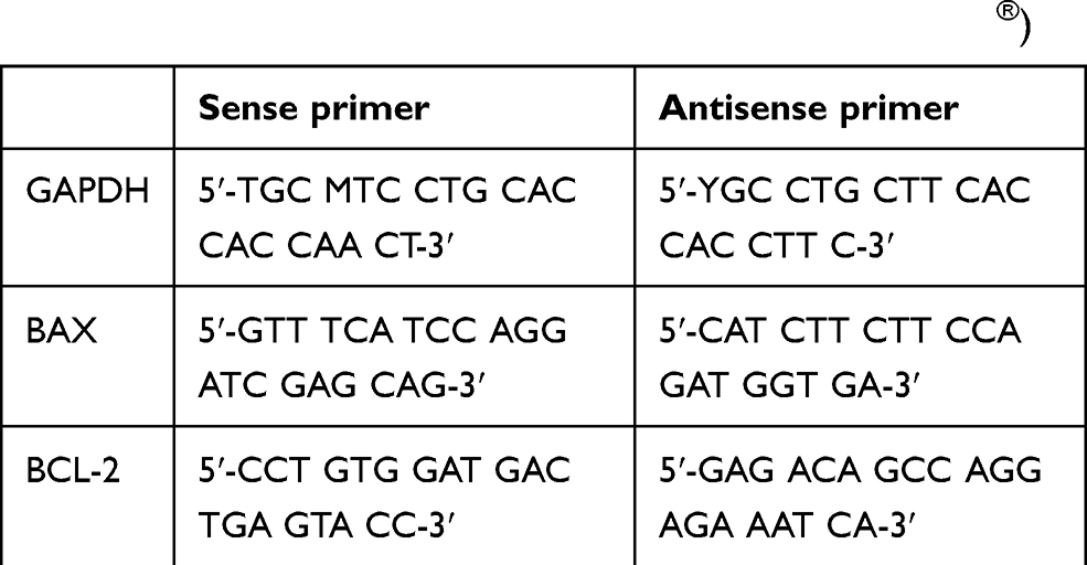 |
Table 1 BAX and BCL-2 multiplex primers (set of SIGMA®) |
Real time qualitative polymerase chain reaction
Kit extraction of GeneAll® was used to extract the testicular DNA. By using PCR kit (NEXpro 1-step RT-qPCR 2x Master Mix, SYBR) the RNA was obtained and multiplied by Bioneer® PCR device. The PCR examination was initialized by the extraction of DNA using extraction kit. Then to obtain normal expression of BAX and BCL-2 we examine control testicle and obtain GAPDH of BAX and BCL-2. Using primer and PCR device we multiplied the RNA of BAX and BCL-2 and obtain the expression value of each group. By using 2−∆∆Ct method we compare the result of control expression of BAX and BCL-2 with each group gene expression. All data that had been obtained from PCR examination was analyzed using Oneway ANOVA to get statistical result.
Results
In this study, all the rats underwent orchiectomy according to the research design, and 48 testicular tissue samples were obtained: 24 from the right testicle and 24 from the left testicle. No rats died during the study. The testicular tissues were analyzed for BAX and BCL-2 gene expression levels as described above.
There was a significant increase in BAX and BCL-2 gene expression levels in both testicles in the absence of lumbrokinase (Table 2). However, lumbrokinase significantly reduced BAX gene expression levels, particularly those in the TDL group compared with those in the TD group (P<0.05). There were no significant changes in BCL-2 gene expression levels after treatment with lumbrokinase.
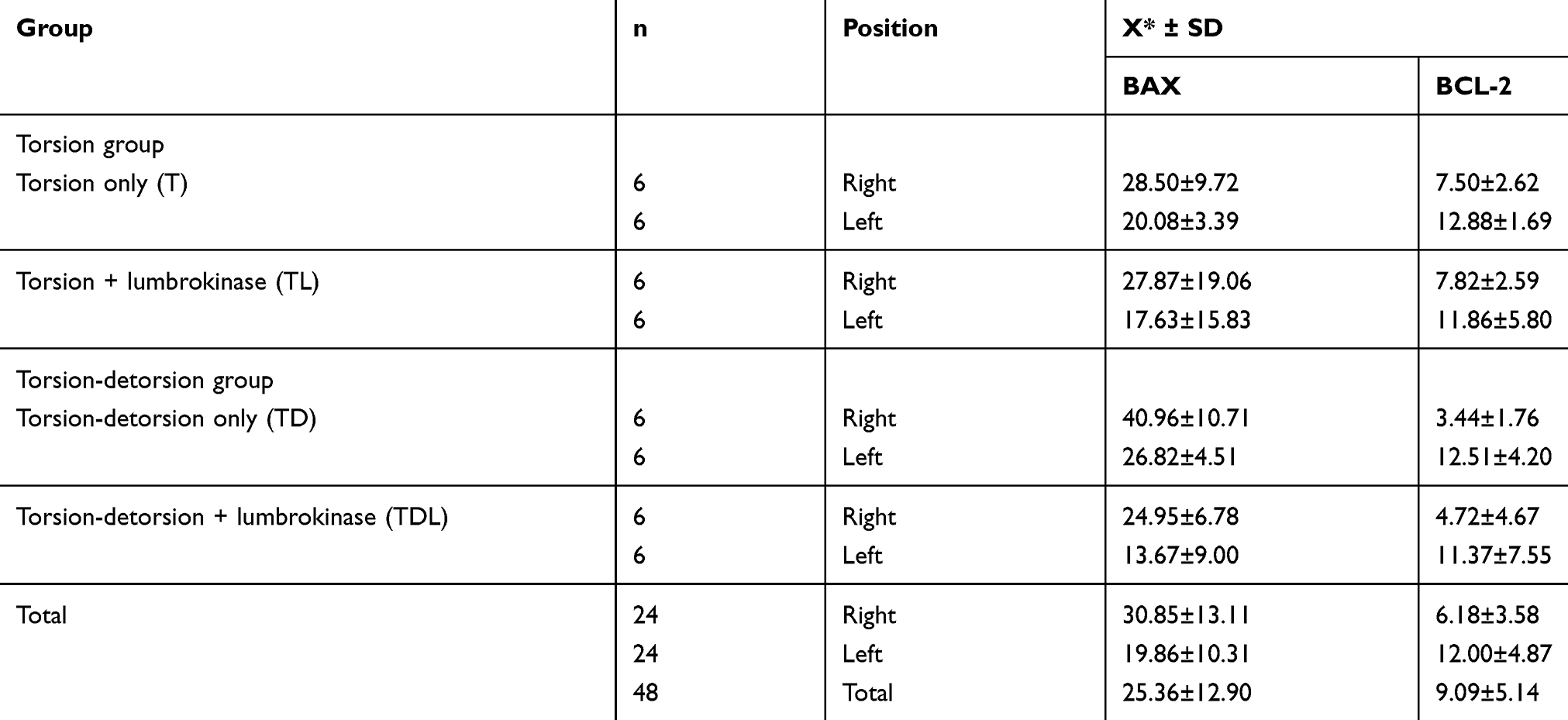 |
Table 2 BAX and BCL-2 gene expression levels in testicular tissue |
The comparison between the groups was performed using ANOVA (Table 3). A significant decrease was observed in BAX gene expression levels in the T groups compared with those in the TD groups (P<0.05). Similar results were observed in the TD group compared with those in the TDL group (P<0.05), with the highest difference shown between TD and TDL. No significant differences were observed in BCL-2 gene expression levels after lumbrokinase treatment.
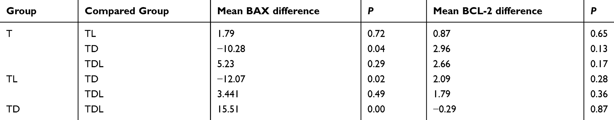 |
Table 3 Comparison between BAX and BCL-2 gene expression levels in each group |
Discussion
IR injury is the main cause of cellular damage from testicular torsion-detorsion.7,11 ROS are upregulated after a testicular IR injury, in which the testes are very sensitive to this type of damage.7 In previous studies of testicular torsion,6,7,11 testicular cells become ischemic, which leads to mitochondrial damage and the activation of apoptosis. During detorsion, the introduction of blood flow and oxygen may increase such damage by the influx of inflammatory agents from the circulation to the testicles, thereby affecting lipids, proteins and DNA and leading to germ cell apoptosis. The emergence of ROS begins with the activation of an intrinsic apoptotic pathway via a mitochondrial pathway, where BAX and BCL-2 genes play leading roles.12
Increased BAX gene expression levels in the mitochondrial membranes induce the release of pro-apoptotic molecules that trigger the activation of effector caspases, resulting in the release of various inflammatory mediators and free radicals. This mechanism is preceded by tissue injuries caused by hypoxia and oxidative stress owing to reperfusion.11–13 BCL-2 is then activated because of an increase in BAX gene expression levels (and subsequent caspase release) to maintain ischemic conditions. BCL-2 inhibits apoptosis by binding Apaf-1 and deactivating caspase 9, thus blocking the apoptotic cascade.13
In this study, BAX and BCL-2 gene expression levels were elevated in all the groups. Similarly, previous studies7,11,14,15 showed that ischemic tissues undergo apoptosis, which results in both reversible and irreversible tissue damage. In addition, BAX gene expression levels are significantly increased in the contralateral testicle, suggesting concomitant apoptosis.7,11 While the resulting damage to the contralateral testicle has not been clearly defined, the following mechanisms have been proposed to explain contralateral testicular damage: autoimmunity to spermatogonia, decrease in testicular blood flow caused by a sympathetic reflex, ROS formation after detorsion and overproduction of nitric oxide.6,7,11 Long-term observations of seminiferous tubule structures revealed a decrease in the germinal cell layer, indicating aberrant spermatogenesis after IR injury.11
Our data showed that pre-operative lumbrokinase treatment of testicular tissues after testicular torsion-detorsion resulted in a significant decrease in BAX gene expression levels but not in BCL-2 gene expression levels. Previous studies7,11,14,15 have examined the protective role of anti-oxidative and anti-inflammatory effects on IR injury. The administration of lumbrokinase to inflamed tissue elicits effects similar to that of non-steroidal anti-inflammatory drugs, which block the stimulation of IR-induced cyclooxygenase (COX)-2 expressions but has greater effects on fibrinolysis.8 Lumbrokinase also elicits effects on inducible nitric oxide synthase and matrix metalloproteinase-9 (MMP-9) via the toll-like receptor 4 (TLR4) signaling pathway.9 MMP-9 is critical for the myocardial expression of chemoattractant proteins that mediate neutrophil infiltration.9 Based on a previous study,10 these effects might reduce apoptosis and inflammation after restoring the blood supply owing to testicular detorsion. There is no previous study that has used lumbrokinase in a testicular torsion model, lumbrokinase has been reported to protect ischemic brains and myocardial cells by inhibiting intercellular adhesion molecule-1 and activating Janus kinase-1/signal transducers and activators of transcription-1 in an experimental cerebral and myocardial IR model.10
The definitive mechanism by which lumbrokinase alters BAX gene expression levels is not completely understood. In our study, the ability of lumbrokinase to decrease inflammation through COX-2 production may play a key role. ROS production decreased in a concentration-dependent manner at 37 °C. Excessive amounts of ROS can induce apoptosis through both extrinsic and intrinsic pathways.13 In the extrinsic pathway of apoptosis, ROS are generated by the Fas ligand during Fas activation via phosphorylation, which is necessary for inducing apoptosis. In the intrinsic pathway, ROS facilitate cytochrome c release by activating BCL-2 and BAX. The intrinsic pathway is also known as the caspase cascade and is induced via mitochondrial damage, which triggers the release of cytochrome c, DNA damage, oxidative stress and loss of mitochondrial membrane potential, thereby stimulating apoptosis.6,7,11,13 These findings are consistent with those of our study, which are more related to the intrinsic pathway owing to the increase in pro-apoptotic BAX and anti-apoptotic BCL-2 gene expression levels.
The main limitation of our study is the method of administering lumbrokinase cause the limitation of laboratory tools, we use sonde tube directly to stomach whereas it will be effective if administered intravenously. The dose calculations are more precise and effective if the drug is intravenously administered through the jugular vein. The study results suggest that testicular injury causes rapid cellular damage via alterations in gene expressions and that such a damage occurs prior to increases in ROS and germ cell damage, which have already been shown to decrease fertility.
Conclusion
The study results show that administering lumbrokinase before orchiectomy can protect against testicular damage by significantly reducing pro-apoptotic gene expression.
Ethics approval and consent to participate
The Ethical Committee of Faculty of Medicine, Universitas Gadjah Mada/Dr. Sardjito Hospital gave approval for this study (KE/FK/0843/EC). We followed the American Veterinary Medical Association (AVMA) guidelines for the welfare of the animals.
Abbreviations
IR, ischemia-reperfusion; T, torsion only; TL, torsion plus lumbrokinase; TD, torsion-detorsion only; TDL, torsion-detorsion plus lumbrokinase; BCL-2, B-cell lymphoma-2 gene; BAX-2, BCL-2-associated X protein gene; ROS, reactive oxygen species; NO, nitrite oxide; PDE5, phosphodiesterase; K-ATP, ATP-sensitive potassium; COX, cyclooxygenase; MMP, matrix metalloproteinase; TLR, toll-like receptor.
Data availability
All data generated or analyzed during this study are included in the submission. The raw data can be requested to the corresponding author.
Acknowledgment
We acknowledgement Department of Publishers and Publications – Universitas Gadjah Mada and Enago for the English language review.
Author contributions
All authors contributed toward data analysis, drafting and revising the paper, gave final approval of the version to be published and agree to be accountable for all aspects of the work.
Disclosure
The authors declare no potential conflicts of interest with respect to the research, authorship, and/or publication of this article.
References
1. Caesar RE, Kaplan GW. Incidence of the bell-clapper deformity in an autopsy series. Urology. 1994;44:114–116. doi:10.1016/s0090-4295(94)80020-0
2. Mansbach JM, Forbes P, Peters C. Testicular torsion and risk factors for orchiectomy. Arch Pediatr Adolesc Med. 2016;159:1167–1171. doi:10.1001/archpedi.159.12.1167
3. Lewis AG, Bukowski TP, Jarvis PD, Wacksman J, Sheldon CA. Evaluation of acute scrotum in the emergency department. J Pediatr Surg. 1995;30:272–277. doi:10.1016/0022-3468(95)90574-X
4. Dresner ML. Torsed appendage diagnosis and management: blue dot sign. Urology. 1973;1:63–66. doi:10.1016/0090-4295(73)90116-7
5. Bartsch G, Frank ST, Marberger H, Mikuz G. Testicular torsion: late results with special regard to fertility and endocrine function. J Urol. 1980;124:375–378. doi:10.1016/s0022-5347(17)55456-7
6. Lysiak JJ, Nguyen QAT, Turner TT. Peptide and nonpeptide reactive oxygen scavengers provide partial rescue of the testis after torsion. J Androl. 2002;23:400–409.
7. Mertoğlu C, Senel U, Cayli S, Tas U, Küskü Kiraz Z, Özyurt H. Protective role of methylprednisolone and heparin in ischaemic-reperfusion injury of the rat testicle. Andrologia. 2016;48:737–744. doi:10.1111/and.12503
8. Mihara H, Sumi H, Yoneta T, et al. A novel fibrinolytic enzyme extracted from the earthworm, Lumbricus rubellus. Jpn J Physiol. 1991;41:461–472.
9. Wang YH, Chen KM, Chiu PS, et al. Lumbrokinase attenuates myocardial ischemia-reperfusion injury by inhibiting TLR4 signaling. J Mol Cell Cardiol. 2017;99:113–122. doi:10.1016/j.yjmcc.2016.08.004
10. Sun H, Ge N, Shao M, et al. Lumbrokinase attenuates diabetic nephropathy through regulating extracellular matrix degradation in streptozotocin-induced diabetic rats. Diabetes Res Clin Pract. 2013;100:85–95. doi:10.1016/j.diabres.2013.01.012
11. Mogilner JG, Elenberg Y, Lurie M, Shiloni E, Coran AG, Sukhotnik I. Effect of dexamethasone on germ cell apoptosis in the contralateral testis after testicular ischemia-reperfusion injury in the rat. Fertil Steril. 2006;85(Suppl 1):1111–1117. doi:10.1016/j.fertnstert.2005.10.021
12. Fuchs Y, Steller H. Programmed cell death in animal development and disease. Cell. 2011;147:742–758. doi:10.1016/j.cell.2011.10.033
13. Sinha K, Das J, Pal PB, Sil PC. Oxidative stress : the mitochondria-dependent and mitochondria-independent pathways of apoptosis. Arch Toxicol. 2013;87:1157–1180. doi:10.1007/s00204-013-1034-4
14. Shimizu S, Tsounapi P, Dimitriadis F, Higashi Y, Shimizu T, Saito M. Testicular torsion-detorsion and potential therapeutic treatments: a possible role for ischemic postconditioning. Int J Urol. 2016;23:454–463. doi:10.1111/iju.13110
15. Ribeiro CT, Milhomem R, De Souza DB, Costa WS, Sampaio FJB, Pereira-Sampaio MA. Effect of antioxidants on outcome of testicular torsion in rats of different ages. J Urol. 2014;191(Suppl 5):1578–1584. doi:10.1016/j.juro.2013.09.066
 © 2019 The Author(s). This work is published and licensed by Dove Medical Press Limited. The full terms of this license are available at https://www.dovepress.com/terms.php and incorporate the Creative Commons Attribution - Non Commercial (unported, v3.0) License.
By accessing the work you hereby accept the Terms. Non-commercial uses of the work are permitted without any further permission from Dove Medical Press Limited, provided the work is properly attributed. For permission for commercial use of this work, please see paragraphs 4.2 and 5 of our Terms.
© 2019 The Author(s). This work is published and licensed by Dove Medical Press Limited. The full terms of this license are available at https://www.dovepress.com/terms.php and incorporate the Creative Commons Attribution - Non Commercial (unported, v3.0) License.
By accessing the work you hereby accept the Terms. Non-commercial uses of the work are permitted without any further permission from Dove Medical Press Limited, provided the work is properly attributed. For permission for commercial use of this work, please see paragraphs 4.2 and 5 of our Terms.