")
Back to Journals » OncoTargets and Therapy » Volume 9
Lower expressed miR-198 and its potential targets in hepatocellular carcinoma: a clinicopathological and in silico study
Authors Huang W, Wang H, Yang H, Ren F, Luo Y, Huang C, Liang Y, Liang H, Chen G , Dang Y
Received 19 March 2016
Accepted for publication 30 June 2016
Published 22 August 2016 Volume 2016:9 Pages 5163—5180
DOI https://doi.org/10.2147/OTT.S108828
Checked for plagiarism Yes
Review by Single anonymous peer review
Peer reviewer comments 4
Editor who approved publication: Dr Jianmin Xu
Wen-Ting Huang,1,* Han-Lin Wang,1,* Hong Yang,2 Fang-Hui Ren,1 Yi-Huan Luo,1 Chun-Qin Huang,1 Yue-Ya Liang,1 Hai-Wei Liang,1 Gang Chen,1 Yi-Wu Dang1
1Department of Pathology, 2Department of Ultrasonography, First Affiliated Hospital of Guangxi Medical University, Nanning, People’s Republic of China
*These authors contributed equally to this work
Purpose: To investigate the clinicopathological value and potential roles of microRNA-198 (miR-198) in hepatocellular carcinoma (HCC).
Methods: Ninety-five formalin-fixed paraffin-embedded HCC and the para-cancerous liver tissues were gathered. Real-time reverse transcription quantitative polymerase chain reaction was applied to determine the miR-198 expression. The association between the miR-198 expression and clinicopathological features was examined. Meanwhile, potential target messenger RNAs of miR-198 in HCC were obtained from 14 miRNA prediction databases and natural language processing method, in which we pooled the genes related to the tumorigenesis and progression of HCC and classified them by their frequency. The selected target genes were finally analyzed in the Gene Ontology analysis and Kyoto Encyclopedia of Genes and Genomes pathway.
Results: miR-198 expression was significantly lower in HCC than that in adjacent noncancerous liver tissues (1.30±0.72 vs 2.01±0.58, P<0.001). Low miR-198 expression was also correlated to hepatitis C virus infection (r=-0.48, P<0.001), tumor capsular infiltration (r=-0.43, P<0.001), metastasis (r=-0.26, P<0.010), number of tumor nodes (r=-0.25, P=0.013), vaso-invasion (r=-0.24, P=0.017), and clinical tumor node metastasis stage (r=-0.23, P=0.024). Altogether, 1,048 genes were achieved by the concurrent prediction from at least four databases and natural language processing indicated 1,800 genes for HCC. Further, 127 overlapping targets were further proceeded with for pathway analysis. The most enriched Gene Ontology terms in the potential target messenger RNAs of miR-198 were cell motion, cell migration, cell motility, and regulation of cell proliferation in biological process; organelle lumen, membrane-enclosed lumen, and nuclear lumen in cellular component; and enzyme binding, protein domain-specific binding, and protein kinase activity in molecular function. Kyoto Encyclopedia of Genes and Genomes analysis showed that these target genes were obviously involved in focal adhesion and pathways in cancer.
Conclusion: Lower expression of miR-198 was related to several clinicopathological parameters in HCC patients. miR-198 might play a regulatory role through its target genes in the development of HCC.
Keywords: miR-198, hepatocellular carcinoma, metastasis, RT-qPCR, in silico
Introduction
Hepatocellular carcinoma (HCC) is one of the most frequent malignancies in the world. The estimate incidence of liver cancer ranked fourth and its mortality ranked third in People’s Republic of China, 2015.1 Currently, several genes have been identified to be related with the tumorigenesis and progression of HCC, but the molecular mechanisms of their functions and interactions have not been clearly studied. Therefore, it is urgent to find the potential function of these genes during the development of HCC.
MicroRNA (miRNA) is classified as endogenous noncoding RNA, repressing protein translation by binding to their target genes.2 From nearly all biological processes, miRNA can exert the function as posttranscriptional regulation.3 Aberrant expression of microRNA-198 (miR-198) has been found to be correlated to the carcinogenesis and disease progression of several classes of cancers, including lung adenocarcinoma,4 esophageal cancer,5 squamous cell carcinoma of tongue,6 retinoblastoma,7 pancreatic adenocarcinoma,8 multiple myeloma,9 colorectal cancer,10 and prostate cancer.11 However, there were only three research papers studying the expression of miR-198 in HCC. Varnholt et al first reported the lower expressed miR-198 in Caucasian patients with hepatitis C virus (HCV) related HCC.12 Tan et al identified the suppressing role of miR-198 in HGF/c-MET pathway that influenced migration and invasion of HCC cells.13 Elfimova et al found another pathway that was repressed by miR-198 in vitro which could negatively regulate cell growth and migration.14 However, whether miR-198 is related to clinicopathological parameters has not been studied, there still remains a mystery regarding miR-198’s role in the key processes of hepatocellular carcinogenesis.
Thus, in this study, we assessed the expression of miR-198 in tumor tissues of HCC patients with their paired noncancerous liver. In addition, the relationship of miR-198 with several clinicopathological parameters was also investigated. Moreover, we predicted the target genes of miR-198 via 14 miRNA databases and filtered in the natural language processing (NLP) to achieve the specific messenger RNAs (mRNA) profiles of miR-198 targets for HCC. Gene Ontology (GO) terms and Kyoto Encyclopedia of Genes and Genomes (KEGG) pathway annotation were further undertaken to explore the function of these mRNAs. We attempted to pioneer the use of real-time reverse transcription quantitative polymerase chain reaction (RT-qPCR) and in silico method to clarify the potential molecular mechanism of miR-198, providing guidelines for the following research on miR-198 in HCC.
Materials and methods
Tissue samples
There were 95 cases of HCCs and their paired paraneoplastic liver formalin-fixed paraffin-embedded (FFPE) tissues included in this retrospective study. The age of the HCC patients ranged from 29 to 82 years old, with a mean age of 52 years. All the clinicopathological features are provided in Table 1. Adjacent noncancerous liver tissues were obtained from the location at least 2 cm away from the tumor node. The adjacent tissue was from the same HCC patient. We designed these paired sample groups in order to eliminate the individual difference. Tumor sizes of HCC ranged from 1 to 11 cm (mean size, 6.4 cm). All cases were from patients who were under initial hepatectomies without any treatment and they were randomly chosen from hepatectomy surgeries in the First Affiliated Hospital of Guangxi Medical University, People’s Republic of China between March 2010 and December 2011. The research protocol was approved by the Ethical Committee of the First Affiliated Hospital of Guangxi Medical University. Written informed consent was signed by the patients for the treatment and sample usage in this study. All samples were diagnosed by three pathologists independently (Dian-zhong Luo, Zhen-bo Feng, Gang Chen).
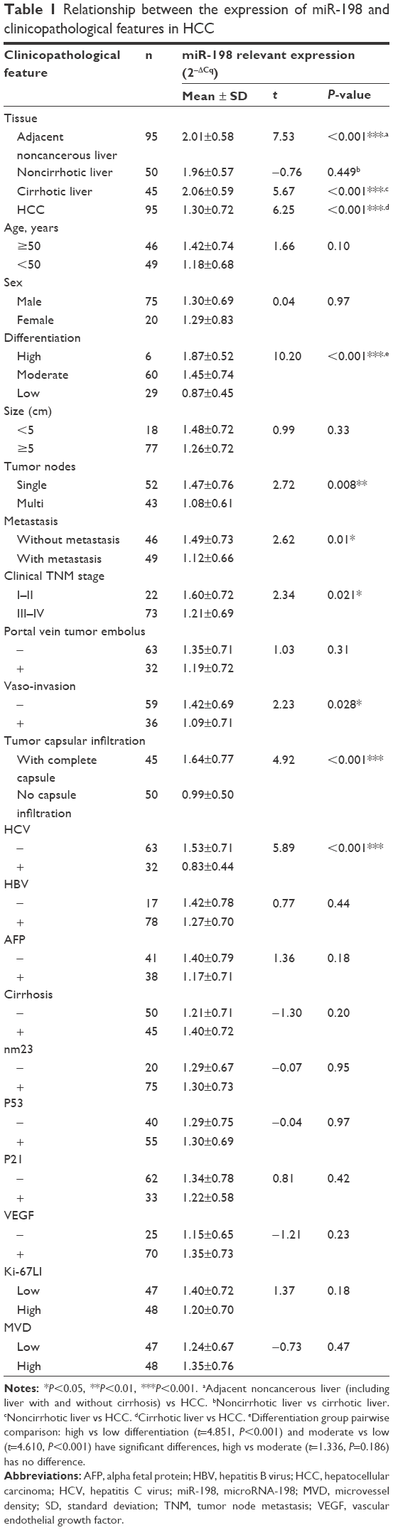 | Table 1 Relationship between the expression of miR-198 and clinicopathological features in HCC |
RT-qPCR
Total RNA including miRNA was extracted from tumor and nontumorous liver sections and the miRNeasy FFPE kit (QIAGEN, KJ Venlo, the Netherlands) was applied according to our previous reports.15–17 RNA concentrations were measured by NanoDrop 2000 (Thermo Fisher Scientific, Wilmington, DE, USA). The OD260/OD280 ratio of the total mRNA (including miRNA) isolated from the FFPE tissues ranged from 1.84 to 2.06, and OD260/OD230 from 1.90 to 2.04. The PCR amplification efficiency of all the quantitative real-time PCR (RT-qPCR) reactions ranged from 91.0% to 95.2%. A combination of RUN6B and RUN48 was chosen as the house-keeping reference for detection of miR-198 expression. The primers of miR-198, as well as RNU6B and RNU48, were provided by TaqMan® MicroRNA Assays (4427975, Applied Biosystems, Life Technologies, Grand Island, NY, USA). Sequence of miRNA and references in the paper were as follows: miR-198 (Applied Biosystems Cat No 4427975-000468): UCCCAAAUGUAGACAAAGCA; RNU6B (Applied Biosystems Cat No 4427975-001093): CGCAAGGAUGACACGCAAAUUCGUGAAGCGUUCCAUAUUUUU; RNU48 (Applied Biosystems Cat No 4427975-001006): GAUGACCCCAGGUAACUCUGAGUGUGUCGCUGAUGCCAUCACCGCAGCGCUCUGACC. The reverse transcription was carried out with TaqMan® MicroRNA Reverse Transcription Kit (4366596, Applied Biosystems, Life Technologies) in a total volume of 10 μL. Real-time qPCR for miRNA trial was performed with Applied Biosystems PCR7900. The normalization of miR-198 quantity in each sample was compared with its internal reference. At last, the formula 2−Δcq was used to calculate the expression of miR-198 in this experiment.15
Potential target mRNAs of miR-198 in HCC
To achieve predicted target mRNAs of miR-198, 14 miRNA in silico databases were used in this study, including TargetScan, miRanda, RNAv22, miRWalk, miRTarBase, PicTar, TargetS, DIANA, miRDB, miRNAMap, PITA, TargetMiner, Human miRNA Targets, and PolymiRTS Database. More than 16,000 target mRNAs were predicted once in total. Only mRNAs that were simultaneously predicted by four or more than four databases were selected for subsequent study. Other validated genes targeted by miR-198 were also added to the study throughout literature review.
NLP analysis of liver cancer
To identify the specific genes that were abnormally expressed in HCC, we conducted a literature search in the PubMed and EMBASE with an attempt to cover all studies available in publication from January 1980 and May 2015 through the following strategy: (liver or hepat*) and (cancer or carcinoma or tumor or neoplasm or malignan* or carcinogenesis or tumorigenesis) and (resistance OR prognosis OR metastasis OR recurrence OR survival OR sorafenib OR bevacizumab) (“1980/01/01” [PDAT]: “2015/05/25” [PDAT]). We then identified all the genes and mRNAs that were associated with the key message, and used a biomedical tool – entity recognizer (ABNER)16 to add them to a list via gene mention tagging.17
Gene-enrichment and functional annotation analysis
To evaluate the function of the potential target mRNAs, we used the GO terms and KEGG pathway annotation for the miR-198 targets based on functional annotation summaries provided by DAVID (Database for Annotation, Visualization, and Integrated Discovery, http://david.abcc.ncifcrf.gov/).18 The GO terms with a modified Fisher exact P-value <0.01 and the pathways with P-value <0.05 were chosen for the next analysis. The enrichment map of annotation analysis was drawn by software Cytoscape 3.3.0.19
Validation of the expression of some potential target genes based on The Cancer Genome Atlas (TCGA) dataset
To further study the potential role of miR-198, additional analyses were performed on the expression of some predicted genes randomly selected from one top pathway generated by the signaling pathway analysis. All gene expressions of 369 patients of liver HCC and 50 adjacent liver tissues were extracted and analyzed from the TCGA RNAseq profiles (http://cancergenome.nih.gov).
Statistical analysis
SPSS 21.0 (IBM Corporation, Armonk, NY, USA) was employed for statistical analysis. Values were shown as the mean ± standard deviation. Student’s paired or unpaired t-test was adopted to analyze the significance between paired and unpaired groups respectively. One-way analysis of variance test was applied to analyze the significance between groups of various differentiations of HCC. Fold change (FC) was calculated to describe the degree of difference on a log 2 scale. Statistical significance was defined at a P<0.05 level.
Results
Expression of miR-198 in HCC
The relative level of miR-198 was 1.23±0.72 in HCC tissues, remarkably lower than that in adjacent noncancerous liver tissues (2.01±0.58, P<0.001) (Table 1, Figure 1A). Furthermore, the area under curve (AUC) of miR-198 accessed by receiver operating characteristic (ROC) curve was 0.775 (95% confidence interval (CI): 0.708–0.841, P<0.001) (Figure 2A). The cutoff value for miR-198 was 1.78. The sensitivity was 66.3% and the specificity was 75.8%. However, there was no significant difference of the miR-198 level between the noncirrhotic liver (1.96±0.57) and cirrhotic liver (2.06±0.59, t=−0.76, P=0.449) (Table 1). And miR-198 expression was markedly downregulated in HCC (1.30±0.72) as compared to noncirrhotic liver (t=5.67, P<0.001) and cirrhotic liver (t=6.25, P<0.001), respectively.
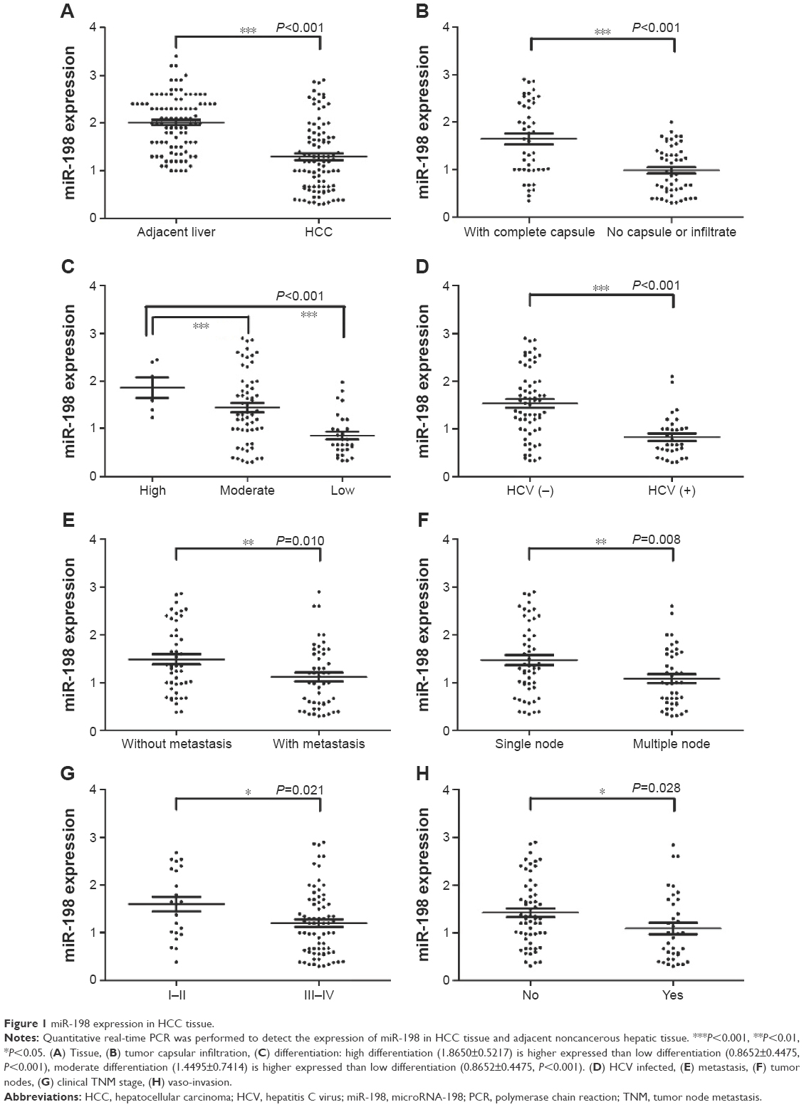 | Figure 1 miR-198 expression in HCC tissue. |
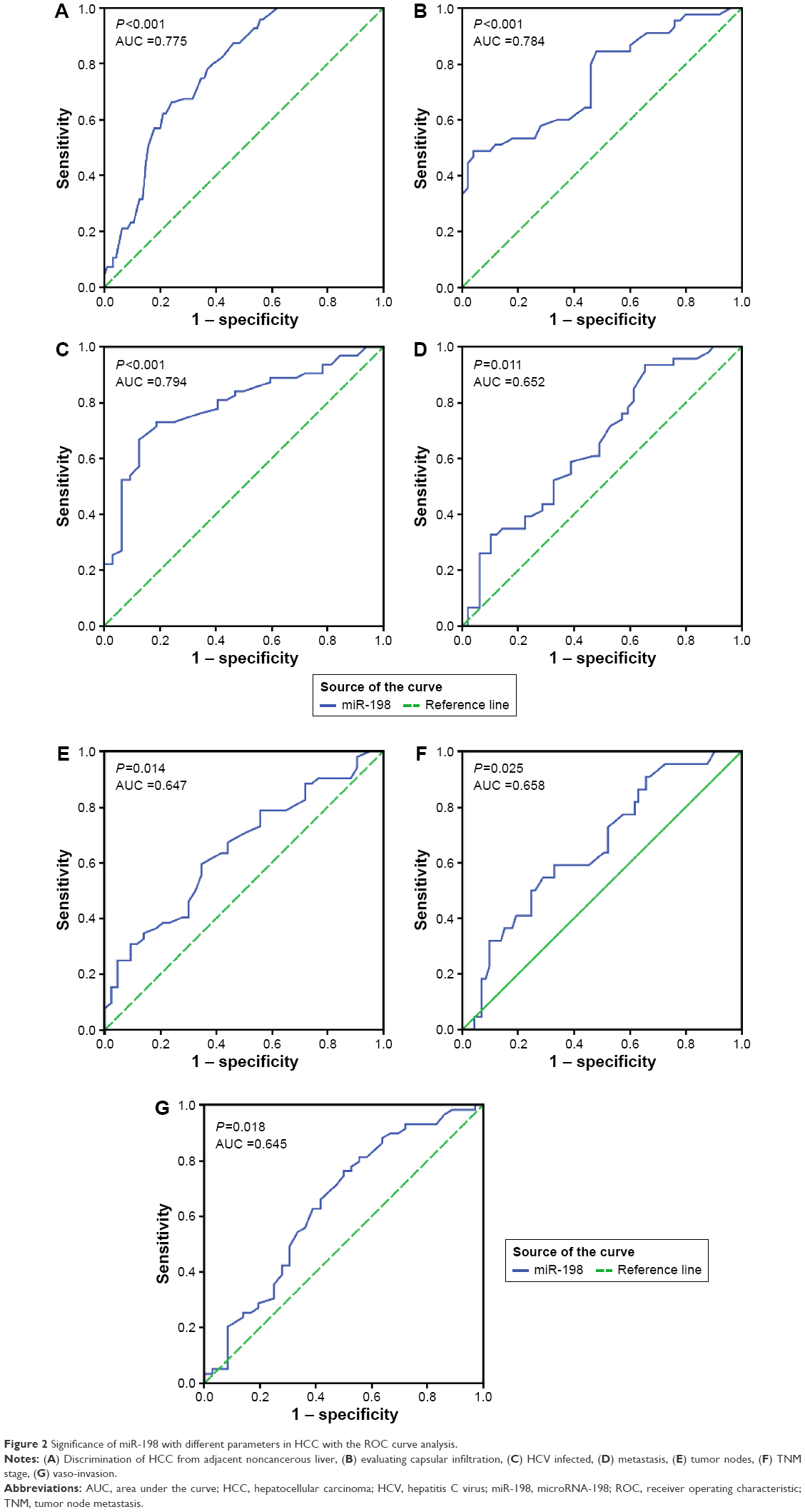 | Figure 2 Significance of miR-198 with different parameters in HCC with the ROC curve analysis. |
Expression of miR-198 with clinicopathological parameters in HCC
Considering correlation between miR-198 expression and clinical features, several clinicopathological parameters were related to miR-198. Remarkably higher expression of miR-198 was found in the tumor with complete capsular (1.64±0.77) than in the tumor with no capsule or with tumor infiltration (0.99±0.50, P<0.001) (Figure 1B). MiR-198 differential expression could be found in tissue differentiation, and significant differences existed between high differentiation (1.87±0.52) and low differentiation (0.87±0.45, P<0.001) (Figure 1C), and similar variation was also found between moderate (1.45±0.74) and low differentiation (0.87±0.45, P<0.001) (Figure 1C). However, high differentiation and moderate one showed no significant difference. HCV positive patients had a noticeably lower miR-198 expression level (0.83±0.44) than negative ones (1.53±0.71, P<0.001) (Figure 1D). In tumor without metastasis, higher miR-198 expression (1.49±0.73) was observed compared with that with metastasis (1.12 ± 0.66, P=0.010) (Figure 2E). Expression of miR-198 was higher in patients with single tumor node (1.47±0.76) than in those with multitumor nodes (1.08±0.61, P=0.008) (Figure 1F). As for clinical tumor node metastasis (TNM) stage, miR-198 expression in early stages (I and II, 1.60±0.72) was higher than that in advanced stages (III and IV, 1.21±0.69, P=0.021) (Figure 1G). Concerning the status of vaso-invasion, expression of miR-198 was lower in positive ones (1.09±0.71) than in negative ones (1.42±0.69, P=0.028) (Figure 1H). Simultaneously, additional Spearman’s correlation test confirmed the relationship between miR-198 expression and the following clinicopathological features: differentiation (r=−0.43, P<0.001) and clinical TNM stage (r=−0.23, P=0.024). However, no significant differences between miR-198 expression and other clinicopathological parameters were noted, such as age, sex, tumor size, portal vein tumor embolus, hepatitis B virus, alpha fetal protein, cirrhosis, expression of other markers: nm23, p53, p21, vascular endothelial growth factor, Ki-67, or microvessel density. The detection of the expression of nm23, p53, p21, vascular endothelial growth factor, Ki-67, and CD34 to present microvessel density was carried out by the immunohistochemistry method (Figure 3A–L). And hematoxylin and eosin staining images of HCC, cirrhosis, and noncirrhotic liver tissues are shown in Figure 4. We also searched for the data of the expression of miR-198 in Gene Expression Omnibus datasets and brought in all the HCC-related miR-198 expression chip data to perform a meta-analysis. Standardized mean difference with 95% CIs were calculated and pooled by using STATA 12.0 (StataCorp LP, College Station, TX, USA). Six microarray datasets with miR-198 expression level were employed. But the results obtained by a random-effects model showed no statistical significance (P>0.05) between the expression of miR-198 in HCC and noncancer tissues (data not shown). So, the more expanded sample size will be needed for the validation of miR-198 expression in the future.
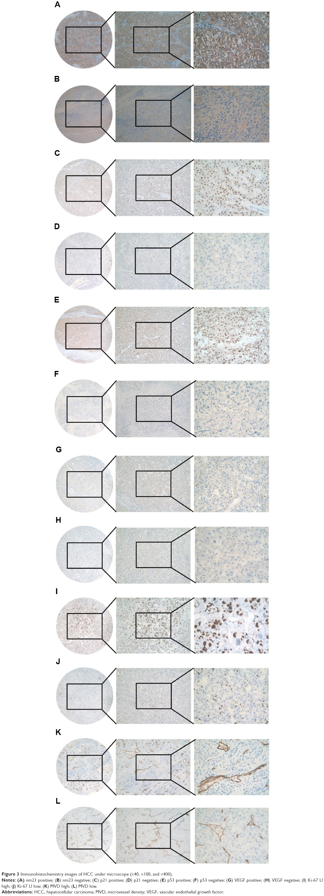 | Figure 3 Immunohistochemistry images of HCC under microscope (×40, ×100, and ×400). |
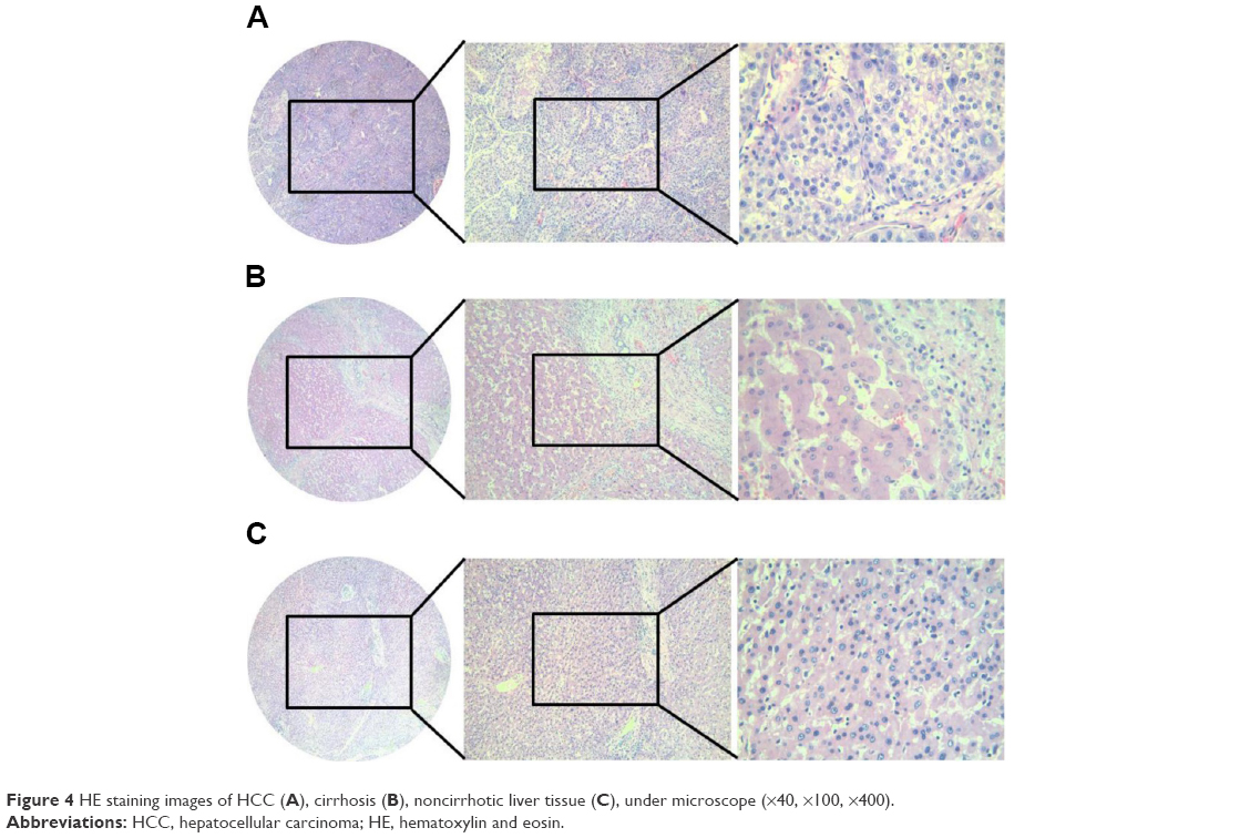 | Figure 4 HE staining images of HCC (A), cirrhosis (B), noncirrhotic liver tissue (C), under microscope (×40, ×100, ×400). |
ROC analysis for miR-198 expression in HCC
ROC curve was also performed to identify the predictive value of miR-198 level in HCC patients for clinicopathological features. The AUC to discriminate HCC from adjacent noncancerous liver accessed by ROC curve was 0.775 (95% CI: 0.708~0.841, P<0.001) (Figure 2A). The cut-off value for miR-198 was 1.78. The sensitivity was 66.3% and the specificity was 75.8%. The AUC to judge the capsule and tumor infiltration was 0.748 (95% CI: 0.649~0.846, P<0.001) (Figure 2B). The cut-off value for miR-198 was 1.73. The sensitivity and specificity were 48.9% and 96%, respectively. The AUC to judge the HCV infected was 0.794 (95% CI: 0.702~0.886, P<0.001) (Figure 2C). The cut-off value for miRNA-198 was 1.12. The sensitivity and specificity were 73% and 81.2%, respectively. The AUC to evaluate without tumor metastasis was 0.652 (95% CI: 0.542–0.761, P=0.011) (Figure 2D). The cutoff value for miR-198 was 0.62. The sensitivity and specificity were 93.5% and 34.7%, respectively. The AUC to predict single tumor node was 0.647 (95% CI: 0.537–0.757, P=0.014) (Figure 2E). The cutoff value for miR-198 was 1.22. The sensitivity and specificity were 59.6% and 65.1%, respectively. The AUC to estimate TNM stage was 0.658 (95% CI: 0.533–0.784, P=0.025) (Figure 2F). The cutoff value for miR-198 was 1.365. The sensitivity and specificity were 59.1% and 67.1%, respectively. The AUC to predict vaso-invasion was 0.645 (95% CI: 0.526–0.763, P=0.018) (Figure 2G). The cutoff value for miR-198 was 0.935. The sensitivity and specificity were 76.3% and 50%, respectively. The sensitivity and specificity were 73% and 81.2%, respectively (Figure 2B). The cutoff value for miR-198 was 1.73. The sensitivity and specificity were 48.9% and 96%, respectively. The AUC in HCV negative patients was 0.794 (95% CI: 0.702–0.886, P<0.001) (Figure 2C). The cutoff value for miR-198 was 1.12. The sensitivity and specificity were 73% and 81.2%, respectively.
Role of miR-198 on recurrence of HCC patients
We further studied the value of miR-198 on patient recurrence. Of the 95 patients studied, 70 had the complete follow-up data: 34 on low miR-198 level (lower than the median level of 1.2000) and 36 on high miR-198 level. There was no significant difference of recurrence between low miR-198 expression group and the high group (P=0.542).
Potential targets of miR-198 in HCC
To get predicted targets of miR-198, we conducted the prediction process online via 14 computational algorithms mentioned above, and attained a total number of 16,381 mRNAs after excluding duplications generated from the prediction process, and subsequently in order to increase reliability of our study we screened 1,048 mRNAs which were considered as more or less likely predictions. Another 37 genes validated to be the targets of miR-198 were added to our study through literature review. In addition, the abstracts or titles of 64,577 studies had been included through data and text mining tools in the process of NLP, and a total of 1,800 HCC-related genes were subsequently identified. Furthermore, we attained the potential target mRNAs of miR-198 in HCC by combining the predicted target list and genes specifically expressed in liver cancer, as shown in Table 2. Altogether, 127 mRNAs were selected for gene-annotation enrichment analysis and KEGG pathway annotation, which could be largely representative of the molecular mechanism of miR-198 in HCC. The flow diagram is shown in Figure 5.
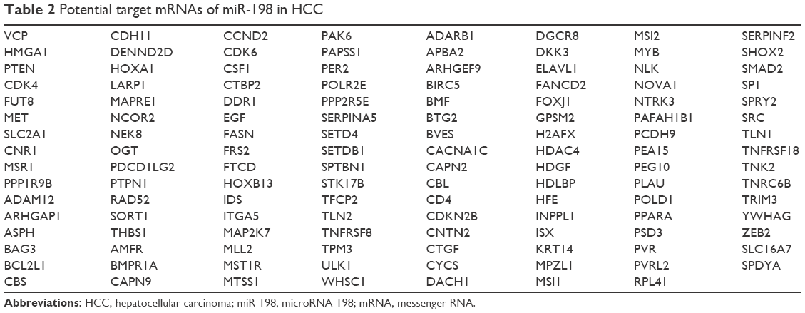 | Table 2 Potential target mRNAs of miR-198 in HCC |
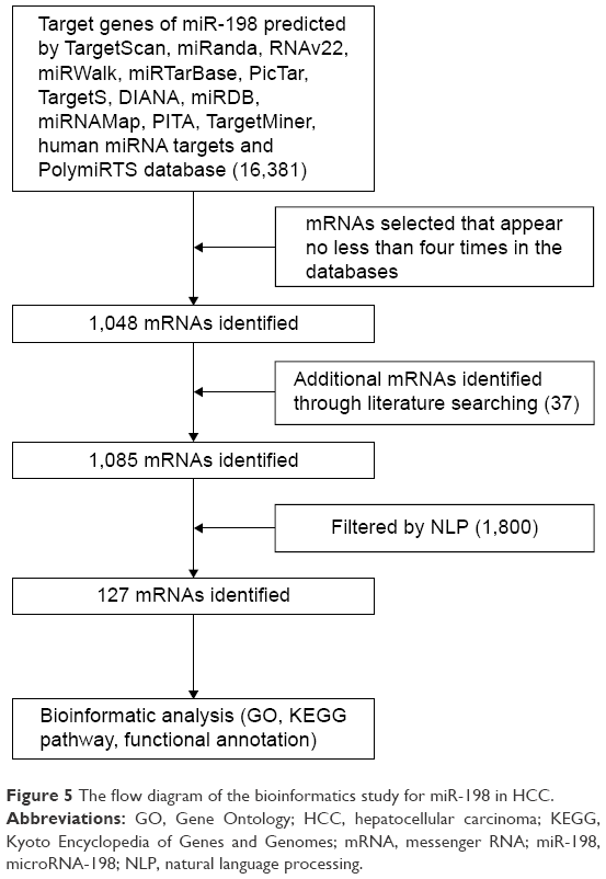 | Figure 5 The flow diagram of the bioinformatics study for miR-198 in HCC. |
Gene-annotation enrichment analysis and KEGG pathway annotation
According to the functional annotation summaries with the background of Homo sapiens listed by DAVID, we gained GO term annotations and KEGG pathways of all the selected target genes of miR-198 in HCC. A modified Fisher’s exact test or the EASE score was used to calculate the P-value. For biological process in GO results, the potential targets of miR-198 were significantly involved in the regulation of cell motion, cell migration, cell motility, regulation of cell proliferation, enzyme-linked receptor protein signaling pathway, and transmembrane receptor protein tyrosine kinase signaling pathway, and so on (P<0.001, Table 3, Table S1); for cellular component, these genes were most enriched in organelle lumen (n=34), membrane-enclosed lumen (n=34), nuclear lumen (n=29), plasma membrane (n=49), nucleoplasm (n=18), cytosol (n=26), ruffle (n=6), and caveola (n=5) (P<0.001, Table 3, Table S2); as for molecular function, the selected mRNAs were markedly involved in enzyme binding, protein domain-specific binding, and protein kinase activity, and so forth (P<0.001, Table 3, Table S3); and for the KEGG pathway analysis, the potential targets of miR-198 were notably associated with focal adhesion, pathways in cancer, p53 signaling pathway, and adheres junction (P<0.005, Table 4). The overview schematic of the analysis is shown in Table 2, Figures 6 and 7.
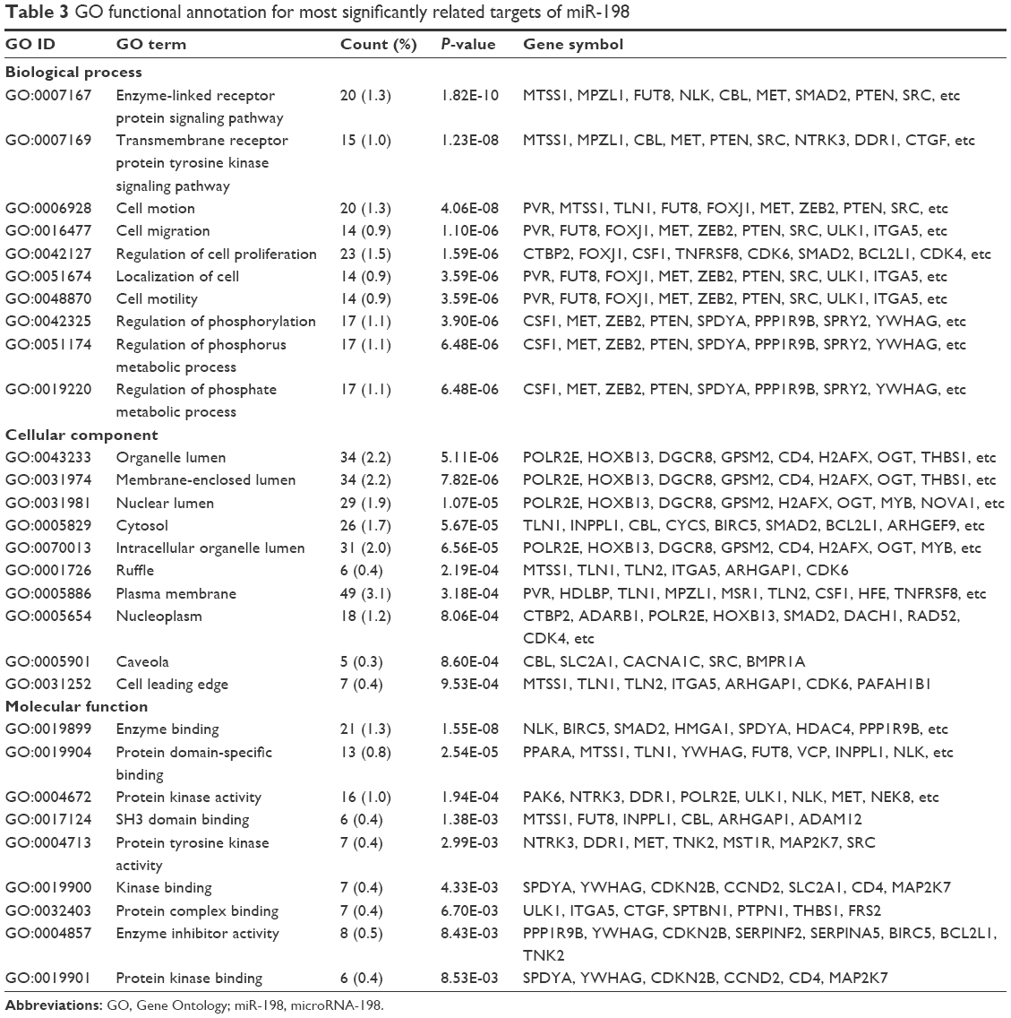 | Table 3 GO functional annotation for most significantly related targets of miR-198 |
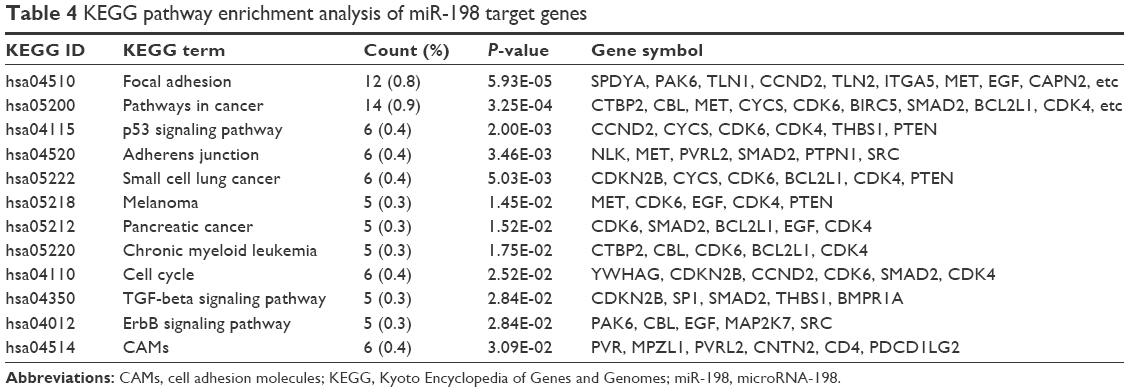 | Table 4 KEGG pathway enrichment analysis of miR-198 target genes |
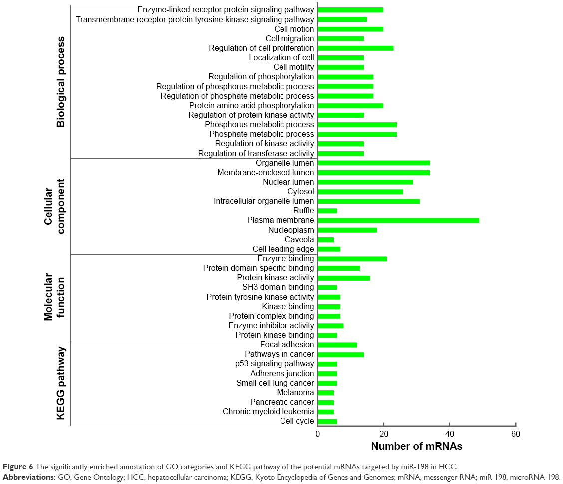 | Figure 6 The significantly enriched annotation of GO categories and KEGG pathway of the potential mRNAs targeted by miR-198 in HCC. |
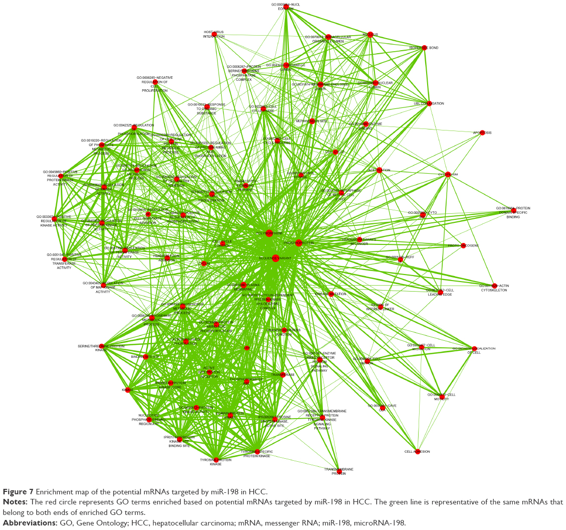 | Figure 7 Enrichment map of the potential mRNAs targeted by miR-198 in HCC. |
Validation of some potential target gene expression based on TCGA dataset
Among all the significant pathways in the current study, the organelle lumen (GO: 0043233) was randomly selected. There were 34 genes involved in this pathway. We then analyzed the relative expression (on a log 2 scale) of these 34 predicted genes of miR-198 by using TCGA LIHC dataset. Eleven genes were downregulated, and they could not be real targets of miR-198 due to same trend of the expression in liver tumor (Table 5). And 22 among the rest 23 genes yielded significant overexpression in liver cancers as compared to that in the adjacent liver tissues (P<0.05, Table 5). More interesting, ten among these 22 genes were extremely overexpressed in liver tumor tissues as their FC value was >1 (P<0.001, Table 5, Figure 8A–J), such as BIRC5, FANCD2, EGF, HMGA1, GPSM2, H2AFX, POLD1, HOXB13, HDAC4, SPDYA.
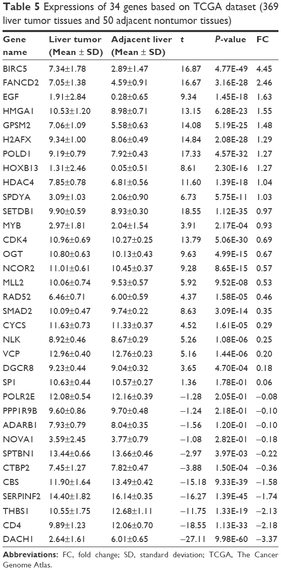 | Table 5 Expressions of 34 genes based on TCGA dataset (369 liver tumor tissues and 50 adjacent nontumor tissues) |
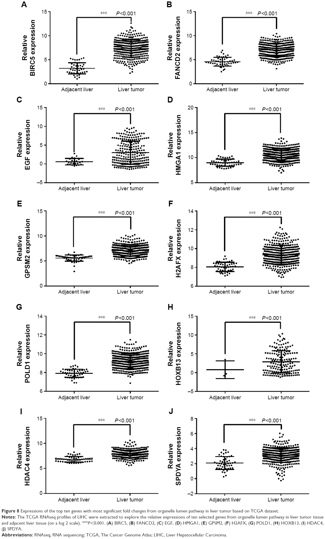 | Figure 8 Expressions of the top ten genes with most significant fold changes from organelle lumen pathway in liver tumor based on TCGA dataset. |
Discussion
In the present study, we found relevant lower expression of miR-198 in HCC, as compared to that in their para-noncancerous liver tissues. Downregulated miR-198 level was closely related to the disease progression of HCC. Our experiment showed that miR-198 had a close relationship with differentiation, tumor nodes, metastasis, clinical TNM stage, vaso-invasion, tumor capsular infiltration, and HCV infection. And these clinicopathological features could reflect a trend of poor prognosis. We found that the expression of miR-198 was not obviously changed in precancerous lesions of HCC, once liver tissue turned into cancerous tissue, the expression level of miR-198 was downregulated remarkably. Thus we deduce that miR-198 could have a key role in the late stage of tumorigenesis of HCC, not the period of development from normal liver to cirrhosis. Furthermore, we also performed in silico prediction for the prospective targets of miR-198 in HCC and found that miR-198 was involved in a few key pathways related to the tumorigenesis and progression of HCC. As for tissue origin, it could have been better if both normal liver tissues and adjacent noncancerous tissues had been collected in this study. Despite the adjacent noncancerous tissue from the same HCC patient, the absence of normal liver tissues is a limitation in our study.
Besides HCC, recent studies have focused on the expression of miR-198 and its regulatory roles in other diseases. Up to now, lower expression of miR-198 has been reported in lung adenocarcinoma,4 pancreatic cancer,20 and colorectal cancer.10 However, higher expressed miR-198 has been confirmed in other malignancies such as retinoblastoma,7 squamous cell carcinoma of tongue,6 esophageal cancer,5 pancreatic adenocarcinoma or ampullary adenocarcinoma,8 prostate cancer,11 and multiple myeloma.9 Overexpression of miR-198 has also been found in noncancerous diseases such as severe preeclampsia,21 lupus nephritis,22 and human anencephaly.23 In lung cancer, Yang et al reported that miR-198 could suppress proliferation and induce apoptosis by targeting fibroblast growth factor receptor 1 in vitro,24 while Wu et al validated that serine hydroxymethyltransferase 1, another target of miR-198, could exert similar functions in lung adenocarcinoma.25 In colorectal cancer, Wang et al demonstrated that miR-198 targeted fucosyltransferase 8 to inhibit tumor growth and metastasis.10 And in pancreatic cancer, Marin-Muller et al identified miR-198 as a crucial tumor suppressor which linked a tumor network signature including mesothelin, OCT-2, pre-B-cell leukemia homeobox 1, and valosin containing protein and prognosticates poor patient outcome.20 miR-198 could play versatile roles in different diseases via influencing various biological processes.
So far, several studies have been conducted on the roles of miR-198 in HCC. Varnholt et al first reported that miR-198 was downregulated in HCV-associated HCC compared to normal hepatic parenchyma in Caucasian patients,12 but the study was confined to the limitation of racial difference. Later, Tan et al13 identified the suppressing role of miR-198 in HGF/c-MET pathway that influenced migration and invasion of HCC cells. Then, Elfimova et al14 found that miR-198 was associated with the repression of mitogenic and motogenic pathways in vitro, which could negatively regulate cell growth and migration, thus classifying miR-198 as a main tumor suppressor miRNA in HCC carcinogenesis. However, no study has been reported on the relationship between miR-198 and its corresponding clinicopathological parameters in HCC in the existing literatures. What consisted in previous studies was that the lower expression of miR-198 was confirmed in 95 HCC tissues, compared to their paired controls. The comparison between HCC and the paired noncancerous liver tissues can effectively avoid individual differences and lead to a reliable experimental result. Furthermore, in this study, we revealed the clinicopathological value of miR-198 in HCC. It was obvious that lower expression of miR-198 could be seen in HCC tissues characterized by HCV positive groups, deficient tumor capsule, tumor with metastasis, multitumor nodes, vaso-invasion positive, clinical TNM stage of III–IV, and low differentiation (compared to high differentiation and moderate differentiation separately). Evidently, lower expression of miR-198 might develop a relationship with the tumorigenesis and disease progression of HCC.
To obtain insight into the potential function of miR-198 in the process of tumorigenesis of HCC, we further focused on the function based on in silico methods. Although some studies have reported the method of miRNA targets prediction through two or three public databases online; however, from the perspective of artificial prediction, there still can be some inevitable insufficiencies in the single computational algorithm. Therefore, in this study, we used 14 computational algorithms online to predict the putative targets of miR-198, and screened the possible targets in a high probability. Moreover, to achieve potential target mRNAs especially expressed in HCC, we further narrowed down the list by performing NLP analysis in HCC. Having identified 127 mRNAs as potential targets for miR-198, we performed GO enrichment analysis and KEGG pathway annotation to further explore the regulatory function of these mRNAs in HCC. Significant results were adopted according to the P-value provided by DAVID.20 We observed that these mRNAs were most enriched in organelle lumen, membrane-enclosed lumen, nuclear lumen, and plasma membrane, which indicated a potential membrane-associated metabolism regulated by these target genes. Besides, for biological process, we found these targets were significantly involved in regulation of cell proliferation, cell migration, cell motion, cell motility, and localization of cell, which may contribute to the clinicopathological manifestation of multiple tumor nodes, vaso-invasion, metastasis, and capsular formation, respectively. As for molecular function, the selected mRNAs were significantly involved in enzyme binding, protein domain-specific binding, and protein kinase activity, which showed high tendency of cellular invasion. Furthermore, in KEGG pathway analysis, we found that the targeted mRNAs were most associated with focal adhesion, pathways in cancer, p53 signaling pathway, and adheres junction, which indicated that the potential miR-198 targets may participate in tumor-related intercellular signaling molecules exchange and signal transduction, thus contributing to the stepwise development of tumorigenesis in HCC.
To better explore the possibility of the target candidates from simple prediction, we further inspected liver data on TCGA dataset and confirmed the overexpression of 23 of 34 genes described in the organelle lumen (GO: 0043233), which was the most significant GO term for cellular component. Especially, ten genes were dramatically upregulated in liver tumor tissues, giving a FC of >1. These genes may have more possibility to be the real targets of miR-198 in HCC due to their noteworthy overexpression in cancer tissues. Of course, the results need to be further confirmed with other experiments.
Conclusion
In this study, we confirmed the lower expression of miR-198 in HCC by using RT-qPCR on paraffin-embedded tissues; potential targets and pathways of miR-198 were achieved through bioinformatics methods, which further clarified the potential role of miR-198 in the processes of hepatocellular carcinogenesis. Additionally, we provided guidelines for clinical reference and scientific research in the future. In conclusion, miR-198 might play essential regulatory roles via different target genes and relevant pathways in the development of HCC. Further investigation of the molecular mechanisms of miR-198 is needed to be explored in malignant phenotype of HCC.
Acknowledgments
The study was supported partly by the Fund of Guangxi Provincial Health Bureau Scientific Research Project (Z2014054), Youth Science Foundation of Guangxi Medical University (GXMUYSF201311), Guangxi University Science and Technology Research Projects (LX2014075), and the Fund of National Natural Science Foundation of China (NSFC 81360327, NSFC81560489). The funders had no role in study design, data collection and analysis, decision to publish, or preparation of the manuscript.
Disclosure
The author reports no conflicts of interest in this work.
References
Chen W, Zheng R, Baade PD, et al. Cancer statistics in China, 2015. CA Cancer J Clin. 2016;66(2):115–132. | ||
Wang J, Raimondo M, Guha S, et al. Circulating microRNAs in pancreatic juice as candidate biomarkers of pancreatic cancer. J Cancer. 2014;5(8):696–705. | ||
Cheng G. Circulating miRNAs: roles in cancer diagnosis, prognosis and therapy. Adv Drug Deliv Rev. 2015;81:75–93. | ||
Han HS, Yun J, Lim SN, et al. Downregulation of cell-free miR-198 as a diagnostic biomarker for lung adenocarcinoma-associated malignant pleural effusion. Int J Cancer. 2013;133(3):645–652. | ||
Qi B, Yao WJ, Zhao BS, et al. Involvement of microRNA-198 overexpression in the poor prognosis of esophageal cancer. Asian Pac J Cancer Prev. 2013;14(9):5073–5076. | ||
Wong TS, Liu XB, Wong BY, Ng RW, Yuen AP, Wei WI. Mature miR-184 as potential oncogenic microRNA of squamous cell carcinoma of tongue. Clin Cancer Res. 2008;14(9):2588–2592. | ||
Zhao JJ, Yang J, Lin J, et al. Identification of miRNAs associated with tumorigenesis of retinoblastoma by miRNA microarray analysis. Childs Nerv Syst. 2009;25(1):13–20. | ||
Schultz NA, Werner J, Willenbrock H, et al. MicroRNA expression profiles associated with pancreatic adenocarcinoma and ampullary adenocarcinoma. Mod Pathol. 2012;25(12):1609–1622. | ||
Bi C, Chung TH, Huang G, et al. Genome-wide pharmacologic unmasking identifies tumor suppressive microRNAs in multiple myeloma. Oncotarget. 2015;6(28):26508–26518. | ||
Wang M, Wang J, Kong X, et al. MiR-198 represses tumor growth and metastasis in colorectal cancer by targeting fucosyl transferase 8. Sci Rep. 2014;4:6145. | ||
Walter BA, Valera VA, Pinto PA, Merino MJ. Comprehensive microRNA profiling of prostate cancer. J Cancer. 2013;4(5):350–357. | ||
Varnholt H, Drebber U, Schulze F, et al. MicroRNA gene expression profile of hepatitis C virus-associated hepatocellular carcinoma. Hepatology. 2008;47(4):1223–1232. | ||
Tan S, Li R, Ding K, Lobie PE, Zhu T. miR-198 inhibits migration and invasion of hepatocellular carcinoma cells by targeting the HGF/c-MET pathway. FEBS Lett. 2011;585(14):2229–2234. | ||
Elfimova N, Sievers E, Eischeid H, et al. Control of mitogenic and motogenic pathways by miR-198, diminishing hepatoma cell growth and migration. Biochim Biophys Acta. 2013;1833(5):1190–1198. | ||
Rong M, He R, Dang Y, Chen G. Expression and clinicopathological significance of miR-146a in hepatocellular carcinoma tissues. Ups J Med Sci. 2014;119(1):19–24. | ||
Settles B. ABNER: an open source tool for automatically tagging genes, proteins and other entity names in text. Bioinformatics. 2005;21(14):3191–3192. | ||
Gao W, Liu L, Xu J, et al. A systematic analysis of predicted MiR-31-targets identifies a diagnostic and prognostic signature for lung cancer. Biomed Pharmacother. 2014;68(4):419–427. | ||
Huang da W, Sherman BT, Lempicki RA. Systematic and integrative analysis of large gene lists using DAVID bioinformatics resources. Nat Protoc. 2009;4(1):44–57. | ||
Shannon P, Markiel A, Ozier O, et al. Cytoscape: a software environment for integrated models of biomolecular interaction networks. Genome Res. 2003;13(11):2498–2504. | ||
Marin-Muller C, Li D, Bharadwaj U, et al. A tumorigenic factor interactome connected through tumor suppressor microRNA-198 in human pancreatic cancer. Clin Cancer Res. 2013;19(21):5901–5913. | ||
Choi SY, Yun J, Lee OJ, et al. MicroRNA expression profiles in placenta with severe preeclampsia using a PNA-based microarray. Placenta. 2013;34(9):799–804. | ||
Lu J, Kwan BC, Lai FM, et al. Glomerular and tubulointerstitial miR-638, miR-198 and miR-146a expression in lupus nephritis. Nephrology. 2012;17(4):346–351. | ||
Zhang Z, Chang H, Li Y, et al. MicroRNAs: potential regulators involved in human anencephaly. Int J Biochem Cell Biol. 2010;42(2):367–374. | ||
Yang J, Zhao H, Xin Y, Fan L. MicroRNA-198 inhibits proliferation and induces apoptosis of lung cancer cells via targeting FGFR1. J Cell Biochem. 2014;115(5):987–995. | ||
Wu S, Zhang G, Li P, et al. miR-198 targets SHMT1 to inhibit cell proliferation and enhance cell apoptosis in lung adenocarcinoma. Tumour Biol. 2015;37(4):5193–5202. |
 © 2016 The Author(s). This work is published and licensed by Dove Medical Press Limited. The full terms of this license are available at https://www.dovepress.com/terms.php and incorporate the Creative Commons Attribution - Non Commercial (unported, v3.0) License.
By accessing the work you hereby accept the Terms. Non-commercial uses of the work are permitted without any further permission from Dove Medical Press Limited, provided the work is properly attributed. For permission for commercial use of this work, please see paragraphs 4.2 and 5 of our Terms.
© 2016 The Author(s). This work is published and licensed by Dove Medical Press Limited. The full terms of this license are available at https://www.dovepress.com/terms.php and incorporate the Creative Commons Attribution - Non Commercial (unported, v3.0) License.
By accessing the work you hereby accept the Terms. Non-commercial uses of the work are permitted without any further permission from Dove Medical Press Limited, provided the work is properly attributed. For permission for commercial use of this work, please see paragraphs 4.2 and 5 of our Terms.