")
Back to Journals » OncoTargets and Therapy » Volume 13
Long Non-Coding RNA NRAD1 and LINC00152 are Highly Expressed and Associated with Prognosis in Patients with Hepatocellular Carcinoma
Authors Wang B, Yang S, Zhao W
Received 26 March 2020
Accepted for publication 11 June 2020
Published 14 October 2020 Volume 2020:13 Pages 10409—10416
DOI https://doi.org/10.2147/OTT.S251231
Checked for plagiarism Yes
Review by Single anonymous peer review
Peer reviewer comments 2
Editor who approved publication: Prof. Dr. Takuya Aoki
Binbin Wang,* Shuxia Yang,* Wei Zhao
Department of Hepatology, Qingdao Sixth People’s Hospital, Qingdao, People’s Republic of China
*These authors contributed equally to this work
Correspondence: Wei Zhao
Department of Hepatology, Qingdao Sixth People’s Hospital, No. 9 Fushun Road, Qingdao 266000, People’s Republic of China
Tel/ Fax +861057830131
Email [email protected]
Background: Hepatocellular carcinoma (HCC) is prevalent throughout the world. The aim of this study is to explore new long non-coding RNAs (lncRNAs) associated with hepatocellular carcinoma and detect their expression levels in hepatocellular carcinoma cell lines and tissues. These results will provide new clues on further function and biomarker studies of HCC-related lncRNAs.
Patients and Methods: All patients were diagnosed as HCC between 30th, March, 2015 and 30th, July, 2018. LncRNA human gene expression microarray was applied to the profiling of lncRNAs in four cancerous tissues and the paired paracancerous tissues.
Results: We retrospectively reviewed 63 patients with primary HCC who underwent a curative liver resection at the Department of Hepatology, Qingdao Sixth People’s Hospital. The expression level of lncRNA NRAD1 and LINC00152 was detected by real-time PCR. Prognostic factors were evaluated using Kaplan–Meier curves and Cox proportional hazards models. By microarray profiling of lncRNAs, 256 lncRNAs were found to be differentially expressed, including 162 upregulated and 94 downregulated (P< 0.05, fold change> 2). Two candidate lncRNAs were determined as the targets in this study, which were NRAD1 (upregulated by 6.35 fold), LINC00152 (upregulated by 4.53 fold). NRAD1 and LINC00152 were downregulated in the normal liver cell lines Chang liver, HL7702, THLE-2, THLE-3, FL62891 and AML12, which were significantly lower than HCC cell lines SMMC-7721, Hep3B, HuH7, MHCC-97H, HCC-LM and SK-Hep-1 (P< 0.05). Overexpression of lncRNA NRAD1 and LINC00152 was associated with decreasing OS rates, respectively (P=0.0263 and P=0.0285). Meanwhile, overexpression of NRAD1 and LINC00152 was associated with decreasing PFS rates, respectively (P=0.0174 and P=0.0041). After adjusting for competing risk factors, we identified that microvascular invasion (P=0.014), tumor size (P=0.026), lncRNA NRAD1 (P=0.001) and LINC00152P9 (P=0.036) expression levels were independent prognostic factors associated with prognosis of patients with HCC.
Conclusion: We found lncRNA NRAD1 and LINC00152 expressed significantly higher in HCC tissues compared with non-tumorous tissues. Overexpression of lncRNA NRAD1 and LINC00152 were independent risk factors associated with the prognosis of patients with HCC.
Keywords: long non-coding RNA, HCC, NRAD1, LINC00152
Introduction
Hepatocellular carcinoma (HCC) is reported as the eleventh most prevalent cancer throughout the world. For males, HCC is much more frequent. In 2011, the incidence of hepatocellular carcinoma in males was 7.68/100 000 in China, with a mortality of 3.03/100 000.1–3 Researchers exerted efforts to determine the potential genes contribution to hepatocellular carcinoma and to understand the molecular mechanism of gene expression regulation in developing novel diagnostic and therapeutic biomarkers.4–6
During recent decades, long non-coding RNA (lncRNA) is the popular research field, which is a type of non-coding RNA with a length varying from 200nt to 100kb. Widely presenting in nuclei and cytoplasm, lncRNA is rarely involved in protein-coding regulation due to the lack of open reading frame.7–9 Many researchers have shown that lncRNAs are involving in several cell processes including cell growth, proliferation, differentiation and apoptosis.10–12 With respect to cancer cells, lncRNAs are reported to have significant roles in tumor growth and distant metastases.13 The database “lncRNA disease” has already included several lncRNAs related to HCC: BLACAT1, H19, MALAT1, AX800134, HULC, HOTAIR and AB074278.14,15 NRAD1 was a novel mediator of cell proliferation and survival within different cancer cells and is considered as a candidate biomarker of HCC. Moreover, another lncRNA (LINC00152), which is an 828 bp lncRNA located at chromosome 2p11.2 and also well reported, was first discovered in HCC and identified as a crucial biomarker for HCC.16,17
Along with the advance in high-throughput screening and bioinformatics, a growing number of important regulatory lncRNAs in cancer have been discovered. A detection on the biological functions of differentially expressed lncRNAs in HCC would bring new dimensions for diagnosis and prognosis prediction for patients who received HCC tumor resection. This study first identified the differentially expressed lncRNAs using lncRNA expression microarray. Several candidate lncRNAs were selected by bioinformatics technique and we further detect the differential expression of lncRNAs in tissues and the relation with diagnosis and prognosis of patients with HCC. Ultimately, new lncRNAs related to the prognosis of HCC were identified.
Patients and Methods
Patients and Tissue Samples
Totally, 63 patients who had primary HCC and received a curative liver resection at the Department of Hepatology, Qingdao Sixth People’s Hospital, were enrolled in this study. All patients in this study were diagnosed in the same hospital as HCC between 30th, March, 2015 and 30th, July, 2018. The study was approved by the Research Ethics Committee of Qingdao Sixth People’s Hospital. Informed consent was obtained from all patients. Tumor staging was determined according to the eighth edition of the tumor-node-metastasis (TNM) classification of the International Union against Cancer. Firstly, matched cancerous and paracancerous normal tissues were collected from four males with HCC in September 2015 for microarray analysis. Differential expression of lncRNAs was analyzed by using dual channel lncRNA microarray technology. These four males were aged 49–76 years old with an average age of 54.3 years. All of them had multiple lesions which were not treated by radiotherapy or chemotherapy. According to postoperative pathology, the four patients were diagnosed as HCC. The tumor and non-tumor tissues were immediately frozen in liquid nitrogen after surgical removal and stored at −80°C until use.
Cell Lines Selection and Cell Culture Conditions
Human HCC cell lines (HuH7, MHCC-97H, HCC-LM, SK-Hep-1, SMMC-7721 and Hep3B cells) were gifted from the Institute of Molecular and Cell Biology, Chinese Academy of Sciences (Beijing, China). MHCC-97H, HCC-LM were cultured with DMEM (SIGMA), both containing 10% fetal bovine serum (FBS), 21% penicillin-streptomycin. Hep3B, HuH7, SMMC-7721, and SK-Hep-1 cell lines were cultured in RPMI-1640 Medium (SIGMA) supplemented with 10% fetal bovine serum (FBS) and 1% penicillin-streptomycin. Meanwhile, the normal liver cell lines (Chang liver, HL7702, THLE-2, THLE-3, FL62891 and AML12 cells) were cultured in BEGM (Bronchial Epithelial Medium, Invitrogen, Carlsbad, CA, USA).
Gene Microarray Analysis
We performed LncRNA and mRNA human gene expression microarray V4.0 (Capitalbio, 4×180K, two-channel, containing about 37 thousand lncRNAs and 34 thousand mRNAs) was applied to the profiling of lncRNAs in four cancerous tissues and the paired paracancerous tissues. Microarray data were normalized and analyzed using Agilent Gene Spring software. Microarray No. were 256740610228_1_1, 256740610227_1_4, 256740610282_1_3 and 256740610282_1_4, respectively.
RNA Isolation and Real-Time Quantitative PCR
Total RNA extraction was performed using Trizol reagent (Life, USA) in accordance with the instructions. cDNA was synthesized by reverse transcription using ReverTra Ace® qPCR RT Kit (TOYOBO, Japan). Expression of lncRNAs in each cell line was determined by RT-PCR using KAPA SYBR Green kit (KAPA, USA). GAPDH was used as the internal reference gene. PCR was conducted under the following conditions: 95°C for 3 min, 95°C for 3 sec, 60°C for 30sec, 40 cycles. Cycle threshold value (Ct) was obtained and the relative expression of lncRNAs in each cell line was calculated using 2−ΔΔCT method. Every sample was calculated with the mean+SD and examined in triplicate. A ratio of 2.0-fold was defined as over-expression and 0.5-fold was defined as under-expression.
Statistical Methods
Unpaired Student’s t-test was used to calculate the Continuous variables with mean ± SD (standard deviation). While categorical variables were compared using χ2 or Fisher analysis. Survival analysis was used as a previous report, which is according to the Kaplan and Meier methodology.18 A Cox proportional hazards regression approach19 was chosen for the evaluation of the prognosis. All statistical evaluations were carried out using SPSS software (Statistical Package for the Social Science, version 15.0, SPSS Inc, Chicago, IL) and GraphPad Prism v6.01 (GraphPad software, lnc.).
Results
Patients’ Characteristics
Totally, 63 patients were recruited into this study, most patients were male and all patients received curative hepatic resection after primary diagnosis. The median follow-up time was 4.5 years (range 3.6 months –8.7 years). Table 1 shows the baseline of all patients and the comparison of differential expression of lncRNAs levels.
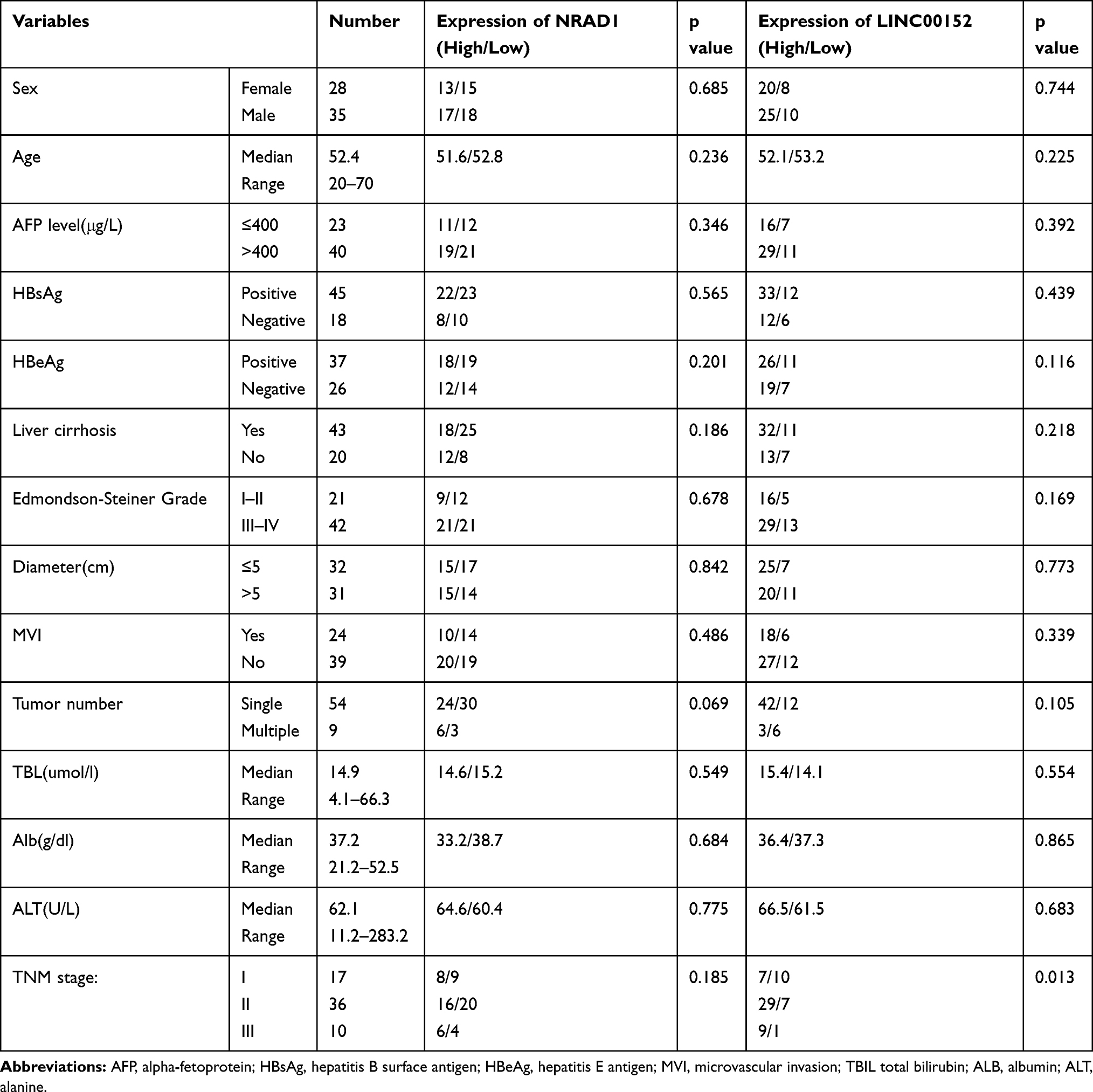 |
Table 1 Patients’ Clinicopathologic Features (n=63) |
Profiling Results and Candidate lncRNAs Screening
By microarray profiling of lncRNAs, 256 lncRNAs were found to be differentially expressed, including 162 upregulated and 94 downregulated (P<0.05, fold change>2). The raw images of the microarray analysis are shown in Figure 1A and B, and the scatter plot was shown in this Figure 1C. Using BLAST program, excluding those subjected to alternative splicing that created difficulty for designing high-specificity PCR primers, lncRNAs with large fold changes were left. Further screening was based on the fold change, gene alignment and analysis of adjacent coding genes. Then after excluding the lncRNAs that have been reported previously, two candidate lncRNAs were determined as the targets in this study, which were NRAD1 (upregulated by 6.35 folds), LINC00152 (upregulated by 4.53 folds).
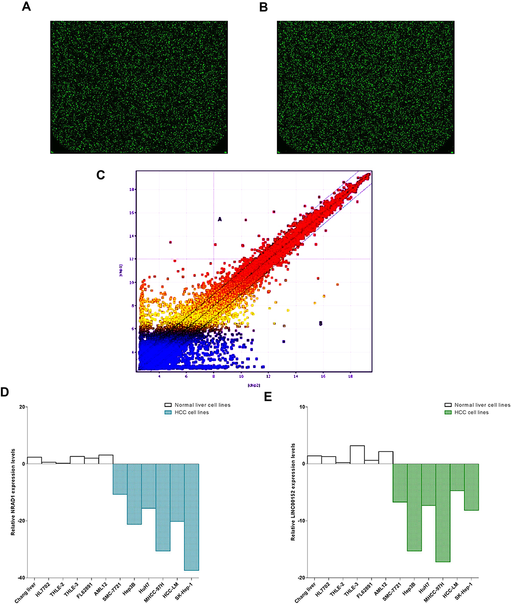 |
Figure 1 Relative expression of lncRNAs. (A, B) The raw images of the microarray analysis were shown; (C) the scatter plot was shown; (D) The comparison of relative expression of NRAD1 in HCC and normal cell lines; (E) The comparison of relative expression of LINC00152 in HCC and normal cell lines. |
Gene Expression Detected by RT-PCR in Each Cell Line
According to the results, NRAD1 and LINC00152 were downregulated in the normal liver cell lines Chang liver, HL7702, THLE-2, THLE-3, FL62891 and AML12, which were significantly lower than HCC cell lines SMMC-7721, Hep3B, HuH7, MHCC-97H, HCC-LM and SK-Hep-1 (P<0.05)(Figure 1D and E).
NRAD1 and LINC00152 Were Over-Expressed in HCC Tissues Compared with the Non-Tumor Tissues
NRAD1 expressed in 47.6% (30 of 63) of all patients in HCC tissues and LINC00152 expressed in 71.4% (45 of 63) of all patients in HCC tissues, which was significantly higher than that in non-tumorous tissues (P<0.001) (Figure 2A and B).
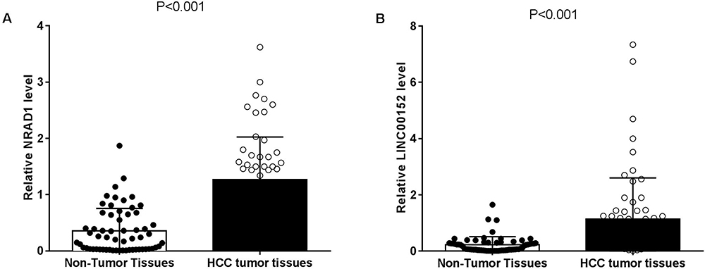 |
Figure 2 NRAD1 and were up-regulated in HCC tissues compared with nontumor tissues. (A) Relative NRAD1 concentration was detected using Real-Time qPCR; (B) Relative LINC00152 concentration was detected using Real-Time qPCR (p<0.001). |
Survival Descriptions of Different Subgroups Divided by lncRNA NRAD1 and LINC00152 Expression Levels
Descriptive survival statistics and Kaplan-Meier curves suggested that the variable of lncRNA NRAD1 and LINC00152 expression level had prognostic significance in this relatively selected cohort. Overexpression of lncRNA NRAD1 and LINC00152 were associated with decreasing OS rates, respectively (P=0.0263 and P=0.0285, Figure 3A and C). Meanwhile, overexpression of NRAD1 and LINC00152 were associated with decreasing PFS rates, respectively (P=0.0174 and P=0.0041, Figure 3B and D).
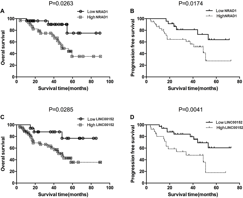 |
Figure 3 Overall survival and progression free survival estimates. (A) OS of patients stratified by NRAD1 expression levels (P = 0.0263); (B) PFS of patients stratified by NRAD1 expression levels (P = 0.0174); (C) OS of patients stratified by expression levels (P = 0.0285); (D) PFS of patients stratified by LINC00152 expression levels (P = 0.0041). |
Cox Proportional Hazard Analysis
Cox proportional hazards model was then used to quantify the prognostic significance of risk factors after multivariable adjustment. Univariable Cox proportional hazards analysis demonstrated that TNM stage (P=0.026), tumor number (P=0.031), AFP>400 (ng/mL) (P=0.008), microvascular invasion (P=0.003), tumor size (P=0.012), lncRNA NRAD1 (P=0.001) and LINC00152 (P=0.008). After performing the univariable analysis, we identified that microvascular invasion (P=0.014), tumor size (P=0.026), lncRNA NRAD1 (P=0.001) and LINC00152 (P=0.036) expression levels were significantly associated with the prognosis of patients with HCC (Table 2).
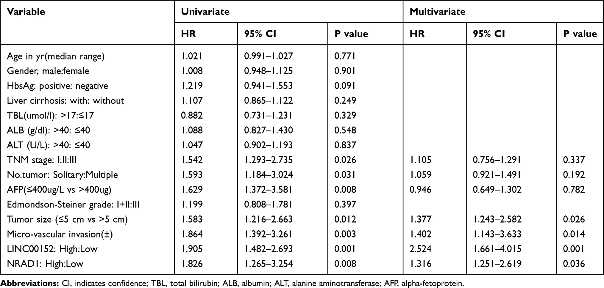 |
Table 2 Cox Proportional Hazard Regression Analyses |
Discussion
The differences of lncRNA expression profile between four cases of HCC tumor tissues besides normal tissues were explored by using dual channel lncRNA microarray technology.20 Previous reports demonstrated that lncRNAs were dysregulated in several kinds of diseases, especially in neoplastic disease.21,22 lncRNAs were identified as novel diagnostic and prognostic biomarkers for different kinds of cancers.23,24 Targeting NRAD1 with antisense oligonucleotides can reduce the cell viability and tumor growth of TNBC cell lines in patient-derived xenografts (PDX). In vitro analysis of residual PDX tumors after treatment showed that there are fewer living cancer cells and reduced breast bulb formation potential. These results are consistent with gene expression analysis. NRAD1 up-regulates genes involved in catabolism and survival, and down-regulates genes involved in differentiation. Functional analysis shows that NRAD1 is nuclear-localized, and its regulatory genes enrich genome-wide chromatin interactions.25–27 LINC00152 has been shown to play a role in the progression of many cancers, including lung cancer, gallbladder cancer, colorectal cancer, gastric cancer, tongue squamous cell carcinoma, and renal cell carcinoma.28–30 In present study, novel lncRNAs with specific upregulation in HCC were identified for the first time, namely, NRAD1 and LINC00152 by lncRNA expression profiling in HCC. We then performed further validation and found that NRAD1 and LINC00152 were overexpression in HCC tissues compared with the adjacent nontumor tissues. After adjusting for competing risk factors, we identified that expression level of lncRNA NRAD1 (P=0.001) and LINC00152 were independent risk factors associated with the prognosis of patients with HCC by Cox proportional hazards analysis.
However, several limitations were observed in this study: (1) in this study, the number of patients was so small and the results should be validated by other studies with large cohort; (2) whether overexpression of lncRNA NRAD1 (P=0.001) and LINC00152 have the optimal specificity and sensitivity for HCC diagnosis and prognosis also needs future confirmation.
In summary, we found lncRNA NRAD1 and LINC00152 expressed significantly higher in HCC tissues compared with non-tumorous tissues. Overexpression of lncRNA NRAD1 (P=0.001) and LINC00152 were independent risk factors associated with the prognosis of patients with HCC. They also have the potential to be served as therapeutic targets for further research into the molecular mechanism behind the development of HCC.
Abbreviations
HCC, Hepatocellular carcinoma; TNM, Tumor-node-metastasis; ROC, Receiver-operating characteristic; MRI, Magnetic resonance imaging; OS, Overall survival; PFS, Progression free survival.
Disclosure
The authors have declared that no competing interests exist.
References
1. Devcic Z, Elboraey M, Vidal L, et al. Individualized ablation of hepatocellular carcinoma: tailored approaches across the phenotype spectrum. Semin Intervent Radiol. 2019;36:287–297. doi:10.1055/s-0039-1698755
2. Zhang C, Liu P, Zhang C. Hepatitis B virus X protein upregulates alpha-fetoprotein to promote hepatocellular carcinoma by targeting miR-1236 and miR-329. J Cell Biochem. 2020;121(3):2489–2499.
3. Hartke J, Johnson M, Ghabril M. The diagnosis and treatment of hepatocellular carcinoma. Semin Diagn Pathol. 2017;34:153–159. doi:10.1053/j.semdp.2016.12.011
4. Guo W, Huang J, Wang N, et al. Integrating network pharmacology and pharmacological evaluation for deciphering the action mechanism of herbal formula zuojin pill in suppressing hepatocellular carcinoma. Front Pharmacol. 2019;10:1185. doi:10.3389/fphar.2019.01185
5. Huang DH, Jian J, Li S, Zhang Y, Liu LZ. TPX2 silencing exerts antitumor effects on hepatocellular carcinoma by regulating the PI3K/AKT signaling pathway. Int J Mol Med. 2019;44:2113–2122. doi:10.3892/ijmm.2019.4371
6. Zhang L, Luo B, Dang YW, et al. Clinical significance of microRNA-196b-5p in hepatocellular carcinoma and its potential molecular mechanism. J Cancer. 2019;10:5355–5370. doi:10.7150/jca.29293
7. C HL, Chen Y. Small and long non-coding RNAs: novel targets in perspective cancer therapy. Curr Genomics. 2015;16:319–326. doi:10.2174/1389202916666150707155851
8. Morceau F, Chateauvieux S, Gaigneaux A, Dicato M, Diederich M. Long and short non-coding RNAs as regulators of hematopoietic differentiation. Int J Mol Sci. 2013;14:14744–14770. doi:10.3390/ijms140714744
9. Uchida S, Bolli R. Short and long noncoding RNAs regulate the epigenetic status of cells. Antioxid Redox Signal. 2018;29(9):832–845.
10. Chen M, Zhuang C, Liu Y, et al. Tetracycline-inducible shRNA targeting antisense long non-coding RNA HIF1A-AS2 represses the malignant phenotypes of bladder cancer. Cancer Lett. 2016;376(1):155–164. doi:10.1016/j.canlet.2016.03.037
11. Yao Y, Ma J, Xue Y, et al. Knockdown of long non-coding RNA XIST exerts tumor-suppressive functions in human glioblastoma stem cells by up-regulating miR-152. Cancer Lett. 2015;359:75–86. doi:10.1016/j.canlet.2014.12.051
12. Wang D, Ding L, Wang L, et al. LncRNA MALAT1 enhances oncogenic activities of EZH2 in castration-resistant prostate cancer. Oncotarget. 2015;6(38):41045–41055. doi:10.18632/oncotarget.5728
13. Qi P, Zhou X-Y, Du X. Circulating long non-coding RNAs in cancer: current status and future perspectives. Mol Cancer. 2016;15(1):39. doi:10.1186/s12943-016-0524-4
14. Li C, Chen J, Zhang K, Feng B, Wang R, Chen L. Progress and prospects of long noncoding RNAs (lncRNAs) in hepatocellular carcinoma. Cell Physiol Biochem. 2015;36:423–434. doi:10.1159/000430109
15. Wang F, Ying HQ, He BS, et al. Upregulated lncRNA-UCA1 contributes to progression of hepatocellular carcinoma through inhibition of miR-216b and activation of FGFR1/ERK signaling pathway. Oncotarget. 2015;6:7899–7917. doi:10.18632/oncotarget.3219
16. Neumann O, Kesselmeier M, Geffers R, et al. Methylome analysis and integrative profiling of human HCCs identify novel protumorigenic factors. Hepatology. 2012;56:1817–1827. doi:10.1002/hep.25870
17. Vidovic D, Huynh TT, Konda P, et al. ALDH1A3-regulated long non-coding RNA NRAD1 is a potential novel target for triple-negative breast tumors and cancer stem cells. Cell Death Differ. 2020;27(1):363–378.
18. Kaplan EL, Meier P. Nonparametric estimations from incomplete observations. J Am Stat Assoc. 1958;53:457–481. doi:10.1080/01621459.1958.10501452
19. DR C. Regression models and life-tables. J Royal Stat Soc B. 1972;34:187–220.
20. Liu Z, Li X, Sun N, et al. Microarray profiling and co-expression network analysis of circulating lncRNAs and mRNAs associated with major depressive disorder. PLoS One. 2014;9(3):e93388. doi:10.1371/journal.pone.0093388
21. Yang L, Lin C, Jin C, et al. lncRNA-dependent mechanisms of androgen-receptor-regulated gene activation programs. Nature. 2013;500(7464):598–602. doi:10.1038/nature12451
22. Zhou X, Ye F, Yin C, Zhuang Y, Yue G, Zhang G. The interaction between MiR-141 and lncRNA-H19 in regulating cell proliferation and migration in gastric cancer. Cell Physiol Biochem. 2015;36(4):1440–1452. doi:10.1159/000430309
23. Wang Y, Gao S, Liu G, Jia R, Fan D, Feng X. Microarray expression profile analysis of long non-coding RNAs in human gastric cardiac adenocarcinoma. Cell Physiol Biochem. 2014;33(4):1225–1238. doi:10.1159/000358692
24. Tang Q, Ni Z, Cheng Z, Xu J, Yu H, Yin P. Three circulating long non-coding RNAs act as biomarkers for predicting NSCLC. Cell Physiol Biochem. 2015;37(3):1002–1009. doi:10.1159/000430226
25. Levy ML, Garnett F, Kuku A, Pertsovskaya I, McKnight E, Haughney J. A review of asthma care in 50 general practices in Bedfordshire, United Kingdom. NPJ Prim Care Respir Med. 2018;28(1):29. doi:10.1038/s41533-018-0093-7
26. Tan X, Zhu F, Wang C, et al. Two-dimensional micro-/nanoradian angle generator with high resolution and repeatability based on piezo-driven double-axis flexure hinge and three capacitive sensors. Sensors. 2017;17:2672.
27. Yashchuk VV, Lacey I, Gevorkyan GS, McKinney WR, Smith BV, Warwick T. Ex situ metrology and data analysis for optimization of beamline performance of aspherical pre-shaped x-ray mirrors at the advanced light source. Rev Sci Instrum. 2019;90:021711. doi:10.1063/1.5057441
28. Liu P, He W, Lu Y, Wang Y. Long non-coding RNA LINC00152 promotes tumorigenesis via sponging miR-193b-3p in osteosarcoma. Oncol Lett. 2019;18:3630–3636. doi:10.3892/ol.2019.10700
29. Wang J, Zhang Y, Lu L, Lu Y, Tang Q, Pu J. Insight into the molecular mechanism of LINC00152/miR-215/CDK13 axis in hepatocellular carcinoma progression. J Cell Biochem. 2019;120:18816–18825. doi:10.1002/jcb.29197
30. Zhao L, Chi WW, Cao H, Meng WX, Cui WN, Wang BS. Expression of long-chain non-coding RNA LINC00152 in laryngeal squamous cell carcinoma and its clinical significance. Lin Chung Er Bi Yan Hou Tou Jing Wai Ke Za Zhi. 2019;33:721–725. doi:10.13201/j.issn.1001-1781.2019.08.010
 © 2020 The Author(s). This work is published and licensed by Dove Medical Press Limited. The full terms of this license are available at https://www.dovepress.com/terms.php and incorporate the Creative Commons Attribution - Non Commercial (unported, v3.0) License.
By accessing the work you hereby accept the Terms. Non-commercial uses of the work are permitted without any further permission from Dove Medical Press Limited, provided the work is properly attributed. For permission for commercial use of this work, please see paragraphs 4.2 and 5 of our Terms.
© 2020 The Author(s). This work is published and licensed by Dove Medical Press Limited. The full terms of this license are available at https://www.dovepress.com/terms.php and incorporate the Creative Commons Attribution - Non Commercial (unported, v3.0) License.
By accessing the work you hereby accept the Terms. Non-commercial uses of the work are permitted without any further permission from Dove Medical Press Limited, provided the work is properly attributed. For permission for commercial use of this work, please see paragraphs 4.2 and 5 of our Terms.