")
Back to Journals » Cancer Management and Research » Volume 12
LncRNA WT1-AS Downregulates Survivin by Upregulating miR-203 in Papillary Thyroid Carcinoma
Authors Le F, Luo P, Ouyang Q, Zhong X
Received 24 September 2019
Accepted for publication 17 December 2019
Published 20 January 2020 Volume 2020:12 Pages 443—449
DOI https://doi.org/10.2147/CMAR.S232294
Checked for plagiarism Yes
Review by Single anonymous peer review
Peer reviewer comments 2
Editor who approved publication: Professor Rudolph Navari
Fei Le, 1 Ping Luo, 2 Qian Ouyang, 3 Xiaoming Zhong 4
1Department of Head and Neck Surgery, Jiangxi Province Cancer Hospital, Nanchang City, Jiangxi Province, 330029, People’s Republic of China; 2Department of Surgical Oncology, Nanchang Third Hospital Surgical Oncology, Nanchang City, Jiangxi Province, 330002, People’s Republic of China; 3Department of Intensive Medicine, Jiangxi Province Cancer Hospital, Nanchang City, Jiangxi Province, 330029, People’s Republic of China; 4Department of Tumor Radiotherapy, Jiangxi Province Cancer Hospital, Nanchang City, Jiangxi Province, 330029, People’s Republic of China
Correspondence: Xiaoming Zhong
Department of Tumor Radiotherapy, Jiangxi Province Cancer Hospital, No. 519 Beijing East Road, Nanchang City, Jiangxi Province 330029, People’s Republic of China
Tel +86 15979192617
Email [email protected]
Objective: This study aimed to assessment the functions of lncRNA WT1-AS in papillary thyroid carcinoma (PTC).
Methods: Expression levels of WT1-AS in PTC and non-tumor tissues from 66 PTC patients were measured and compared by performing qPCR and paired t test, respectively. Cell proliferation (CCK-8) assay was performed to evaluate the effects of the overexpression of WT1-AS, miR-203 and survivin on the proliferation of IHH-4 (a human PTC cell line) cells.
Results: We found that WT1-AS was significantly downregulated in PTC and associated with clinical stages. In PTC tissues, WT1-AS was negatively correlated with survivin but positively correlated with miR-203. In PTC cells, WT1-AS overexpression led to significantly upregulated miR-203 and downregulated survivin. MiR-203 overexpression failed to affect WT1-AS but downregulated survivin. Cell proliferation assay showed that overexpression of WT1-AS and miR-203 led to decreased, while survivin overexpression led to increased proliferation of PTC cells. In addition, survivin overexpression attenuated the effects of WT1-AS and miR-203 overexpression.
Conclusion: Therefore, WT1-AS may downregulate survivin by upregulating miR-203 in PTC to inhibit cancer cell proliferation.
Keywords: WT1-AS, papillary thyroid carcinoma, survivin, miR-203
Introduction
According to the latest GLOBOCAN statistics, thyroid cancer in 2018 only affects 567,233, which accounts for about 3.1% of all cancer patients.1 During the same time period, thyroid cancer only caused 41,071 deaths, which accounts for about 0.4% of all cancer mortality.1 With the development of novel diagnostic techniques, such as ultrasonography, early diagnosis of thyroid cancer has been significantly improved in past several decades.2,3 Combined with the slow development nature of thyroid cancer, prognosis of patients with this disease is generally satisfactory.4 However, tumor metastasis is inevitable in some cases and survival of these patients are still poor.5 Therefore, novel therapeutic approaches are still needed to further improve the survival of patients with PTC.
Papillary thyroid carcinoma (PTC) is the most common form of thyroid cancer. Recent studies have identified altered signaling pathways involved in the development and progression of PTC.6,7 In effects, functional analysis of molecular players involved in PTC provided novel insights to the development of targeted therapies.6,7 Survivin is a well-established oncogene in many types of cancers including PTC.8 Survivin inhibits apoptosis protein family to block cell death, thereby promoting cancer growth and progression.8 Therefore, inhibition of survivin is a potential approach for cancer therapies.9 It is well known that certain miRNAs, such as miR-203, can directly target survivin to inhibit cancer growth.10 WT1-AS is a recently identified tumor suppressor in cervical and gastric cancers.11–13 It is known that WT1-AS can suppress cancer progression by interacting with p53, which has crosstalk with miR-203.14 Therefore, WT1-AS may also interact with miR-203. This study aimed to explore the potential interactions between WT1-AS and miR-203 in PTC and analyze their roles in regulating survivin.
Materials and Methods
PTC Patients
The research subjects of this study were 66 PTC patients (37 males and 29 females, 39 to 66 years, 52.2±6.9 years) selected form the 117 PTC cases diagnosed in Jiangxi province cancer hospital between May 2017 and May 2019. This study passed the review board of Ethics Committee of aforementioned hospital. Inclusion criteria: 1) new PTC cases; 2) willing to received biopsy (fine needle aspiration); 3) no therapies were initiated. Exclusion criteria: 1) recurrent PTC; 2) other clinical disorders, such as other malignancies, heart diseases and diabetes, were observed. The patients were staged based on clinical findings. According to AJCC system, the 66 patients included 18, 21, 15 and 12 cases at stages I–IV, respectively. According to pathological grade, there were 18 and 48 at high (aggressive histologic variants) and low grade, respectively. All patients were informed with the principle of this study and the potential publication of this paper. All the 66 PTC provided written informed consent.
PTC Tissues and Cell Line
Before the initiation of therapies, all patients were subjected to biopsy, which was performed under the guidance of MRI. During biopsy, PTC and non-tumor (thyroid tissues within the area 3 cm around tumors) tissues were collected from each patient. The weight of each tissue sample ranged from 0.05 to 0.09g. Based on the results of histopathological tests, all PTC tissues contained more than 95% cancer cells, and all non-tumor tissues contained less than 4% cancer cells.
Human PTC cell line IHH-4 (AJCC, USA) was used in this study. Cell culture medium was a mixture of 90% DMEM and 10% FBS. Cells were cultivated under the following conditions: 95% humidity, 37°C and 5% CO2.
Transient Cell Transfection
Vector construction service was provided by Sangon (Shanghai, China). Expression vectors of WT1-AS and survivin were constructed using pcDNA3.1 vector. Negative control (NC) miRNA and miR-203 mimic were synthesized by Invitrogen (Shanghai, China). To perform transient transfections, IHH-4 cells were harvested at 70–80% confluence. All transfections were mediated by lipofectamine 2000 (Sangon). Briefly, lipofectamine 2000 was first mixture with vectors (empty vector as NC group) or miRNAs (NC miRNA as NC group), followed by incubating with 106 cells for 6 hrs at 37°C. After that, cells were washed with fresh cell culture medium to terminate cell transfections. Control (C) cells for all transfections were untransfected cells. All subsequent experiments were performed using cells harvested at 96 hrs.
Total RNA Extraction
Tissue samples from PTC patients were ground in fine powder. IHH-4 cells were harvested at 24 hrs post-transfection and cells were counted. Total RNAs in 0.03g tissue sample or 105 cells were extracted using Tirzol reagent (Invitrogen, USA) with all steps performed following manufacturer’s instruction. It is worth noting that RNA samples were precipitated using 85% ethanol to retain miRNAs in the sample. All RNA samples were digested with DNase I (Sigma-Aldrich) to remove genomic DNA. Thermo Scientific NanoDrop 2000 spectrophotometer was used to measure RNA concentration.
qPCR
All RNA samples were digested with DNase I for 2 hrs at 37°C to remove genomic DNAs. Total RNA transcriptions were performed using Tetro Reverse Transcriptase (Bioline) and qPCR mixtures were prepared using SYBR® Green Master Mix (Bio-Rad). The expression levels of WT1-AS and survivin mRNA were measured using GAPDH as endogenous control.
Addition of poly(A) to mature miRNAs, miRNA reverse transcriptions and qPCR assays were performed using All-in-One™ miRNA qRT-PCR Reagent Kit (GeneCopoeia). With U6 as endogenous control, expression levels of miR-203 were measured.
All Ct values were normalized using 2−ΔΔCT, and all PCR reactions were performed in 3 replicates.
Western Blot
To measure the expression levels of survivin at 24 hrs post-transfection, total proteins in 105 cells of each transfection group were extracted using RIPA solution (Sangon). Protein concentrations were measured using a BCA kit (Sangon). Protein samples were denatured in boiling water for 8 mins, followed by electrophoresis performed using 12% SDS-PAGE. Protein samples were transferred to PVDF membranes, and blocking was performed in PBS containing 5% non-fat milk for 2 hrs at 22°C. After that, rabbit primary antibodies of survivin (1:2000, ab469, Abcam) and endogenous control GAPDH (1:2000, ab37168, Abcam) were used to incubate with the membranes for 12 hrs at 4°C. Following that, HRP (IgG) (1:2000; ab6721; Abcam) goat secondary antibody was used to further incubate with the membranes for 2 hrs at 22°C. Finally, membranes were incubated with ECL (Sigma-Aldrich) for 5 mins at 22°C to develop signals. Image J v1.46 software was then used to normalize signals.
Cell Proliferation Assay
Cell counting kit-8 (CCK-8, Sigma-Aldrich) was used to evaluate the effects of overexpression of WT1-AS, survivin and miR-203 on the proliferation of IHH-4 cells. Briefly, 3×10⁴ cells were mixture with 1mL medium. Cells were cultivated in a cell culture plate (96-well, 0.1 mL per well) under aforementioned conditions. CCK-8 solution (10 μL) was added into each well at 4h before the termination of cell culture. After that 10 μL DMSO was added into each well and OD values were measured at 450 nm.
Statistical Analysis
Mean values of 3 independent biological replicates were calculated and were used for all data analysis, which was performed using GraphPad Prism 6 software. Exploration of differences between tissue types (PTC vs non-tumor) and among multiple cell transfection and patient groups (clinical stages) was performed by paired t test and ANOVA (one-way) in combination with Tukey’s test, respectively. Comparison between two groups of patients (high and low grade) was performed by unpaired t test. Correlations were analyzed by linear regression. p<0.05 was statistically significant.
Results
Downregulation of WT1-AS in PTC Was Associated with Clinical Stages and Pathological Grades
Expression levels of WT1-AS in PTC and non-tumor tissues from PTC patients were measured and compared by performing qPCR and paired t test, respectively. It was observed that, compared to non-tumor tissue group, significantly higher expression levels of WT1-AS were detected in PTC tissue group (Figure 1A, p<0.05). Expression levels of WT1-AS in PTC tissues were compared among 4 clinical stages by performing ANOVA (one-way) in combination with Tukey’s test. Significantly decreased expression levels of WT1-AS were observed with the increase in clinical stages (Figure 1B, p<0.05). Expression levels of WT1-AS in PTC tissues were compared between 2 pathological grades by performing unpaired t test. It can be observed that expression levels of WT1-AS in PTC were significantly lower in high-grade group than in low-grade group (Figure 1C, p<0.05).
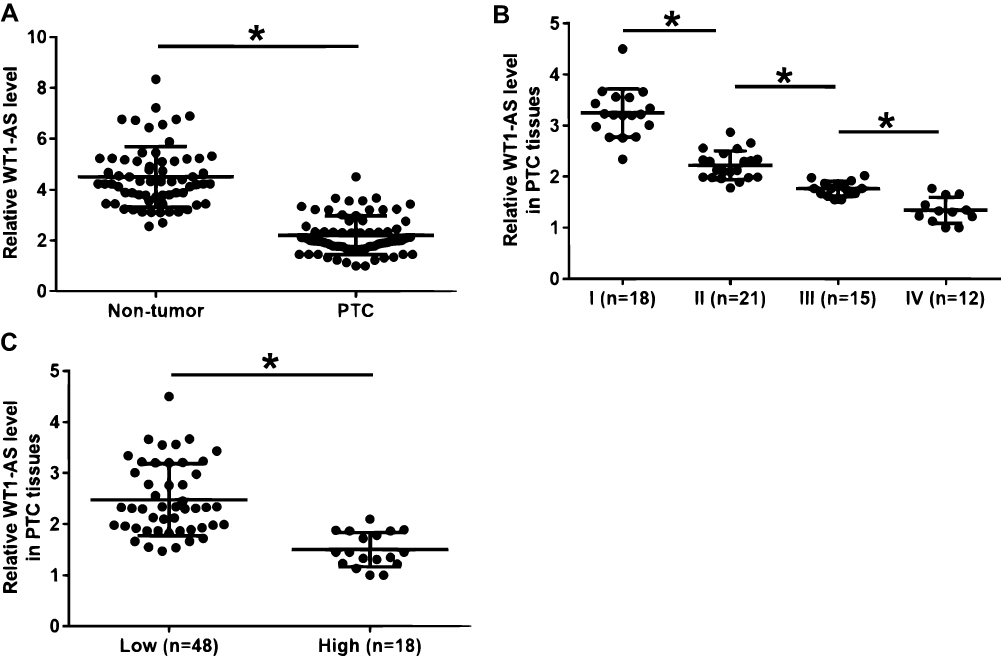 |
Figure 1 Downregulation of WT1-AS in PTC was associated with clinical stages and pathological grades. Expression levels of WT1-AS in PTC and non-tumor tissues from PTC patients were measured and compared by performing qPCR and paired t test, respectively (A). Expression levels of WT1-AS in PTC tissues were compared among 4 AJCC clinical stages by performing ANOVA (one-way) in combination with Tukey’s test (B). Expression levels of WT1-AS in PTC tissues were compared between 2 pathological grades (high and low) by performing unpaired t test (C). Mean values of 3 biological replicates were presented, *p<0.05. |
Levels of WT1-AS Expression Were Significantly Correlated with Expression Levels of miR-203 and Survivin Across PTC Tissues
Expression levels of miR-203 and survivin mRNA in PTC and non-tumor tissues from PTC patients were also measured and compared by performing qPCR and paired t test, respectively. It was observed that, compared to non-tumor tissue group, significantly higher expression levels of miR-203 (Figure 2A) and significantly lower expression levels of survivin mRNA (Figure 2B) were detected in PTC tissue group (p<0.05). Correlations between WT1-AS and miR-203/survivin mRNA were analyzed by linear regression. It was observed that expression levels of WT1-AS were significantly and positively correlated with expression levels of miR-203 (Figure 2C). In addition, expression levels of WT1-AS were significantly and negatively correlated with expression levels of survivin mRNA (Figure 2D).
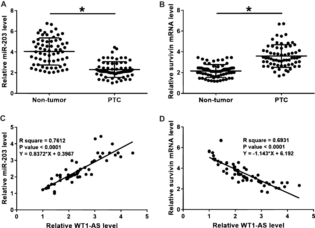 |
Figure 2 Levels of WT1-AS expression were significantly correlated with expression levels of miR-203 and survivin across PTC tissues. Expression levels of miR-203 (A) and survivin mRNA (B) in PTC and non-tumor tissues from PTC patients were also measured and compared by performing qPCR and paired t-test, respectively. Correlations between WT1-AS and miR-203 (C) survivin mRNA (D) in PTC tissues were analyzed by linear regression. Mean values of 3 biological replicates were presented, *p<0.05. |
WT1-AS Overexpression Downregulate Survivin in IHH-4 Cells by Upregulating miR-203
To further analyze the interactions among WT1-AS, miR-203 and survivin, WT1-AS and survivin vectors as well as miR-203 mimic were transfected into IHH-4 cells. Overexpression of WT1-AS, miR-203 and survivin was confirmed by qPCR at 24 hrs after transfections. Compared to NC (cells transfected with NC miRNA or empty vector) and C (untransfected cell) groups, significantly upregulated expression levels of WT1-AS, miR-203 and survivin were observed after transfections (Figure 3A, p<0.05). Compared to NC and C groups, WT1-AS overexpression led to downregulated miR-203, while miR-203 overexpression failed to affect WT1-AS (Figure 3B, p<0.05). Moreover, WT1-AS overexpression led to downregulated survivin but miR-203 overexpression led to downregulated surviving (Figure 3C, p<0.05).
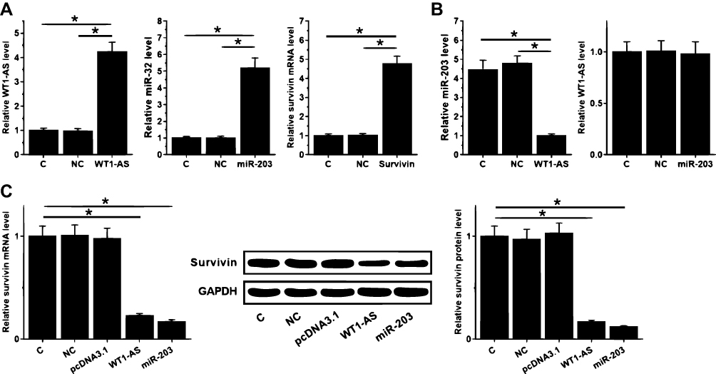 |
Figure 3 WT1-AS overexpression downregulate survivin in IHH-4 cells by upregulating miR-203. To further analyze the interactions among WT1-AS, miR-203 and survivin, WT1-AS and survivin vectors as well as miR-203 mimic were transfected into IHH-4 cells. Overexpression of WT1-AS, miR-203 and survivin was confirmed by qPCR at 24 hrs after transfections (A). Interactions between WT1-AS and miR-203 were analyzed by qPCR (B). The effects of WT1-AS and miR-203 overexpression on the expression of survivin mRNA and protein were analyzed by qPCR and Western blot, respectively (C). Mean values of 3 biological replicates were presented. NC, cells transfected with NC miRNA or empty vector; C, untransfected cell. *p<0.05. |
WT1-AS Suppressed IHH-4 Cell Proliferation Through miR-203 and Survivin
Cell proliferation assay was performed to assessment the effects of the overexpression of WT1-AS, miR-203 and survivin on the proliferation of IHH-4 cells. Compared to C and NC groups, cell proliferation assay showed that overexpression of WT1-AS and miR-203 overexpression led to decreased rates of cell proliferation, while survivin overexpression led to increased proliferation of PTC cells. In addition, survivin overexpression attenuated the effects of WT1-AS and miR-203 overexpression (Figure 4, p<0.05).
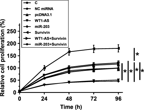 |
Figure 4 WT1-AS suppressed IHH-4 cell proliferation through miR-203 and survivin. Cell proliferation assay was performed to assessment the effects of the overexpression of WT1-AS, miR-203 and survivin on the proliferation of IHH-4 cells. Mean values of 3 biological replicates were presented, *p<0.05. |
Discussion
This paper is the first to investigate the roles of WT1-AS in PTC. We found that WT1-AS was downregulated in PTC and played a tumor-suppressive role by downregulating survivin through the upregulation of miR-203.
The functions of WT1-AS have only been investigated in gastric and cervical cancer.11–13 In gastric cancer, WT1-AS is downregulated and its downregulation accelerated cell proliferation and migration.11 In cervical cancer, WT1-AS is also downregulated and can regulate p53 and miR-203a-5p/FOXN2 to inhibit the aggressiveness of cancer cells.12,13 All these previous studies revealed the tumor-suppressive roles of WT1-AS in cancer biology. Our study is the first to report the downregulation of WT1-AS in PTC and the decreased PTC proliferation rate after WT1-AS overexpression. Therefore, our data suggested that WT1-AS was also a tumor-suppressive lncRNA in PTC.
Cancer stages and pathological grades are closely correlated with the survival of PTC patients.14 Clinical studies have shown that patients at advanced clinical stages and with high pathological grade usually will experience high mortality rate.14 This study failed to perform survival analysis. However, our study showed that expression levels of WT1-AS were negatively correlated with clinical stages and pathological grades. Therefore, low WT1-AS expression levels may be correlated with the poor survival of PTC patients. Our future studies will perform more explorations. Dai et al reported that low levels of WT1-AS were closely correlated with the poor survival of cervical cancer patients.13 Interestingly, in another study, Cao et al showed that the loss of WT1-AS is associated with good survival of cervical cancer patients at early stage.15 Therefore, more studies are needed to further explore the prognostic applications of WT1-AS for cancer patients.
It is known that lncRNAs may sponge miRNAs to attenuate their effects on their downstream genes.16 However, this study showed that WT1-AS can upregulate miR-203 to downregulate survivin. Therefore, WT1-AS is unlikely a sponge of miR-203. It is known that both miR-203 and WT1-AS have crosstalk with p53,12,14 and p53 is dysregulated in PTC. Therefore, the altered p53 may mediate the interaction between them.
In conclusion, WT1-AS is downregulated in PTC, and WT1-AS may downregulate survivin by upregulating miR-203, thereby suppressing cancer cell proliferation.
Ethical Statement
The patient consent was written informed consent, and the study was conducted in accordance with the Declaration of Helsinki.
Data Sharing Statement
The analyzed data sets generated during the study are available from the corresponding author on reasonable request.
Author Contributions
All authors contributed to data analysis, drafting or revising the article, gave final approval of the version to be published, and agree to be accountable for all aspects of the work.
Disclosure
The authors report no conflicts of interest in this work.
References
1. Bray F, Ferlay J, Soerjomataram I, et al. Global cancer statistics 2018: GLOBOCAN estimates of incidence and mortality worldwide for 36 cancers in 185 countries. CA Cancer J Clin. 2018;68(6):394–424. doi:10.3322/caac.21492
2. Hankey BF, Feuer EJ, Clegg LX, et al. Cancer surveillance series: interpreting trends in prostate cancer-part I: evidence of the effects of screening in recent prostate cancer incidence, mortality, and survival rates. J Natl Cancer Inst. 1999;91(12):1017–1024. doi:10.1093/jnci/91.12.1017
3. Vaccarella S, Franceschi S, Bray F, et al. Worldwide thyroid-cancer epidemic? The increasing impact of overdiagnosis. N Engl J Med. 2016;375(7):614–617. doi:10.1056/NEJMp1604412
4. Konturek A, Barczyński M, Stopa M, et al. Trends in prevalence of thyroid cancer over three decades: a retrospective cohort study of 17,526 surgical patients. World J Surg. 2016;40(3):538–544. doi:10.1007/s00268-015-3322-z
5. Cabanillas ME, McFadden DG, Durante C. Thyroid cancer. Lancet. 2016;388(10061):2783–2795. doi:10.1016/S0140-6736(16)30172-6
6. Liang J, Cai W, Feng D, et al. Genetic landscape of papillary thyroid carcinoma in the Chinese population. J Pathol. 2018;244(2):215–226. doi:10.1002/path.2018.244.issue-2
7. He H, Li W, Liyanarachchi S, et al. Genetic predisposition to papillary thyroid carcinoma: involvement of FOXE1, TSHR, and a novel lincRNA gene, PTCSC2. J Clin Endocrinol Metab. 2015;100(1):E164–E172. doi:10.1210/jc.2014-2147
8. Selemetjev S, Savin S, Paunovic I, et al. Concomitant high expression of survivin and vascular endothelial growth factor-C is strongly associated with metastatic status of lymph nodes in papillary thyroid carcinoma. J Cancer Res Ther. 2018;14(8):114–119. doi:10.4103/0973-1482.163675
9. Soleimanpour E, Babaei E. Survivin as a potential target for cancer therapy. Asian Pac J Cancer Prev. 2015;16(15):6187–6191. doi:10.7314/APJCP.2015.16.15.6187
10. Bian K, Fan J, Zhang X, et al. MicroRNA-203 leads to G1 phase cell cycle arrest in laryngeal carcinoma cells by directly targeting survivin. FEBS Lett. 2012;586(6):804–809. doi:10.1016/j.febslet.2012.01.050
11. Du T, Zhang B, Zhang S, et al. Decreased expression of long non-coding RNA WT1-AS promotes cell proliferation and invasion in gastric cancer. Biochim Biophys Acta. 2016;1862(1):12–19. doi:10.1016/j.bbadis.2015.10.001
12. Cui LJ, Nai MM, Zhang K, et al. lncRNA WT1-AS inhibits the aggressiveness of cervical cancer cell via regulating p53 expression via sponging miR-330-5p. Cancer Manag Res. 2019;11:651–667. doi:10.2147/CMAR.S176525
13. Dai SG, Guo LL, Xia X, et al. Long non-coding RNA WT1-AS inhibits cell aggressiveness via miR-203a-5p/FOXN2 axis and is associated with prognosis in cervical cancer. Eur Rev Med Pharmacol Sci. 2019;23(2):486–495. doi:10.26355/eurrev_201901_16860
14. Li J, Chen Y, Zhao J, et al. miR-203 reverses chemoresistance in p53-mutated colon cancer cells through downregulation of Akt2 expression. Cancer Lett. 2011;304(1):52–59. doi:10.1016/j.canlet.2011.02.003
15. Cao J, Wang H, Xu P, et al. Loss of long noncoding RNA WT1-AS is associated with better survival in early stage of cervical cancer. Int J Clin Exp Med. 2018;11(8):8158–8163.
16. Militello G, Weirick T, John D, et al. Screening and validation of lncRNAs and circRNAs as miRNA sponges. Brief Bioinform. 2016;18(5):780–788.
 © 2020 The Author(s). This work is published and licensed by Dove Medical Press Limited. The full terms of this license are available at https://www.dovepress.com/terms.php and incorporate the Creative Commons Attribution - Non Commercial (unported, v3.0) License.
By accessing the work you hereby accept the Terms. Non-commercial uses of the work are permitted without any further permission from Dove Medical Press Limited, provided the work is properly attributed. For permission for commercial use of this work, please see paragraphs 4.2 and 5 of our Terms.
© 2020 The Author(s). This work is published and licensed by Dove Medical Press Limited. The full terms of this license are available at https://www.dovepress.com/terms.php and incorporate the Creative Commons Attribution - Non Commercial (unported, v3.0) License.
By accessing the work you hereby accept the Terms. Non-commercial uses of the work are permitted without any further permission from Dove Medical Press Limited, provided the work is properly attributed. For permission for commercial use of this work, please see paragraphs 4.2 and 5 of our Terms.