")
Back to Journals » International Journal of General Medicine » Volume 14
Liquid–Liquid Phase Separation-Related Genes Associated with Tumor Grade and Prognosis in Hepatocellular Carcinoma: A Bioinformatic Study
Authors Fang ZS , Zhang Z, Liang ZJ, Long ZR , Xiao Y, Liang ZY, Sun X, Li HM, Huang H
Received 11 October 2021
Accepted for publication 30 November 2021
Published 14 December 2021 Volume 2021:14 Pages 9671—9679
DOI https://doi.org/10.2147/IJGM.S342602
Checked for plagiarism Yes
Review by Single anonymous peer review
Peer reviewer comments 2
Editor who approved publication: Dr Scott Fraser
Zhao-Shan Fang,1,* Zhi Zhang,1,* Zhi-Jie Liang,2 Zhong-Rong Long,1 Yi Xiao,1 Zhi-Yin Liang,1 Xing Sun,1 Hong-Mian Li,3 Hai Huang4
1Department of Hepatobiliary Surgery, The Fifth Affiliated Hospital of Guangxi Medical University, Nanning, Guangxi, 530022, People’s Republic of China; 2Department of Wound Repair Surgery, The Fifth Affiliated Hospital of Guangxi Medical University, Nanning, Guangxi, 530022, People’s Republic of China; 3Department of Medical Laboratory Center, The Fifth Affiliated Hospital of Guangxi Medical University, Guangxi, 530022, People’s Republic of China; 4Department of Hepatobiliary Surgery, Wuming Hospital of Guangxi Medical University, Nanning, Guangxi, 530199, People’s Republic of China
*These authors contributed equally to this work
Correspondence: Hong-Mian Li
Department of Medical Laboratory Center, The Fifth Affiliated Hospital of Guangxi Medical University, Guangxi, 530022, People’s Republic of China
Email [email protected]
Hai Huang
Department of Hepatobiliary Surgery, Wuming Hospital of Guangxi Medical University, Nanning, Guangxi, 530199, People’s Republic of China
Email [email protected]
Aim: The aim of the present study was to identify the association between tumor grade and liquid–liquid phase separation (LLPS)-related genes, and to generate a LLPS-related gene-based risk index (LLPSRI) as a prognostic tool for hepatocellular carcinoma (HCC).
Methods: Weighted gene correlation network analysis was performed to test whether the LLPS-related gene modules were associated with tumor grade of HCC. The candidate modules were subjected to functional enrichment analysis. We generated a LLPSRI using the expression profiles of the hub genes among the candidate modules in order to identify patients at high risk. Then, the biological characteristics of the high-risk patients were revealed using gene set enrichment analysis. Additionally, an independent external data set was used to validate the LLPSRI.
Results: Four gene modules showed a significant positive correlation with tumor grade and involved various cancer-related pathways. Among the hub genes, six were selected to generate the LLPSRI, which was significantly associated with prognosis of HCC patients. The LLPSRI could successfully divide patients with HCC into high- and low-risk groups, and patients in the high-risk group showed shorter overall survival than those in the low-risk group. E2F, MYC, and mTORC1 signaling may be important determinants of survival in the high-risk group. The prognostic value of the LLPSRI was validated with the independent external data set.
Conclusion: We identified LLPS-related gene modules that are associated with HCC tumor grade. The LLPSRI may be useful as a prognostic marker of HCC, and it may reliably stratify patients into groups at low or high risk of worse survival. Our analysis also suggests that certain biological characteristics of HCC may be associated with high risk of worse survival.
Keywords: hepatocellular carcinoma, liquid–liquid phase separation, WGCNA, prognostic biomarker
Introduction
Hepatocellular carcinoma (HCC) is the most common type of malignant tumor, and it is a global health challenge whose incidence is growing worldwide.1,2 Infection with hepatitis B virus (HBV) or hepatitis C virus (HCV) as well as cirrhosis are considered risk factors for HCC development.3 Our understanding of the pathophysiology of HCC has improved in recent decades, but this knowledge has yet to be translated into clinical practice. Novel concepts are required to reveal the complex mechanisms underlying this disease. In recent years, liquid–liquid phase separation (LLPS) has emerged as a new concept to explain how cells perform their vital functions.4 LLPS refers to the generation of membraneless intracellular condensates where essential processes occur, including chromatin organization, X-chromosome inactivation, transcription, DNA damage response, autophagy, and tumorigenesis.5–7 Aberrant LLPS is associated with various diseases, including cancer.8–11 For example, LLPS underlies the formation of nucleoli, whose morphology is considered an indicator of tumor malignancy and differentiation.12 Numerous genes encoding proteins or RNA have been implicated in LLPS.9 The field of LLPS-mediated regulation of biological processes is still in an early stage, and much more needs to be learned about the molecular mechanisms involved and what they mean for cells and organisms.
At the same time, HCC is a highly heterogeneous disease,3 and certain patients with early-stage disease may be at higher risk of worse survival. A “one-size-fits-all” method to predict patients prognosis is unreliable, so novel prognostic models are urgently needed. Considering that LLPS is associated with HCC development, we hypothesized that the expression pattern of LLPS-related genes may be associated with prognosis of HCC. In the present study, we identified modules of LLPS-related genes that are associated with tumor grade, and from a subset of hub genes we created an index to predict prognosis of patients with HCC.
Materials and Methods
Data Processing
The TCGA-LIHC data set, including RNA sequencing (RNA-seq) expression profiles (displayed as read counts) and clinical information of HCC patients in The Cancer Genome Atlas (https://www.cancer.gov/), were downloaded to perform weighted gene correlation network analysis (WGCNA)13 and to generate an LLPS-related gene-based risk index (LLPSRI) in HCC. The RNA-seq profiles were normalized using the voom method in the limma package.14 To be included in our study, both gene expression profiles and clinical information had to be available, and the tumor had to be HCC. Data for healthy liver tissues, samples from patients with combined HCC and cholangiocarcinoma, and HCC samples of unknown tumor grade were excluded from our analysis. Another processed HCC data set (LIRI-JP) from the JP Project of the International Cancer Genome Consortium (https://dcc.icgc.org/) was downloaded from the HCCDB database15 and used to validate the LLPSRI. The LLPS-related genes were downloaded from PhaSepDB (http://db.phasep.pro/).16 The workflow of our study is shown in Figure 1. Our institution waived the requirement for ethical approval for our study because it retrospectively meta-analyzed data already deposited in public databases.
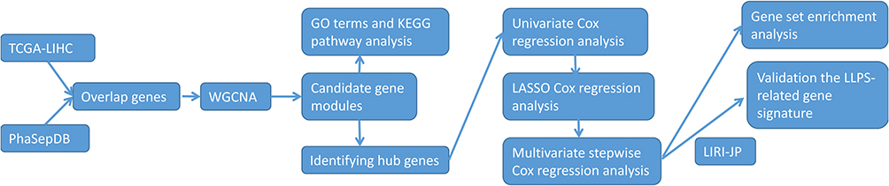 |
Figure 1 Workflow of the present study. Abbreviations: TCGA-LIHC, the Cancer Genome Atlas-liver hepatocellular carcinoma; HCC, hepatocellular carcinoma; WGCNA, weighted gene correlation network analysis; GO, Gene Ontology; KEGG, Kyoto Encyclopedia of Genes and Genomes; LLPS, liquid–liquid phase separation; LASSO, least absolute shrinkage and selection operator. |
Identification of Modules Associated with Tumor Grade Using WGCNA
A total of 2158 LLPS-related genes were found in the RNA-seq expression profiles of the TCGA-LIHC data set. We extracted the expression profiles of LLPS-related genes in order to perform WGCNA using the WGCNA package. First, hierarchical clustering analysis was performed, and the samples with significantly different gene expression patterns were removed. Then, the soft thresholding power value was screened during module construction. A suitable power value was identified if the degree of independence was > 0.9. The minimum module size was set to 30, and the individual module was assigned a unique color. If a module showed a significant positive correlation with tumor grade (P < 0.05), it was considered a candidate module.
Functional Enrichment Analysis of Candidate Modules
The clusterProfiler package17 was used to perform functional enrichment analysis to reveal the potential biological functions of candidate modules, based on Gene Ontology (GO) terms and Kyoto Encyclopedia of Genes and Genomes (KEGG) pathways. Results were considered significant if they were associated with P < 0.01 after adjustment using the Benjamini & Hochberg method.
Hub Genes and the LLPSRI
In WGCNA, module membership (MM) was defined as the association between a gene and its module. In the present study, a gene with MM > 0.8 was considered a hub gene.18 The expression profiles of the hub genes were used to generate the LLPSRI. Briefly, univariate Cox regression analysis was carried out to identify overall survival (OS)-associated genes. A gene with P < 0.05 was considered significant. The gene expression profiles of the OS-associated genes were used to perform least absolute shrinkage and selection operator (LASSO) Cox regression analysis using the “glmnet” package.19 The features with non-zero coefficients were selected in 10-fold cross-validation for multivariate stepwise Cox regression, and the LLPSRI was defined as
LLPSRI=Exprgene1*Coefgene1+Exprgene2*Coefgene2+Exprgene3*Coefgene3+...
where “Expr” represents the expression value of the candidate genes in the multivariate Cox regression analysis, and “Coef” is the corresponding estimated regression coefficient. Then, patients with HCC were divided into low- or high-risk groups based on the median LLPSRI. OS was compared between the low- and high-risk groups using the log-rank method.
Gene Set Enrichment Analysis (GSEA)
We performed GSEA20,21 to explore the biological characteristics of high-risk patients. The hallmark gene set and the Kyoto Encyclopedia of Genes and Genomes (KEGG) pathway gene sets from the Molecular Signatures Database (version 7.2)20,22 were used as the reference gene set. GSEA was carried out using the GSEA software written in JAVA (http://www.broadinstitute.org/gsea). We set the cutoff criteria as a false discovery rate value < 0.05.
Validation of the LLPSRI
As we did with patients in the TCGA-LIHC dataset, we assigned each patient in the JP-ICGC an LLPSRI according to the above formula. Then, patients with HCC were divided into low- or high-risk groups based on the median PRS. OS was compared between the low- and high-risk groups using the log-rank method.
Results
Multiple LLPS-Related Gene Modules are Associated with HCC Tumor Grade
Four samples of HCC were removed as they showed significantly different expression patterns from the other samples (Figure 2A). In the end, 352 HCC samples were used for WGCNA. The power was 4, which was the lowest value for the scale with an independence degree of up to 0.90 (Figure 2B). WGCNA identified nine modules (Figure 2C), among which the black, turquoise, brown, and yellow modules showed a significant positive correlation with tumor grade and were therefore selected as candidate modules for subsequent analysis (Figure 2D). The green module, in contrast, negatively correlated with tumor grade. Gene significance (GS) was defined by WGCNA as the correlation of a gene with tumor grade. GS and MM correlated significantly with each other in these four candidate modules (Figure 2E-H).
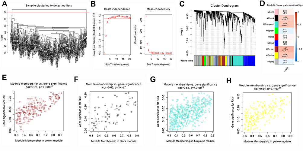 |
Figure 2 Weighted correlation network analysis. (A) Four samples show significantly different expression patterns. (B) Network topologies at different soft-thresholding powers, indicated by the red numbers. (C) Dendrogram of genes clustered based on a dissimilarity measure (1-Topological Overlap Matrix). Each color represents a module in the gene co-expression network constructed by WGCNA. (D) Heatmap of the correlation between module eigengenes and tumor grade. (E–H) Gene significance and module membership correlated significantly with each other in the brown, black, turquoise, and yellow modules. |
Tumor Grade-Related Gene Modules are Involved in Various Pathways
Analysis of GO indicated the genes of these four candidate modules were involved mainly in GOs related to nucleic acid metabolism (Figure 3A). KEGG pathway enrichment analysis showed that the genes were involved tumor-related pathways, such as the p53 signaling pathway, cell cycle, and FoxO signaling pathway (Figure 3B).
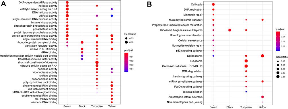 |
Figure 3 Functional enrichment analysis. (A) Gene Ontology analysis and (B) Kyoto Encyclopedia of Genes and Genomes pathway analysis of the four modules. |
The LLPSRI May Be a Prognostic Indicator in HCC
Forty-three LLPS-related genes were considered as OS-associated genes in univariate Cox analysis (Table 1), and LASSO analysis showed nine of them to have non-zero coefficients (Figure 4A and Table 1). A subset of six genes (CDCA8, CENPA, KPNA2, NCL, PRC1, and RAD51) was selected by multivariate stepwise Cox regression to generate the LLPSRI (Table 1). The LLPRRI was significantly associated with OS [hazard ratio (HR) 2.718, 95% confidence interval (CI) 2.160–3.420, P < 0.001]. The patients were divided into high- and low-risk groups according to the median LLPSRI (Figure 4B), and patients in the high-risk group had shorter OS than those in the low-risk group (Figure 4C). In fact, the LLPSRI was associated with OS at an even greater significance level than some clinicopathological characteristics, including sex, age, American Joint Committee on Cancer stage, and vascular invasion (Figure 4D).
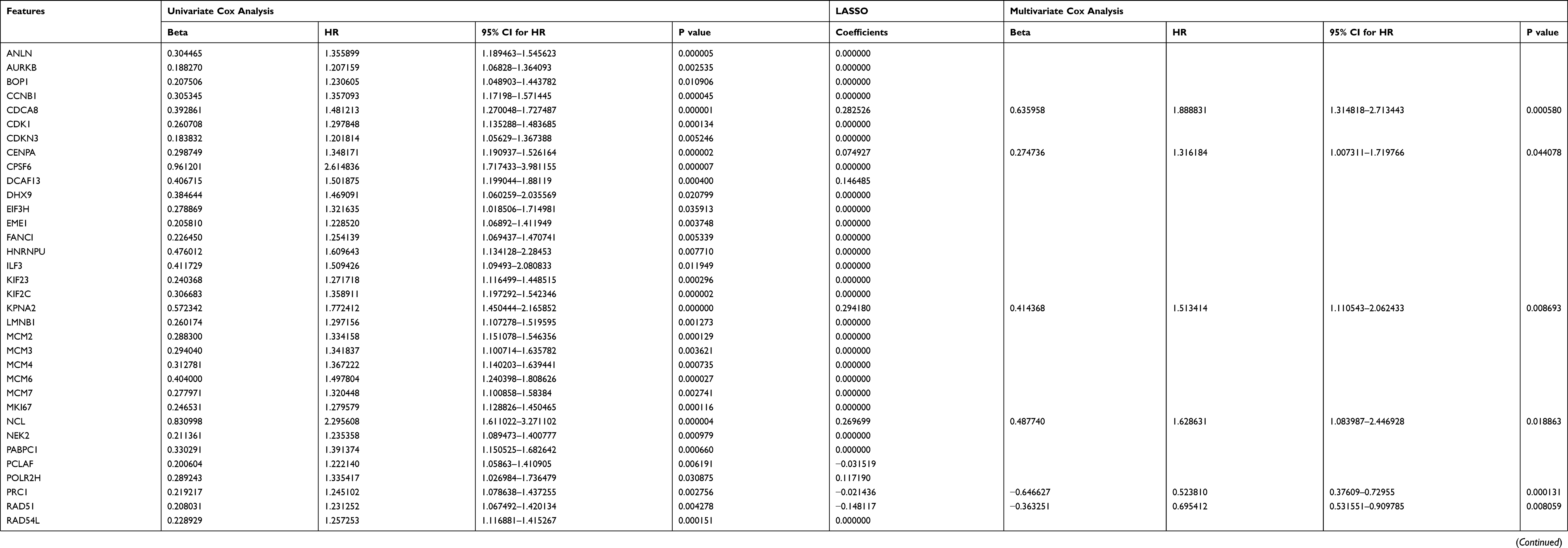 |
Table 1 The Liquid-Liquid Phase Separation-Related Overall Survival-Associated Genes and Coefficients |
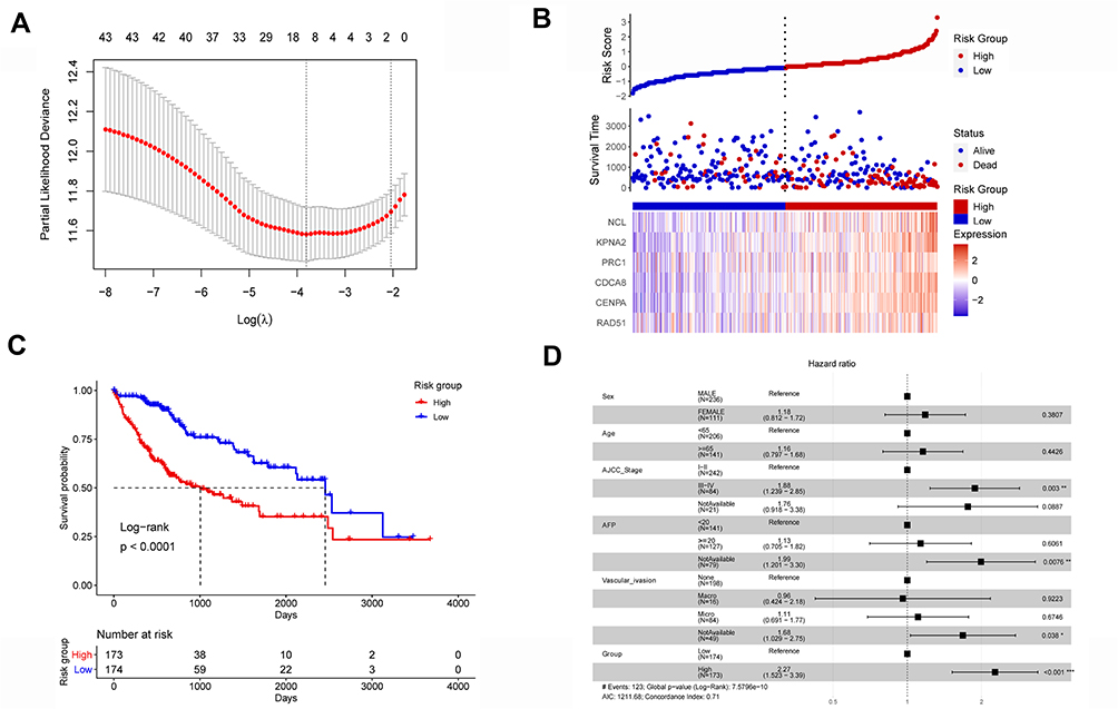 |
Figure 4 The liquid–liquid phase separation-related gene-based risk index (LLPSRI) in the TCGA-LIHC data set. (A) Nine genes were identified as optimal features in LASSO analysis. (B) Risk factor correlation diagram. Top panel: risk predictions for each patient. The horizontal dotted line represents the median risk value, which distinguished a group at low risk (blue) and a group at high risk (red). Middle panel: patients ranked by predicted risk value, showing slightly longer survival time among patients at low risk than among those at high risk. Blue dots represent living patients; red dots, deceased patients. Bottom panel: expression of candidate genes. (C) The high-risk group showed worse overall survival than the low-risk group. (D) The LLPSRI is a prognostic factor independent of routine clinicopathological characteristics. |
Biological Characteristics of Patients in the High-Risk Group
Five hallmark gene sets, hallmark E2F targets, hallmark G2M checkpoint, hallmark MTORC1 signaling, hallmark MYC targets v1, and hallmark unfold protein response, were significantly enriched in the samples of high-risk group (Figure 5A). Two pathways, cell cycle and spliceosome, were significantly enriched in the samples of high-risk group (Figure 5B).
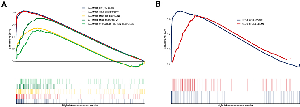 |
Figure 5 Gene set enrichment analysis. (A) Five hallmark and (B) two Kyoto Encyclopedia of Genes and Genomes gene sets were significantly enriched in the high-risk samples. |
Validation of the LLPSRI Using an Independent Data Set
All HCC patients in the LIRI-JP data set were assigned LLPSRIs using the above formula. The LLPRRI was significantly associated with OS (HR 2.939, 95% CI 1.886–4.578, P < 0.001). As we did with patients in the TCGA-LIHC data set, we divided the LIRI-JP patients into high- and low-risk groups according to the median LLPSRI (Figure 6A). Patients in the high-risk group had shorter OS than those in the low-risk group (Figure 6B).
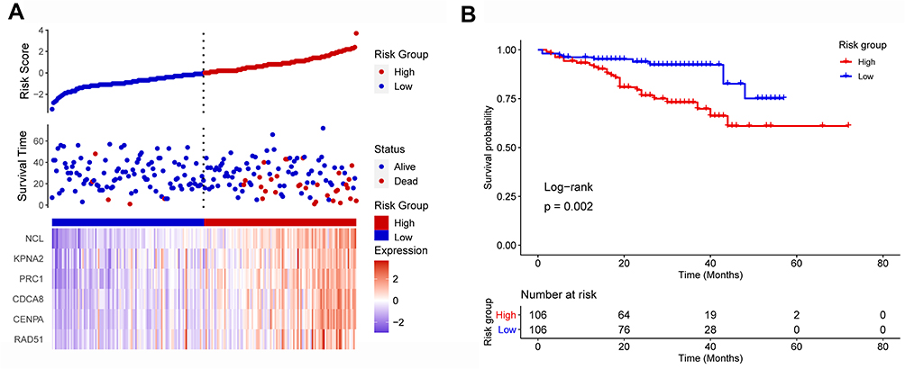 |
Figure 6 Validation of the prognostic value of the liquid–liquid phase separation-related gene-based risk index using an external data set. (A) Patients with HCC were divided into high- and low-risk groups, whose risk score, survival and gene expression were compared. (B) The overall survival rate was higher among low-risk patients than among high-risk patients. |
Discussion
Many gene-based signatures have been proposed for prognostic stratification of HCC.23–25 Lin et al proposed a twenty gene-based prognostic stratification in HCC.18 Lv et al proposed a seven gene-based risk index for predicting early recurrence in HCC.26 Huang et al reported a RNA binding protein-related prognostic signature for HCC.27 These studies have usually not started from subsets of genes with particular biological significance. The present study, in contrast, started from the newly introduced concept of LLPS as important in health and disease. Focusing on genes related to LLPS, we defined several gene modules that were associated with HCC tumor grade. Our LLPSRI was generated using the hub genes in the modules and was found to be associated with OS. Furthermore, its prognostic value was validated with a second, independent data set. These results suggest that LLPS may be a useful concept for explaining the pathogenesis of HCC. The LLPSRI may provide a valuable reference for the personalized management of HCC patients.
According to our GSEA, the target genes of transcription factors E2F and MYC are significantly enriched in high-risk samples, so the networks regulated by these two transcription factors are likely related to LLPS. Previous studies suggested critical roles for E2F and MYC in hepatocarcinogenesis,28–30 which the present analysis supports in the case of HCC. Our results justify further work to elucidate how E2F and MYC are involved in LLPS. In addition, we found that high-risk patients may show greater mTORC1 signaling, which is considered a major tumor-initiating pathway in HCC.31 This suggests that mTOR inhibitors may benefit our HCC patients in the high-risk group, but not those in the low-risk group. Thus, the LLPSRI may be useful for screening patients for potential response when conducting prospective trials of mTOR inhibitors.
Our LLPSRI consists of six LLPS-related genes, some of which have previously been associated with HCC. Aberrant expression of KPNA2 correlates with early recurrence and poor prognosis in patients with small HCC.32 PRC1 promotes early recurrence of HCC in association with the Wnt/β-catenin signalling pathway.33 Silencing CDCA8 can suppress HCC growth and stemness.34 RAD51 appears to influence the onset, progression and drug resistance of various cancers.35 NCL and CENPA should be explored further to confirm and clarify their involvement in HCC.
The present study may provide new insights into the associations of LLPS with HCC tumor grade and prognosis, but several limitations should be noted. Most importantly, our analysis is based exclusively on bioinformatics, so our findings should be verified and extended in experimental studies, ultimately prospective studies with patients. Second, some of the genes that we identified here have yet to be linked to HCC. Indeed, whether the LLPSRI is linked to prognosis through causality or simple correlation needs to be explored.
Conclusion
We identified LLPS-related gene modules that are associated with HCC tumor grade. The LLPSRI may be useful as a prognostic marker of HCC, and it may reliably stratify patients into groups at low or high risk of worse survival. Our bioinformatic analysis also identified some biological characteristics of HCC that may be linked to worse survival. In these ways, our study generates numerous testable hypothesis to help guide future research into the mechanisms and personalized treatment of HCC.
Data Sharing Statement
The raw analyses from this study can be obtained from the corresponding author upon reasonable request.
Funding
This work was supported by the Senior Talent Launch Fund of Wuming Hospital (2019-17), the Self-financing Research Project of the Guangxi Province Health and Family Planning Commission (Z20180668), the Project of the Nanning Scientific Research and Technology Development Plan (20153012), and the Guangxi Graduate Education Innovation Project (YCBZ2018041).
Disclosure
Zhao-Shan Fang and Zhi Zhang are co-first authors for this study. The authors declare that the research was conducted in the absence of any commercial or financial relationships that could be construed as a potential conflict of interest.
References
1. Llovet JM, Zucman-Rossi J, Pikarsky E, et al. Hepatocellular carcinoma. Nat Rev Dis Primers. 2016;2:16018. doi:10.1038/nrdp.2016.18
2. Villanueva A. Hepatocellular Carcinoma. N Engl J Med. 2019;380(15):1450–1462. doi:10.1056/NEJMra1713263
3. Llovet JM, Kelley RK, Villanueva A, et al. Hepatocellular carcinoma. Nat Rev Dis Primers. 2021;7(1):6. doi:10.1038/s41572-020-00240-3
4. Brangwynne CP, Eckmann CR, Courson DS, et al. Germline P granules are liquid droplets that localize by controlled dissolution/condensation. Science. 2009;324(5935):1729–1732. doi:10.1126/science.1172046
5. Shin Y, Brangwynne CP. Liquid phase condensation in cell physiology and disease. Science. 2017;357(6357). doi:10.1126/science.aaf4382
6. Banani SF, Lee HO, Hyman AA, Rosen MK. Biomolecular condensates: organizers of cellular biochemistry. Nat Rev Mol Cell Biol. 2017;18(5):285–298. doi:10.1038/nrm.2017.7
7. Nozawa RS, Yamamoto T, Takahashi M, et al. Nuclear microenvironment in cancer: control through liquid-liquid phase separation. Cancer Sci. 2020;111(9):3155–3163. doi:10.1111/cas.14551
8. Alberti S, Carra S. Quality control of membraneless organelles. J Mol Biol. 2018;430(23):4711–4729. doi:10.1016/j.jmb.2018.05.013
9. Aguzzi A, Altmeyer M. Phase separation: linking cellular compartmentalization to disease. Trends Cell Biol. 2016;26(7):547–558. doi:10.1016/j.tcb.2016.03.004
10. Alberti S, Hyman AA. Are aberrant phase transitions a driver of cellular aging? Bioessays. 2016;38(10):959–968. doi:10.1002/bies.201600042
11. Alberti S, Dormann D. Liquid-liquid phase separation in disease. Annu Rev Genet. 2019;53:171–194. doi:10.1146/annurev-genet-112618-043527
12. Shimazui T, Koiso K, Uchiyama Y. Morphometry of nucleoli as an indicator for grade of malignancy of bladder tumors. Virchows Arch B Cell Pathol Incl Mol Pathol. 1990;59(3):179–183. doi:10.1007/BF02899403
13. Langfelder P, Horvath S. WGCNA: an R package for weighted correlation network analysis. BMC Bioinform. 2008;9:559. doi:10.1186/1471-2105-9-559
14. Ritchie ME, Phipson B, Wu D, et al. limma powers differential expression analyses for RNA-sequencing and microarray studies. Nucleic Acids Res. 2015;43(7):e47. doi:10.1093/nar/gkv007
15. Lian Q, Wang S, Zhang G, et al. HCCDB: a database of Hepatocellular carcinoma expression atlas. Genomics Proteomics Bioinformatics. 2018;16(4):269–275. doi:10.1016/j.gpb.2018.07.003
16. You K, Huang Q, Yu C, et al. PhaSepDB: a database of liquid-liquid phase separation related proteins. Nucleic Acids Res. 2020;48(D1):D354–D359. doi:10.1093/nar/gkz847
17. Yu G, Wang LG, Han Y, He QY. clusterProfiler: an R package for comparing biological themes among gene clusters. OMICS. 2012;16(5):284–287. doi:10.1089/omi.2011.0118
18. Lin Y, Liang R, Ye J, et al. A twenty gene-based gene set variation score reflects the pathological progression from cirrhosis to hepatocellular carcinoma. Aging (Albany NY). 2019;11(23):11157–11169. doi:10.18632/aging.102518
19. Friedman JH, Hastie T, Tibshirani R. Regularization paths for generalized linear models via coordinate. Descent. 2010;33(1):22. doi:10.18637/jss.v033.i01
20. Subramanian A, Tamayo P, Mootha VK, et al. Gene set enrichment analysis: a knowledge-based approach for interpreting genome-wide expression profiles. Proc Natl Acad Sci U S A. 2005;102(43):15545–15550. doi:10.1073/pnas.0506580102
21. Mootha VK, Lindgren CM, Eriksson KF, et al. PGC-1alpha-responsive genes involved in oxidative phosphorylation are coordinately downregulated in human diabetes. Nat Genet. 2003;34(3):267–273. doi:10.1038/ng1180
22. Liberzon A, Subramanian A, Pinchback R, Thorvaldsdottir H, Tamayo P, Mesirov JP. Molecular signatures database (MSigDB) 3.0. Bioinformatics. 2011;27(12):1739–1740. doi:10.1093/bioinformatics/btr260
23. Gu JX, Zhang X, Miao RC, et al. Six-long non-coding RNA signature predicts recurrence-free survival in hepatocellular carcinoma. World J Gastroenterol. 2019;25(2):220–232. doi:10.3748/wjg.v25.i2.220
24. Wang K, Chen X, Jin C, et al. A novel immune-related genes prognosis biomarker for hepatocellular carcinoma. Aging (Albany NY). 2020;13(1):675–693. doi:10.18632/aging.202173
25. Lei J, Zhang D, Yao C, Ding S, Lu Z. Development of a predictive immune-related gene signature associated with hepatocellular carcinoma patient prognosis. Cancer Control. 2020;27(1):1073274820977114. doi:10.1177/1073274820977114
26. Lv Y, Wei W, Huang Z, et al. Long non-coding RNA expression profile can predict early recurrence in hepatocellular carcinoma after curative resection. Hepatol Res. 2018;48(13):1140–1148. doi:10.1111/hepr.13220
27. Huang Y, Chen S, Qin W, et al. A novel RNA binding protein-related prognostic signature for Hepatocellular Carcinoma. Front Oncol. 2020;10:580513. doi:10.3389/fonc.2020.580513
28. Zhan L, Huang C, Meng XM, et al. Promising roles of mammalian E2Fs in hepatocellular carcinoma. Cell Signal. 2014;26(5):1075–1081. doi:10.1016/j.cellsig.2014.01.008
29. Xanthoulis A, Tiniakos DG. E2F transcription factors and digestive system malignancies: how much do we know? World J Gastroenterol. 2013;19(21):3189–3198. doi:10.3748/wjg.v19.i21.3189
30. Zimonjic DB, Popescu NC. Role of DLC1 tumor suppressor gene and MYC oncogene in pathogenesis of human hepatocellular carcinoma: potential prospects for combined targeted therapeutics (review). Int J Oncol. 2012;41(2):393–406. doi:10.3892/ijo.2012.1474
31. Bhat M, Sonenberg N, Gores GJ. The mTOR pathway in hepatic malignancies. Hepatology. 2013;58(2):810–818. doi:10.1002/hep.26323
32. Jiang P, Tang Y, He L, et al. Aberrant expression of nuclear KPNA2 is correlated with early recurrence and poor prognosis in patients with small hepatocellular carcinoma after hepatectomy. Med Oncol. 2014;31(8):131. doi:10.1007/s12032-014-0131-4
33. Chen J, Rajasekaran M, Xia H, et al. The microtubule-associated protein PRC1 promotes early recurrence of hepatocellular carcinoma in association with the Wnt/beta-catenin signalling pathway. Gut. 2016;65(9):1522–1534. doi:10.1136/gutjnl-2015-310625
34. Jeon T, Ko MJ, Seo YR, et al. Silencing CDCA8 suppresses Hepatocellular carcinoma growth and stemness via restoration of ATF3 tumor suppressor and inactivation of AKT/beta-catenin signaling. Cancers (Basel). 2021;13:5. doi:10.3390/cancers13051055
35. Laurini E, Marson D, Fermeglia A, Aulic S, Fermeglia M, Pricl S. Role of Rad51 and DNA repair in cancer: a molecular perspective. Pharmacol Ther. 2020;208:107492. doi:10.1016/j.pharmthera.2020.107492
 © 2021 The Author(s). This work is published and licensed by Dove Medical Press Limited. The full terms of this license are available at https://www.dovepress.com/terms.php and incorporate the Creative Commons Attribution - Non Commercial (unported, v3.0) License.
By accessing the work you hereby accept the Terms. Non-commercial uses of the work are permitted without any further permission from Dove Medical Press Limited, provided the work is properly attributed. For permission for commercial use of this work, please see paragraphs 4.2 and 5 of our Terms.
© 2021 The Author(s). This work is published and licensed by Dove Medical Press Limited. The full terms of this license are available at https://www.dovepress.com/terms.php and incorporate the Creative Commons Attribution - Non Commercial (unported, v3.0) License.
By accessing the work you hereby accept the Terms. Non-commercial uses of the work are permitted without any further permission from Dove Medical Press Limited, provided the work is properly attributed. For permission for commercial use of this work, please see paragraphs 4.2 and 5 of our Terms.