")
Back to Journals » International Journal of Nanomedicine » Volume 13 » T-NANO 2014 Abstracts
Liposomal formulation of vitamin A for the potential treatment of osteoporosis
Authors Sachaniya J, Savaliya R, Goyal R, Singh S
Received 14 October 2016
Accepted for publication 21 November 2016
Published 15 March 2018 Volume 2018:13(T-NANO 2014 Abstracts) Pages 51—53
DOI https://doi.org/10.2147/IJN.S124707
Checked for plagiarism Yes
Review by Single anonymous peer review
Peer reviewer comments 2
Editor who approved publication: Professor Lei Yang
Jainy Sachaniya, Reema Savaliya, Ramesh Goyal, Sanjay Singh
Institute of Life Sciences, School of Science and Technology, Ahmedabad University, Ahmedabad, Gujarat, India
Abstract: Vitamin A targeting to bones is a suitable treatment for osteoporosis. In this study, we have developed vitamin A-encapsulated liposomes that can be useful to deliver vitamin A to the bones in a selective manner. This liposomal formulation of vitamin A has been found to be stable for >6 months as no significant change in size and charge occurred. Vitamin A liposomes-induced cell proliferation in SaOS-2 (human osteosarcoma cell line) and release kinetics study concluded that the liposomal formulation of vitamin A gives a controlled release of vitamin A in comparison to the free vitamin. The MTT assay showed the proliferation of SaOS-2 cells after their treatment with vitamin A liposomes.
Keywords: SaOS-2, nanoliposomes, osteoporosis
Introduction
Osteoporosis is a bone degenerative disease triggered by imbalance in bone remodeling process. There are two types of bone cells involved in bone remodeling: osteoblasts (bone forming) and osteoclasts (bone resorbing). Osteoporosis is generally observed in postmenopausal women and aged persons. Approximately 25 million of Indian population is affected by osteoporesis and the number is increasing continuously. Several studies have shown beneficial effects of vitamin A on bone health.1 It has been reported that excessive, as well as insufficient levels of retinol intake may be associated with compromised bone health.2 In postmenopausal women, it has been shown that TTR limits the transport and absorption of vitamin A in bones.3 This creates a need to target the delivery of vitamin A to bones. This research work endeavors toward the development of vitamin A-liposomal formulation which prevents the toxicity caused by high serum level of free vitamin A. Also, it provides a controlled release of vitamin A and bone cell proliferation.
Materials and methods
Methods
Liposomes (blank and vitamin A-loaded) were prepared by the lipid film hydration method followed by sonication and extrusion. The ratio of vitamin A to phosphatidylcholine per milliliter of 0.9% NaCl was 1:100, 1:10, 1:1, and their substitutes. The so-prepared formulations were analyzed for hydrodynamic size, zeta potential release kinetics, and bone cell proliferation.
Release kinetics
Five milliliters of vitamin A solution (20 mg/mL) in 0.9% NaCl or 5 mL of vitamin A-encapsulated liposomes (1:10 of vitamin A:phosphotidylcholine) were enclosed in a dialysis bag and kept in 400 mL of 0.9% NaCl and stirred on a magnetic stirrer. The samples were withdrawn at different time intervals, that is, 0, 0.5, 1, 2, 3, 6, and 24 hours and were analyzed by recording the absorbance of vitamin A at 325 nm.
Cell culture study
Effects of vitamin A in the free form and its liposomal formulation along with the blank liposomes were assessed on osteoblast cell line by performing the MTT assay.4 The SaOS-2 cells were commercially purchased from National Centre for Cell Sciences, Pune, India. SaOS-2 (human osteosarcoma cell line) cells were seeded into a 96-well plate at a density of 1.0×10⁴ cells/well. After 24 hours, treatment of vitamin A in free and liposomal formulation was given to the cells using 0.78, 1.56, 3.125, 6.25, 12.5, 25, 50, and 100 μg/mL vitamin A. The cell proliferation study was conducted for 24 and 72 hours.
Results and discussion
The liposomes were prepared with different ratios of vitamin A and phosphatidylcholine. Among all the formulations, 1:100 and 1:10 ratios were found to be very stable. The formulation prepared by 1:1 ratio of vitamin A:phosphatidylcholine was found to be turbid which may be due to the formation of emulsion and/or due to lack of total vitamin A encapsulation. Some developed formulation showed aggregation after some time even at 4°C. Further optimization of ratios was done using a 32 full factorial design, by formulating several substitutes of the 1:10 ratio in the range of 0.2–0.6:1–3 mg of vitamin A:phosphatidylcholine. The design showed that the size of liposomes is dependent on the amount of vitamin A but independent of the amount of phosphatidylcholine, whereas the charge was independent of the amount of vitamin A and phosphatidylcholine. All these nine formulations were stable for at least 1 month at 4°C with nonsignificant change in size and zeta potential. For further analysis, the 1:10 ratio was selected as it has more vitamin A encapsulation per amount of phosphatidylcholine as compared to the 1:100 ratio. The blank liposomes showed a hydrodynamic size of 88.88 d.nm and zeta potential of −13.7 mV, whereas vitamin A liposomes showed an increase in size (151 d.nm) which could be due to the encapsulation of vitamin A in liposomes (Figure 1A). No significant change in zeta potential suggests that the encapsulation of vitamin A do not alter the charge on liposomes and thus stability of the liposome even after encapsulation of vitamin A.
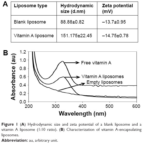 | Figure 1 (A) Hydrodynamic size and zeta potential of a blank liposome and a vitamin A liposome (1:10 ratio). (B) Characterization of vitamin A-encapsulating liposomes. |
A further study was conducted by following the absorbance pattern of vitamin A-encapsulated liposomes using a ultraviolet–visible spectrophotometer. As it is evident from Figure 1B that pure vitamin A, suspended in isopropyl alcohol, shows an absorbance at ~320 nm. Similarly, vitamin A-encapsulating liposomes also show an absorbance at ~320 nm, which further suggests the possible incorporation of vitamin A in liposomes. The empty liposome did not show any absorbance around 320 nm region, which suggests that the vitamin A is encapsulated in liposomes.
Release kinetics of free vitamin A showed that about 95% vitamin A was released within 24 hours, whereas in the case of liposomal formulation of vitamin A, the release was controlled and only 50% of vitamin A was released. Further, ~80% of free vitamin A was released within 5 hours; however, the encapsulated vitamin A showed only 40% release (Figure 2).
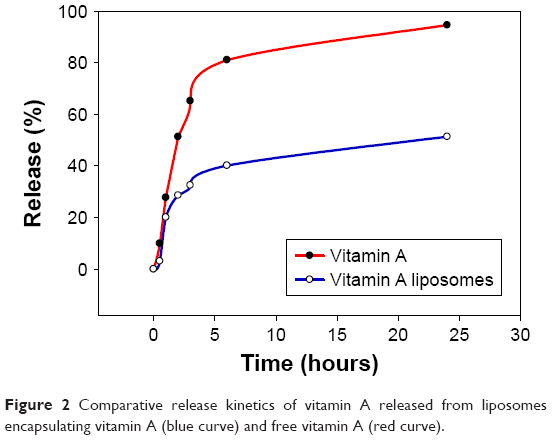 | Figure 2 Comparative release kinetics of vitamin A released from liposomes encapsulating vitamin A (blue curve) and free vitamin A (red curve). |
The effect of vitamin A over the proliferation of osteoblast cells was estimated by the MTT assay (Figure 3). Different concentrations of vitamin A (25, 50, and 100 μg/mL) were exposed to SaOS-2 cells. As evident from Figure 2, after 24 hours vitamin A (25 μg/mL) alone did not show much cell proliferation; however, the same concentration of vitamin A encapsulated in liposome showed an ~1.2-fold increase in cell proliferation. Interestingly, after 72 hours the vitamin A liposome induced ~1.5-fold increase in cell proliferation than the corresponding concentration of free vitamin A. Further, the blank liposomes did not induce any decrease in cell proliferation, suggesting that the blank liposomes are nontoxic.
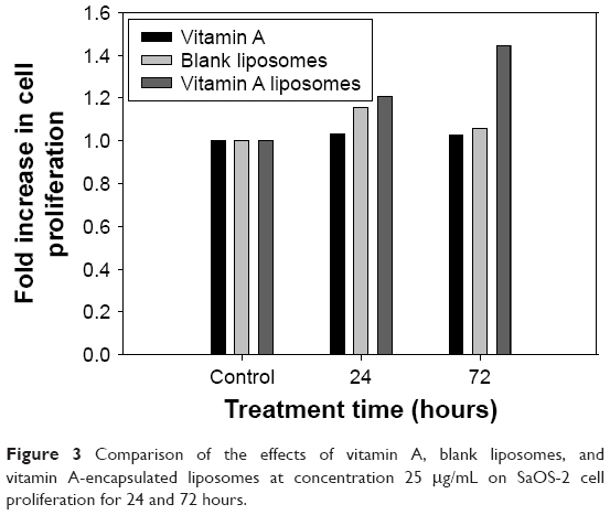 | Figure 3 Comparison of the effects of vitamin A, blank liposomes, and vitamin A-encapsulated liposomes at concentration 25 μg/mL on SaOS-2 cell proliferation for 24 and 72 hours. |
Conclusion
The synthesized vitamin A liposomes (151 d.nm, −13.7 mV) were found to be stable for >1 month at 4°C. Liposomes provided a controlled release of vitamin A which was almost 50% slower than the free form. Osteoblast cells exposed to vitamin A liposomes showed a 45%–50% increase in the growth of SaOS-2 cells than free vitamin A after 72 hours.
Acknowledgments
The financial assistance for the Centre for Nanotechnology Research and Applications (CENTRA) by The Gujarat Institute for Chemical Technology is gratefully acknowledged. RS would like to thank University Grant Commission (UGC), New Delhi, India, for the award of Junior Research Fellowship. The manuscript contains ILS-manuscript No 044.
Disclosure
The authors report no conflicts of interest in this work.
References
Tanumihardjo SA. Vitamin A and bone health: the balancing act. J Clin Densitom. 2013;16(4):414–419. | ||
Ahmadieh H, Arabi A. Vitamins and bone health: beyond calcium and vitamin D. Nutr Rev. 2011;69(10):584–598. | ||
Chupeerach C, Harnroongroj T, Phonrat B, et al. Decreased retinol transport proteins in Thai post-menopausal women with osteoporosis. Southeast Asian J Trop Med Public Health. 2011;42(6):1515–1520. | ||
Mosmann T. Rapid colorimetric assay for cellular growth and survival: application to proliferation and cytotoxicity assays. J Immunol Methods. 1983;65(1–2):55–63. |
 © 2018 The Author(s). This work is published and licensed by Dove Medical Press Limited. The full terms of this license are available at https://www.dovepress.com/terms.php and incorporate the Creative Commons Attribution - Non Commercial (unported, v3.0) License.
By accessing the work you hereby accept the Terms. Non-commercial uses of the work are permitted without any further permission from Dove Medical Press Limited, provided the work is properly attributed. For permission for commercial use of this work, please see paragraphs 4.2 and 5 of our Terms.
© 2018 The Author(s). This work is published and licensed by Dove Medical Press Limited. The full terms of this license are available at https://www.dovepress.com/terms.php and incorporate the Creative Commons Attribution - Non Commercial (unported, v3.0) License.
By accessing the work you hereby accept the Terms. Non-commercial uses of the work are permitted without any further permission from Dove Medical Press Limited, provided the work is properly attributed. For permission for commercial use of this work, please see paragraphs 4.2 and 5 of our Terms.