")
Back to Journals » International Journal of Nanomedicine » Volume 11
Inhibition of E. coli and S. aureus with selenium nanoparticles synthesized by pulsed laser ablation in deionized water
Authors Guisbiers G, Wang Q, Khachatryan E, Mimun L, Mendoza-Cruz R, Larese-Casanova P, Webster T , Nash K
Received 12 February 2016
Accepted for publication 10 May 2016
Published 8 August 2016 Volume 2016:11 Pages 3731—3736
DOI https://doi.org/10.2147/IJN.S106289
Checked for plagiarism Yes
Review by Single anonymous peer review
Peer reviewer comments 3
Editor who approved publication: Professor Carlos Rinaldi
G Guisbiers,1 Q Wang,2 E Khachatryan,1 LC Mimun,1 R Mendoza-Cruz,1 P Larese-Casanova,3 TJ Webster,2,4,5 KL Nash1
1Department of Physics and Astronomy, The University of Texas at San Antonio, San Antonio, TX, 2Department of Bioengineering, 3Department of Civil and Environmental Engineering, 4Department of Chemical Engineering, Northeastern University, Boston, MA, USA; 5Center of Excellence for Advanced Materials Research, King Abdulaziz University, Jeddah, Saudi Arabia
Abstract: Nosocomial diseases are mainly caused by two common pathogens, Escherichia coli and Staphylococcus aureus, which are becoming more and more resistant to conventional antibiotics. Therefore, it is becoming increasingly necessary to find other alternative treatments than commonly utilized drugs. A promising strategy is to use nanomaterials such as selenium nanoparticles. However, the ability to produce nanoparticles free of any contamination is very challenging, especially for nano-medical applications. This paper reports the successful synthesis of pure selenium nanoparticles by laser ablation in water and determines the minimal concentration required for ~50% inhibition of either E. coli or S. aureus after 24 hours to be at least ~50 ppm. Total inhibition of E. coli and S. aureus is expected to occur at 107±12 and 79±4 ppm, respectively. In this manner, this study reports for the first time an easy synthesis process for creating pure selenium to inhibit bacterial growth.
Keywords: nosocomial disease, bacteria, antibiotics resistant, cytotoxicity
Introduction
Escherichia coli is a gram-negative bacteria that is commonly present in the intestines of humans and animals, while Staphylococcus aureus is a gram-positive bacteria frequently found in the human respiratory tract and on the skin. These bacteria are the two main pathogens responsible of nosocomial diseases,1,2 which are also called hospital-acquired infections and defined as infections occurring within 48 hours of hospital admission, 3 days of discharge, or 30 days after an operation. Infections due to bacteria causing nosocomial diseases are difficult to treat, as they are drug resistant to a large group of antibiotics.3,4 In the US, two million people are infected each year by nosocomial diseases, of whom, 5% die.5 Therefore, it is becoming increasingly necessary to develop new strategies to fight nosocomial infections and the solution may come from nanotechnology, where the particle size is <100 nm, exhibiting a large surface-to-volume ratio and therefore especially adapted to interact with bacteria.6 The benefit of using selenium as a therapeutic drug is that selenium is already present in our body as a trace element7 and has very interesting biological assets, such as anticancer8,9 and antibacterial properties.10 It belongs to the oxygen family (group 16 in the periodic table); therefore, it does not oxidize in air and is insoluble in water. Pulsed laser ablation in liquids (PLAL) has several advantages compared to the other conventional methods such as wet chemistry routes, physical vapor deposition, or chemical vapor deposition to synthesize selenium.11 Indeed, the surface of the synthesized nanoparticles is absent of unnecessary adducts and byproducts, and the produced nanoparticles are easy to collect and store as a colloidal solution, and the setup is simple. Furthermore, it does not require any vacuum chamber or clean room environment.
Only four papers in the literature report the synthesis of selenium nanoparticles by PLAL.12–15 The first indication of the antibacterial properties of selenium nanoparticles synthesized by laser ablation was reported by our group using a neodymium-doped yttrium aluminum garnet laser at 355, 532, and 1,064 nm.15 During that study, we could not decrease the E. coli density by a significant percentage due to a low concentration of selenium nanoparticles used in solution (1.35 ppm). Therefore, a new vessel/target combination has been created to increase selenium concentration. The goal of this communication is to report a ~46% decrease in E. coli and a ~63% decrease in S. aureus densities by using pure selenium nanoparticles synthesized by pulsed laser ablation in deionized (DI) water.
Materials and methods
Materials
Selenium pellets (Se, diameter <5 mm, purity ≥99.99% based on trace metals analysis) and sodium hydroxide (NaOH, American Chemical Society reagent, ≥97.0%, pellets) were purchased from Sigma-Aldrich (St Louis, MO, USA).
Synthesis of selenium nanoparticles
The selenium nanoparticles were produced by irradiating pure selenium pellet placed at the bottom of a 1.5 mL microcentrifuge tube filled with 0.5 mL of DI water. The laser used for the irradiation was the neodymium-doped yttrium aluminum garnet NT342B (EKSPLA, Vilnius, Lithuania) with a pulse duration of 3.6 ns and a 20 Hz repetition rate. Each pulse, having a top hat profile, delivers ~20 mJ/pulse. The laser beam was focused on the surface of the selenium pellet, which corresponds to a fluence of 2.5 J/cm2. The irradiation time was fixed at 15 minutes, and the ultraviolet wavelength was chosen to be at 355 nm to produce a more stable colloidal solution compared to visible or infrared wavelengths.15 The conical shape of the cuvette helps to reduce the amount of water required in the vessel compared to flat squared cuvettes used previously,15 while at the same time, maintaining enough height of water above the target and preventing evaporation during irradiation.
Concentration analysis
The selenium concentration was determined by dissolving 1 mL of the colloidal solution into 10 M NaOH and then diluting it 100× before being analyzed by inductively coupled plasma mass spectrometry (ICP-MS, Bruker Aurora M90; Bruker Corporation, Billerica, MA, USA). Previously, it has been reported by Van Overschelde and Guisbiers16 that the production of nanoparticles was larger for a bulk target compared to a powder target.
Morphology
The size distribution and zeta potential were determined after decantation by dynamic light scattering (DLS, Zetasizer Nano ZS from Malvern Instruments, Malvern, UK) at 25°C. Transmission electronic microscope (TEM, JEOL 1230, Tokyo, Japan at 120 kV) images were taken to determine the size and shape of the selenium nanoparticles. Energy dispersive X-ray analysis was done using the scanning electron microscope (SEM, Hitachi STEM S5500; Hitachi Ltd., Tokyo, Japan) at 30 kV.
Cytotoxicity assay
A stock solution of selenium nanoparticles was diluted to the desired concentrations ranging from 50 to 1 ppm in growth medium and subsequently added into 96-well plates containing ARPE-19 cells (5×10⁴ cells/well). Microtiter plates were incubated at 37°C in a 5% CO2 air humidified atmosphere for 24 hours. Assessment of cell viability was carried out using a CellTiter-Glo® Luminescent Cell Viability Assay (Promega Corporation, Fitchburg, WI, USA).
E. coli and S. aureus culture and treatment
Bacterial cell lines of biofilm-producing E. coli and S. aureus were obtained in freeze-dried form from the American Type Culture Collection (8739 and 25923, respectively; ATCC, Manassas, VA, USA). The cells were propagated in 30 mg/mL of tryptic soy broth (TSB) (MP Biomedicals, Solon, OH, USA). Once the second passage of bacteria reached its stationary phase, the second passage was frozen in one part TSB and one part 50% glycerol (Sigma-Aldrich). All experiments were conducted from this frozen stock. One day before bacterial seeding, a sterile 10 μL loop was used to withdraw bacteria from the frozen stock and streaked onto a TSB agar plate and incubated at 37°C for 16 hours. Bacteria from a single colony were then collected using a sterile loop and inoculated in a test tube containing 3 mL of TSB overnight. The test tube was agitated in a shaking incubator at 37°C, 250 rpm to achieve a bacterial solution at the exponential phase of growth.
Antibacterial tests
The antibacterial test was performed on E. coli and S. aureus by using the broth dilution method.17 Various concentrations of selenium nanoparticles and bacteria were added to the plate and incubated for either 4, 8, or 24 hours. The survival bacteria numbers were then calculated by measuring the optical density of the bacterial solution at 562 nm using a standard curve correlating optical densities and bacterial concentrations.
Results
Selenium nanoparticles were synthesized by the PLAL of selenium pellets within a 1.5 mL conical microcentrifuge tube filled with 0.5 mL of DI water using a 3.6 ns pulse neodymium-doped yttrium aluminum garnet laser at 20 Hz in the ultraviolet wavelength of 355 nm. This configuration allowed us to produce a 50 ppm solution compared to a previous study where only 1.35 ppm was prepared.15 The stability of the colloidal solution was evaluated by measuring its zeta potential, that is, the electric potential surrounding the particle. If the zeta potential is higher than the absolute value of ±30 mV, then the solution is considered stable, while below this value, it is unstable and tends to flocculate. In this article, the zeta potential of the selenium nanoparticles was measured at approximately −45.6 mV, which is very stable (Figure 1). The average size of the nanoparticle was determined by DLS to be around 115±38 nm (inset, Figure 1). From our TEM analysis, just after synthesis, the nanoparticles were observed to be spherical with a high polydispersity (Figure 2A). The discrepancy observed between TEM and DLS size distributions can be explained as follows: the size measured by DLS (called hydrodynamic diameter) is representative of the size of a hypothetical sphere that diffuses at the same rate as the particles being measured, while the size measured from TEM observations really informed the physical size of the particle. Indeed, the Rayleigh approximation tells us that the scattered intensity is proportional to d6 where d is the particle diameter and, therefore, the light scattered by the largest particles may hide the light scattered by the smallest ones. As an example, if we consider two particles, one with a diameter ten times larger than the other one, the d6 factor tell us that the largest particle will scatter 106 more light than the smallest one. Moreover, the purity of the selenium nanoparticles produced by PLAL was confirmed by carrying out energy dispersive X-ray analysis (line scans) through those particles (Figure 2B).
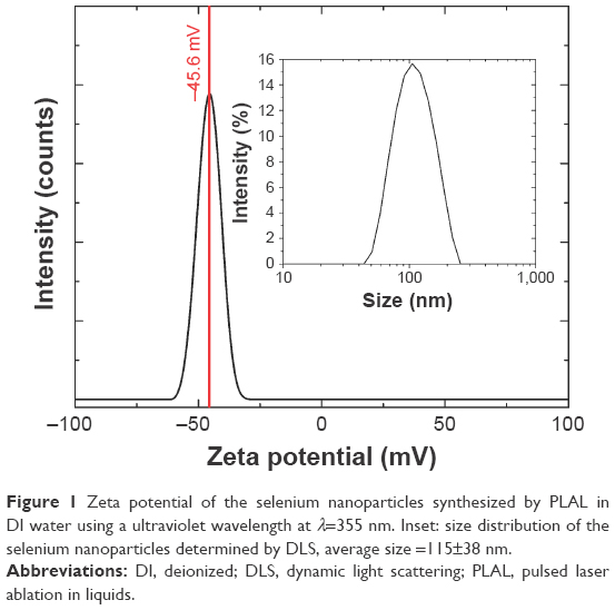 | Figure 1 Zeta potential of the selenium nanoparticles synthesized by PLAL in DI water using a ultraviolet wavelength at λ=355 nm. Inset: size distribution of the selenium nanoparticles determined by DLS, average size =115±38 nm. |
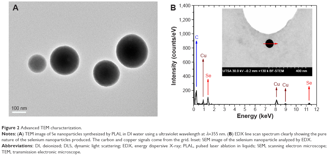 | Figure 2 Advanced TEM characterization. |
Various concentrations of selenium nanoparticles were subjected into E. coli and S. aureus bacteria plated and incubated for either 4, 8, or 24 hours. The most concentrated selenium sample (50 ppm) exhibited the highest inhibition rate with 46% and 63% of E. coli and S. aureus growth inhibition after 24 hours, respectively (Figure 3). To survive, bacteria have evolved a sophisticated and complex cell envelope that protects them, but allows for the selective passage of nutrients from the outside and waste products from the inside. A possible mechanism toward inhibiting E. coli bacteria is that selenium nanoparticles attach by chemisorption18 and penetrate the outer membrane that contains lipopolysaccharides, linked by a covalent bond to the cell’s peptidoglycan by Braun’s lipoprotein.19 The canonical biosynthetic pathway of lipoproteins in the E. coli bacteria involves three enzymes, including preprolipoprotein diacylglyceryl transferase, prolipoprotein signal peptidase, and apolipoprotein N-acyltransferase, which have been shown to play an essential role in the survival of E. coli.20 Therefore, selenium nanoparticles inhibit E. coli by modifying the role of these enzymatic conveyors. By contrast, in gram-positive bacteria, such as S. aureus, the cell wall structure is different, ie, it has a thicker peptidoglycan membrane without any outer lipopolysaccharide membrane. Consequently, selenium penetrates much more easily into the S. aureus bacteria by chemisorption, where the lipoproteins involved are of the diacyl and triacyl forms.20 Therefore, it can be concluded that the cell wall and its polysaccharide components constitute a barrier that reduces the penetration of selenium nanoparticles into the cell interior. Indeed, from Figure 3A, the total inhibition of E. coli and S. aureus is expected to occur at 107±12 and 79±4 ppm, respectively. It is confirmed that selenium nanoparticles can inhibit both gram-negative and gram-positive bacteria with a higher efficiency against gram-positive bacteria (slope_E. coli =0.92±0.08 and slope_S. aureus =1.23±0.04). Finally, the minimal concentration required to ~50% bacterial inhibition (E. coli or S. aureus) after 24 hours should be at a minimum 50 ppm (Figure 3B and 3C).
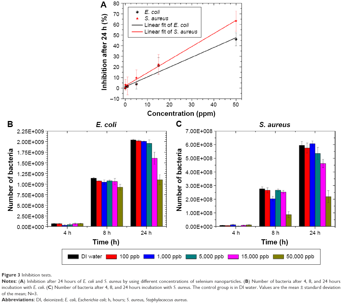 | Figure 3 Inhibition tests. |
Discussion
Our results agree with a recent study made by Tran et al21 stipulating that selenium nanoparticles affect S. aureus in a more efficient way than E. coli. However, disagreement arises with the claim that selenium nanoparticles do not show any significant inhibitory effect on E. coli where we obtained 46% inhibition after 24 hours of incubation. The difference may be due to the selenium synthesis process, since their selenium nanoparticles were produced by a chemical reduction of sodium selenite, whereas we produced selenium nanoparticles by PLAL in DI water. Therefore, the surface contamination that may happen during chemical synthesis could affect their surface and consequently their efficiency.
Apart from selenium, silver is also reported to have excellent antibacterial properties. However, silver is not present naturally in the human body, and the toxicity of silver nanoparticles toward bacteria and human cells is similar;22 while selenium being a trace element found naturally in the body has been demonstrated to be much less toxic. Indeed, our cytotoxicity tests on ARPE-19 cells demonstrate that viable cells decrease slightly by increasing the dose of Se nanoparticles, but >70% of the cells were still alive at concentrations ~50 ppm (Figure 4). This result is in agreement with another study where a cytotoxicity test on 3T3 fibroblasts demonstrated that >70% of cells were still alive at a concentration as high as ~128 ppm.21
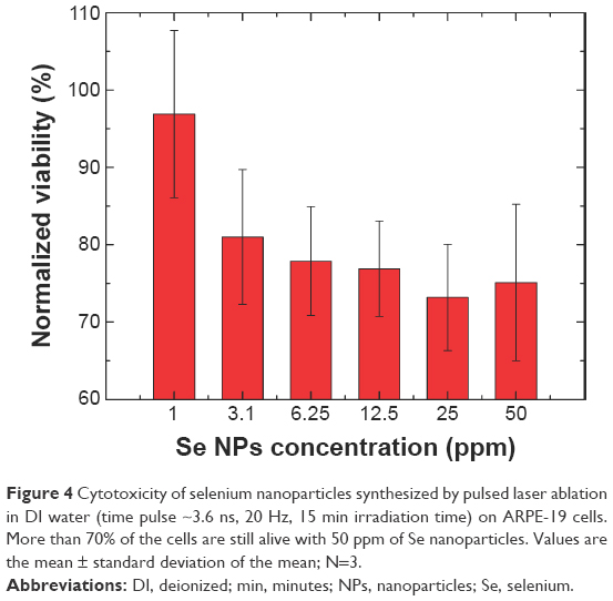 | Figure 4 Cytotoxicity of selenium nanoparticles synthesized by pulsed laser ablation in DI water (time pulse ~3.6 ns, 20 Hz, 15 min irradiation time) on ARPE-19 cells. More than 70% of the cells are still alive with 50 ppm of Se nanoparticles. Values are the mean ± standard deviation of the mean; N=3. |
Conclusion
Based on the results of bacteria assays after 4, 8, and 24 hours, selenium nanoparticles significantly decreased the number of E. coli and S. aureus bacteria. This study showed that selenium nanoparticles, synthesized by pulsed laser ablation in DI water, can be used efficiently as an antibacterial therapeutic. Most importantly, these results demonstrate that decreasing the density of bacteria can be achieved without the use of antibiotics. Nevertheless, further work is still in progress to confirm those results by using the disc diffusion method.
Acknowledgments
The authors (GG and KLN) would like to acknowledge grant support from the Biomedical Research Grants Program of the San Antonio Area Foundation. The authors (EK and LCM) would like to acknowledge support from the Air Force Office of Scientific Research (FA9550-15-1-0109). Brandy Vincent is greatly acknowledged for her help in conducting the cytotoxicity tests.
Disclosure
The authors report no conflicts of interest in this work.
References
Inweregbu K, Dave, J, Pittard A. Nosocomial infections. Cont Educ Anaesth Crit Care Pain. 2005;5(1):14–17. | ||
Jarvis WR, Martone WJ. Predominant pathogens in hospital infections. J Antimicrob Chemoth. 1992;29(Suppl A):19–24. | ||
Pollack A. Rising threat of infections unfazed by antibiotics. New York Times; 2010. | ||
Chambers HF, Deleo FR. Waves of resistance: Staphylococcus aureus in the antibiotic era. Nat Rev Microbiol. 2009;7(9):629–641. | ||
Kaufmann SH. The contribution of immunology to the rational design of novel antibacterial vaccines. Nat Rev Microbiol. 2007;5(7):491–504. | ||
Zhu X, Radovic-Moreno AF, Wu J, Langer R, Shi J. Nanomedicine in the management of microbial infection – overview and perspectives. Nano Today. 2014; 9(4):478–498. | ||
Rayman MP. Selenium and human health. Lancet. 2012;379(9822): 1256–1268. | ||
Rayman MP. Selenium in cancer prevention: a review of the evidence and mechanism of action. Proc Nutr Soc. 2005;64(4):527–542. | ||
Ramamurthy Ch, Sampath KS, Arunkumar P, et al. Green synthesis and characterization of selenium nanoparticles and its augmented cytotoxicity with doxorubicin on cancer cells. Bioprocess Biosyst Eng. 2013;36(8):1131–1139. | ||
Tran PA, Webster TJ. Antimicrobial selenium nanoparticle coatings on polymeric medical devices. Nanotechnology. 2013;24(15): 155101. | ||
Zeng H, Du X-W, Singh SC, et al. Nanomaterials via laser ablation/irradiation in liquid: a review. Adv Funct Mater. 2012;22:1333–1353. | ||
Singh SC, Mishra SK, Srivastava, RK, Gopal R. Optical properties of selenium quantum dots produced with laser irradiation of water suspended Se nanoparticles. J Phys Chem C. 2010;114:17374–17384. | ||
Kuzmin PG, Shafeev GA, Voronov VV, et al. Bioavailable nanoparticles obtained in laser ablation of a selenium target in water. Quant Electron. 2012;42(11):1042–1044. | ||
Van Overschelde O, Guisbiers G, Snyders R. Green synthesis of selenium nanoparticles by excimer pulsed laser ablation in water. APL Mater. 2013;1:042114. | ||
Guisbiers G, Wang Q, Khachatryan E, et al. Anti-bacterial selenium nanoparticles produced by UV/VIS/NIR pulsed nanosecond laser ablation in liquids. Laser Phys Lett. 2015;12(1):016003. | ||
Van Overschelde O, Guisbiers G. Photo-fragmentation of selenium powder by excimer laser ablation in liquids. Opt Laser Technol. 2015;73:156–161. | ||
Wiegand I, Hilpert K, Hancock RE. Agar and broth dilution methods to determine the minimal inhibitory concentration (MIC) of antimicrobial substances. Nat Protoc. 2008;3(2):163–175. | ||
Kieliszek M, Błażejak S, Gientka I, Bzducha-Wróbel A. Accumulation and metabolism of selenium by yeast cells. Appl Microbiol Biotechnol. 2015;99(13):5373–5382. | ||
Silhavy TJ, Kahne D, Walker S. The bacterial cell envelope. Cold Spring Harb Perspect Biol. 2010;2(5):a000414. | ||
Nakayama H, Kurokawa K, Lee BL. Lipoproteins in bacteria: structures and biosynthetic pathways. FEBS J. 2012;279(23):4247–4268. | ||
Tran PA, O’Brien-Simpson N, Reynolds EC, Pantarat N, Biswas DP, O’Connor AJ. Low cytotoxic trace element selenium nanoparticles and their differential antimicrobial properties against S. aureus and E. coli. Nanotechnology. 2016;27(4):045101. | ||
Greulich C, Braun D, Peetsch A, et al. The toxic effect of silver ions and silver nanoparticles towards bacteria and human cells occurs in the same concentration range. RSC Adv. 2012;2(17):6981–6987. |
 © 2016 The Author(s). This work is published and licensed by Dove Medical Press Limited. The full terms of this license are available at https://www.dovepress.com/terms.php and incorporate the Creative Commons Attribution - Non Commercial (unported, v3.0) License.
By accessing the work you hereby accept the Terms. Non-commercial uses of the work are permitted without any further permission from Dove Medical Press Limited, provided the work is properly attributed. For permission for commercial use of this work, please see paragraphs 4.2 and 5 of our Terms.
© 2016 The Author(s). This work is published and licensed by Dove Medical Press Limited. The full terms of this license are available at https://www.dovepress.com/terms.php and incorporate the Creative Commons Attribution - Non Commercial (unported, v3.0) License.
By accessing the work you hereby accept the Terms. Non-commercial uses of the work are permitted without any further permission from Dove Medical Press Limited, provided the work is properly attributed. For permission for commercial use of this work, please see paragraphs 4.2 and 5 of our Terms.