")
Back to Journals » Infection and Drug Resistance » Volume 12
High rate of multiresistant Klebsiella pneumoniae from human and animal origin
Authors Yang F , Deng B, Liao W, Wang P, Chen P, Wei J
Received 12 June 2019
Accepted for publication 16 August 2019
Published 3 September 2019 Volume 2019:12 Pages 2729—2737
DOI https://doi.org/10.2147/IDR.S219155
Checked for plagiarism Yes
Review by Single anonymous peer review
Peer reviewer comments 2
Editor who approved publication: Dr Joachim Wink
Fan Yang1,2, Baoguo Deng1, Wei Liao3, Peizhen Wang1, Ping Chen1, Jidong Wei1
1Department of Microbiology, School of Basic Medical Science, Xinxiang Medical University, Xinxiang, People’s Republic of China; 2Xinxiang Key Laboratory of Pathogenic Biology, Xinxiang, People’s Republic of China; 3Department of Clinical Laboratory, The Affiliated People’s Hospital of Xinxiang Medical University, Xinxiang, People’s Republic of China
Correspondence: Fan Yang
Department of Microbiology, School of Basic Medical Science, Xinxiang Medical University, JinSui Road, Xinxiang city, Henan province, People’s Republic of China
Tel +86 373 302 9130
Email [email protected]
Purpose: The main objectives of the present study were to detect the antimicrobial susceptibility and molecular characteristics of Klebsiella pneumoniae isolated from different hosts and to investigate the possibility of K. pneumoniae transmission between animals and humans.
Materials and methods: A total of 189 nonduplicate K. pneumoniae isolates were collected from hospitals and four species of animals in Henan Province, China. The disk diffusion method was used for antimicrobial susceptibility testing, and resistance and virulence genes were screened by polymerase chain reaction (PCR). The molecular types were identified through multilocus sequence typing (MLST), and the hypermucoviscous (HMV) phenotype was identified using the “string-forming test”. Pearson’s parameters were used to determine the potential link among the molecular types and resistance and virulence genes of all K. pneumoniae strains.
Results: The resistance rates of the 189 K. pneumoniae isolates against 15 antibiotics ranged from 11.6% to 77.8%. The highest multidrug resistance rate was detected in the pig strains (93.6%), followed by the human strains (90.4%), chicken strains (88.9%), cow strains (52.0%) and sheep strains (50.0%). Forty-eight (25.4%) K. pneumoniae strains presented the HMV phenotype. entB, fimH-1 and mrkD were the most prevalent of the detected virulence genes, and magA and rmpA were the least prevalent genes in all the isolates. The MLST analysis revealed 24 unique sequence types (STs) among from the 189 isolates. ST11, ST235 and ST258 were common STs among the five isolates of host origin. ST258 exhibited significantly positive correlations with blaNDM, magA and the HMV phenotype and a negative correlation with qnrB.
Conclusion: K. pneumoniae strains from different hosts, including humans and animals, have common molecular types and similar phenotypes, and these strains can potentially be transmitted between humans and animals.
Keywords: K. pneumoniae, antimicrobial resistance, virulence genes, sequence types
Introduction
Klebsiella pneumoniae is an important facultative pathogen that causes hospital and community infections.1 According to data provided by the Chinese antimicrobial resistance surveillance system (CARSS), K. pneumoniae is the second most prevalent (20.2%) among the isolated gram-negative pathogens.2 An increasing number of studies have shown that K. pneumoniae also causes a variety of animal diseases, including pneumonia, bacteremia, and septicemia.3–5 Due to the extensive use and abuse of antimicrobial agents for promoting growth and treating diseases in animals, K. pneumoniae has become severely resistant to most antibiotic agents. In particular, the emergence of pan-resistant strains, hypervirulent strains and multidrug resistant (MDR) strains has caused great challenges for the prevention and treatment of infections caused by K. pneumoniae.6–10 The aims of this study were to obtain a better understanding of the molecular epidemiological characteristics of K. pneumoniae and to assess the likelihood of K. pneumoniae transmission between humans and animals. Here, we report the drug-resistance profile and molecular characteristics of K. pneumoniae isolated from different host origins in Henan Province, China.
Materials and methods
Bacterial isolates and identification
A total of 137 strains of K. pneumoniae were isolated from cows (n=25), pigs (n=47), sheep (n=20) and chickens (n=45) at large-scale farms and animal hospitals in Henan Province, China, from March to December 2017. Moreover, 52 strains of nosocomial K. pneumoniae were isolated from the teaching hospital of Xinxiang Medical University, and they were part of the routine hospital laboratory procedure of the hospital. All isolated strains were identified through staining, biochemical tests and the VITEK-2 compact system (bioMerieux, Craponne, France). K. pneumoniae ATCC 700603 was used as the quality control strain for species identification.
Detection of the mucous phenotype and extended-spectrum beta-lactamase (ESBL) bacterial phenotype
The hypermucoviscous (HMV) phenotype was tested using the “string-forming test” as previously described.11 Briefly, K. pneumoniae was inoculated onto Columbia blood agar plates, which were incubated overnight at 37 °C, and a loop bacterial colony was used for stretching. If the length of the viscous string was greater than 5 mm, the sample was considered positive for the HMV phenotype. The ESBL phenotype was detected by a double disk synergy test according to the description provided by the Clinical and Laboratory Standards Institute (CLSI 2018).
Antimicrobial susceptibility testing
The antibiotic susceptibility of K. pneumoniae was tested using the diffusion method on Mueller-Hinton agar according to the recommendations provided by the Clinical and Laboratory Standards Institute (CLSI 2018). The following 15 commonly used antibiotic agents were tested: amikacin (AK), ampicillin (AMP), amoxicillin-clavulanic acid (AMC), ceftazidime (CAZ), cefotaxime (CTX), kanamycin (KAN), clindamycin (CLI), azithromycin (AZM), erythromycin (ERY), ciprofloxacin (CIP), gatifloxacin (GAT), tetracycline (TCY), imipenem (IMP), meropenem (MEM), and vancomycin (VAN). The results of the antibiotic susceptibility tests were interpreted according to the guidelines provided by the CLSI (2018). Escherichia coli ATCC 25922 was used as the quality control strain for antimicrobial susceptibility testing.
Detection of resistance- and virulence-associated genes
Genomic DNA from all the isolates was extracted using a bacterial genomic DNA mini prep kit (Tiangen Biotech, Beijing, China) according to the manufacturer’s instructions. Polymerase chain reaction (PCR) was performed to amplify the resistance genes blaKPC, blaNDM, blaCTX-M, blaTEM, blaSHV, qnrA and qnrB and five virulence genes, namely, magA, rmpA, MrkD, fimH-1 and entB. The primers for the resistance and virulence genes were designed as previously described,12–14 and the positive PCR products were sequenced by Majorbio Bio-Pharm Technology Co. (Shanghai, China). The sequencing results were analyzed and compared using NCBI BLAST (http://www.ncbi.nlm.nih.gov/Blast).
Multilocus sequence typing (MLST)
Seven housekeeping genes (rpoB, gapA, mdh, pgi, phoE, infB and tonB) from 189 K. pneumoniae isolates were amplified by PCR, and MLST was performed according to the protocol on the MLST website (https://bigsdb.pasteur.fr/klebsiella/klebsiella.html).
Results
Antimicrobial susceptibility testing
In general, the resistance rates of the 189 K. pneumoniae isolates against the 15 tested antibiotics ranged from 11.6% to 77.8%. The nosocomial strains exhibited higher resistance to the 15 antibiotics than the strains from animal sources. The nosocomial strains showed the highest resistance to AMP (80.8%), followed by CIP (75.0%), TCY and KAN (69.2%), and the lowest resistance to AZM and MEM (38.5%). Among the strains of animal origin, the resistance rates of the isolates from pigs and chickens were higher than those of the isolates from cows and sheep. The isolates from cows and sheep showed resistance rates greater than 50.0% for CIP and TCY, and the strains isolated from pigs showed resistance rates greater than 50.0% for all antibiotics with the exception of AZM, GAT, IMP, MEM and VAN. Among the chicken isolates, the highest resistance rate was found for CIP (82.2%), followed by TCY (73.3%), AMP (71.1%), ERY (64.4%) and KAN (62.2%). Most of the strains isolated from animals were sensitive to carbapenems (IMP and MEM), whereas only three isolates from pigs and two isolates from chickens showed resistance to these two antibiotics. However, the resistance rates of the nosocomial isolates against IMP and MEM reached 42.3% and 38.5%, respectively. In the present study, we considered strains resistant to at least three classes of antibiotics as MDR strains.15 The highest multidrug resistance rates were found among the K. pneumoniae strains from pigs, reaching values of 93.6%, followed by the human strains (90.4%), chicken strains (88.9%), cow strains (52.0%) and sheep strains (50.0%) (Table 1).
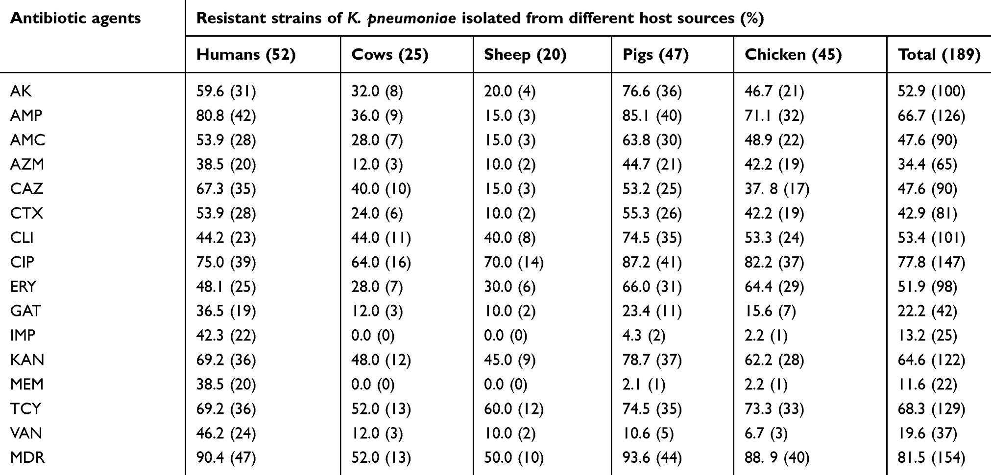 |
Table 1 Antibiotic resistance profiles of K. pneumoniae isolates from different host origins |
Mucous phenotype and virulence genes of K. pneumoniae
The results of the string test showed that 48 (25.4%) K. pneumoniae strains presented the HMV phenotype. Of the 48 string-test-positive isolates, 20 (38.5%) were isolated from humans, 15 (31.9%) were isolated from pigs, nine (20.0%) were isolated from chickens, three (12.0%) were isolated from cows, and only one (5.0%) was isolated from sheep (Table 2). Table 2 characterizes the distribution of the virulence genes among K. pneumoniae strains isolated from different hosts. The siderophore gene entB (enterobactin) and the fimbrial adhesin genes fimH-1 and mrkD were present in 96.8%, 91.5% and 81.5% of the isolates, respectively, and were the most prevalent virulence genes. The mucoviscosity-associated genes magA and rmpA were less prevalent in all the isolates, with prevalence rates of 6.9% and 10.6%, respectively. However, none of the strains isolated from cows and sheep contained the magA and rmpA genes.
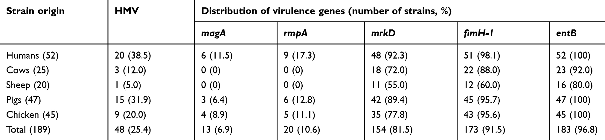 |
Table 2 Distribution of virulence genes among K. pneumoniae isolates from different hosts |
ESBL phenotype and resistance genes of K. pneumoniae
A total of 67 K. pneumoniae strains produced ESBLs. Of the 67 ESBL-positive isolates, 25 (48.1%) were isolated from humans, 21 (44.7%) were isolated from pigs, 14 (31.1%) were isolated from chickens, five (20.0%) were isolated from cows, and two (10.0%) were isolated from sheep (Table 3). Table 3 characterizes the distribution of the antibiotic resistance genes among K. pneumoniae strains isolated from different hosts. In general, tolC (64.0%) and qnrA (63.0%) were the most commonly detected antibiotic resistance genes. Among the genes encoding beta-lactamases, blaKPC (30.2%), blaTEM (29.1%) and blaSHV (26.5%) exhibited the highest prevalence, and blaNDM (2.7%) showed the lowest prevalence among all detected antibiotic resistance genes in K. pneumoniae strains isolated from different hosts. However, different distributions of resistance genes were detected in K. pneumoniae strains from different host sources. blaKPC (51.1%) showed the highest prevalence in pig isolates, followed by human isolates (42.3%), chicken isolates (20.0%), sheep isolates (5.0%) and cow isolates (4.0%). blaSHV was exhibited the highest prevalence in human isolates (51.9%), followed by chicken strains (28.9%), pig strains (14. 9%), cow strains (8.0%) and sheep strains (5.0%). No blaNDM genes were detected in K. pneumoniae strains isolated from cows and sheep.
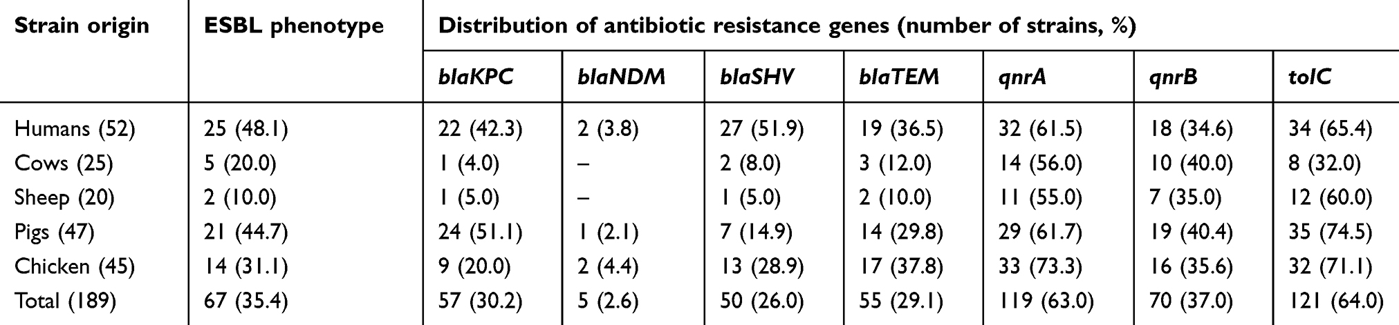 |
Table 3 Distribution of antibiotic-resistance genes of K. pneumoniae isolates from different hosts |
MLST
The MLST analysis revealed that 24 unique sequence types (STs) were identified from the 189 isolates. The unique STs were identified in the human (7), pig (8), chicken (8), cow (5) and sheep strains (5) (Table 4). As clearly shown in the table, seven STs were associated with human strains, eight STs were associated with pig and chicken strains, and five STs were associated with cow and sheep strains. The most frequent ST in K. pneumoniae was ST11, which was commonly detected among the isolates originating from five hosts and was present in human (34.6%), pig (36.2%), chicken (15.6%), cow (28.0%) and sheep (30.0%). ST235 and ST258 were also common STs of K. pneumoniae isolated from human (46.2%), pig (42.6%) and chicken strains (57.8%). ST187 was the most frequent ST among K. pneumoniae strains from cows, with a prevalence of at 52.0%, and ST237 was the most frequent ST among K. pneumoniae isolated from sheep, with a prevalence of 50.0%.
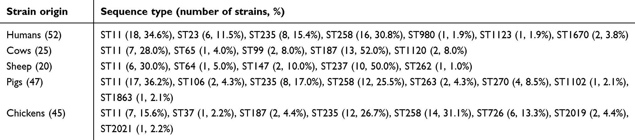 |
Table 4 Multilocus sequence types of K. pneumoniae isolates from different host origins |
Analysis of the correlations among STs and resistance and virulence genes
We performed Pearson’s correlation analysis to assess the existence of potential correlations among the molecular types and resistance and virulence genes of all K. pneumoniae strains. Figure 1 suggests that the HMV phenotype of K. pneumoniae was significantly positively correlated with rmpA (R=0.51, p<0.001), magA (R=0.46, p<0.001), blaKPC (R=0.68, p<0.001), blaTEM (R=0.37, p<0.001) and blaNDM (R=0.28, p<0.001). The STs displayed positive or negative correlations with several resistance and virulence genes. For instance, ST258 exhibited significant positive correlations with blaNDM (R=0.31, p<0.001), magA (R=0.25, p<0.001), and HMV (R=0.24, p<0.001) and a negative correlation with qnrB (R=−0.15, p<0.05). ST237 was obviously negatively correlated with fimH-1 (R=−0.43, p<0.001) and entB (R=−0.36, p<0.001). However, no potential links were found among the other ST types and any resistance and virulence genes of the K. pneumoniae strains. As shown in Figure 1, the virulence gene rmpA displayed a strong positive correlation with magA (R=0.72, p<0.001) and a positive association with the resistance genes blaKPC (R=0.0.49, p<0.001), blaNDM (R=0.26, p<0.001) and blaTEM (R=0.25, p<0.001).
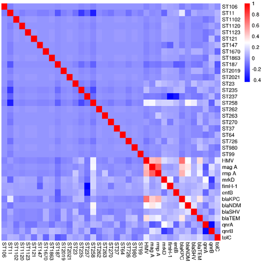 |
Figure 1 Pearson’s correlation analysis of the ST types, drug-resistance genes and virulence genes of the 189 K. pneumoniae isolates. |
Discussion
K. pneumoniae is an important opportunistic pathogen responsible for both human and animal infections, and the emergence of MDR K. pneumoniae has made it difficult to control this pathogen worldwide.1 An increasing number of studies have isolated that MDR K. pneumoniae strains from a variety of animals as well as humans,16,17 but few studies have assessed the molecular relationships among K. pneumoniae isolates from livestock and human infections.18 The present study was performed to assess the prevalence distribution of antibiotic-resistant genotypes, phenotypes, and virulence genes among K. pneumoniae isolated from different hosts, including cows, sheep, pigs, chickens and humans. The antibiotic susceptibility results showed that the resistance profiles of the strains isolated from different hosts were obviously different. Nosocomial K. pneumoniae strains exhibited a higher prevalence of resistance than animal strains. Fifty-two nosocomial K. pneumoniae strains showed high resistance to 15 antimicrobials, with resistance rates ranging from 36.5% to 80.8%, and resistance rates of 42.3% and 38.5% were obtained after the inclusion of imipenem and meropenem among the carbapenem antibiotics. These results are consistent with previous findings.19 Among the animal isolates, the prevalence of resistant isolates from pigs and chickens was higher than that of resistant isolates from cows and sheep. The highest resistance rates among the isolates from chickens and pigs (82.2% and 87.2%, respectively) were found against ciprofloxacin. Quinolones are broad-spectrum antimicrobial agents that have been widely used to treat food-producing animals, including chickens and pigs, in China.20 Some data have shown that 80.0% of isolates from chickens are resistant to ciprofloxacin,21 and similar findings have reported that the incidence of ciprofloxacin resistance among K. pneumoniae from Chinese pigs is 68.9%.22 However, another study found that more than 90% of 61 K. pneumoniae isolates from retail foods in China are susceptible to quinolone antibiotics, including ciprofloxacin,20 which disagrees with our results. K. pneumoniae isolates from different hosts have different antibiotic resistance profiles, and we speculate that these differences might be due to the environmental selective pressures exerted on bacteria. Many antibiotics are often excessively and unreasonably used in animal clinics for the treatment of multiple infections,23 which increases the selective pressure for antibiotic and multidrug resistance. In modern livestock production systems, antimicrobials are heavily used for treating diseases and promoting animal growth, which has resulted in an environment conducive to the amplification of antibiotic resistance. The extensive use and abuse of antimicrobials are common in large-scale pig farms and chicken farms in China, but antibiotics are less commonly used in cows and sheep farms, which might explain the lower prevalence of antibiotic resistance among the K. pneumoniae strains isolated from cows and sheep compared that found among the strains isolated from pigs and chickens. Some expensive and newly synthesized antibiotics are rarely used in animal agriculture,24 and thus, bacteria exhibit less drug resistance against these antibiotics than against traditional antibiotics. For example, GAT, IMP and MEM are rarely used for the treatment of animal infections. In the present study, we found that most K. pneumonia isolates from animals were susceptible to GAT, IMP and MEM, and similar findings revealed that all K. pneumoniae isolates from food animals are susceptible to IMP and MEM.16 In addition, multiresistant strains increase the risk of infections caused by treatment failure in humans and animals. Multiresistant K. pneumonia isolates have emerged in many countries, including Northwest Iran, Turkey, Australia and China.25–28 Our study also showed a high occurrence of multiresistant K. pneumonia among humans and different animals, which suggest that these multiresistant strains can potentially be transmitted among humans and animals.
The pathogenicity of K. pneumoniae is due to various virulence factors, including capsule production, hypermucoviscosity, the presence of lipopolysaccharides and iron acquisition systems, all of which contribute to overcoming the innate immunity of mammalian hosts and to maintaining infections in these hosts.29,30 K. pneumoniae strains with a hypermucoviscous phenotype are considered hypervirulent strains.31 The molecular identification of these strains is related to the presence of the rmpA and magA genes. rmpA is the plasmid gene regulator of the mucoid phenotype A gene, which is a regulator of extracapsular polysaccharide synthesis.31 magA, the chromosomally encoded hypermucoviscosity gene A, encodes the K1 mucoviscous serotype. Although most K. pneumoniae strains with the hypermucoviscous phenotype harbored magA, some magA-negative strains harboring the rmpA gene also exhibited this phenotype.32 In the present study, the overall frequency of the hypermucoviscous phenotype was 25.2%, which was similar to those found in other studies33 but higher than those found in other investigations conducted in European countries.34 Interestingly, the identification of virulence genes performed in our study showed that rmpA (10.6%) and magA (6.9%) were present in the hypermucoviscous isolates. Cubero et al also reported that half of the hypermucoviscous isolates included in their study were magA- and rmpA-negative.35 This finding suggests that the string test (hypermucoviscosity) is not a reliable indicator of the presence of virulent clones with magA and rmpA. We speculate that other factors are involved in the development of the hypermucoviscosity phenotype. In fact, some researchers have suggested that variations in the composition of lipopolysaccharides plays an important role in the development of a hypermucoviscous phenotype that might resemble that found in virulent strains.36 Our results also showed that more K. pneumoniae isolates with the hypermucoviscous phenotype were isolated from humans than from animals, which suggests that the hypermucoviscous characteristics of K. pneumoniae might be associated with the host.18 In addition, the entB gene is a siderophore-associated gene of K. pneumonia. Iron is essential for bacterial survival but is absent in the host plasma. K. pneumoniae mainly acquires iron via the secretion of siderophores, which have a higher affinity for iron than host transport proteins.37 In the present study, the detection rate of the entB gene was 96.8%, which agrees with other reports.37 FimH and mrkD are also important virulent factors that are essential in pili formation and are important for attachment to acellular surfacesen formatting biofilms. In the present study, the detection rates of the fimH and mrkD genes were 91.5% and 81.5%, respectively, which is consistent with the results obtained in a previous study.37,38
ST11 is a common MDR ST that is mainly found in Asia and South America39–41 and is the main ST in hospitals and animals in China.38,42,43 The present results were consistent with these reports. Our data also showed that ST11 was an ST of K. pneumoniae isolates from both humans and animals (including pigs, cows, sheep and chickens), which suggests that ST11 K. pneumoniae can spread between humans and animals. Moreover, ST235 and ST258 were prevalent in the isolates obtained from humans, pigs and chickens, which indicated that these STs might be associated with transmission between humans and animals. The analysis of the correlations among STs, drug-resistance genes, virulence genes and phenotypic characteristics showed that some molecular types exhibited significant correlations with some resistance and virulence genes. As we previously described, the K. pneumoniae strains with the HMV phenotype harbored magA and rmpA genes. Our correlation analysis also showed that the HMV phenotype was significantly positively correlated with magA (R=0.46) and rmpA (R=0.51), which was consistent with other reports (17). The major mechanism underlying the resistance of K. pneumoniae to carbapenems involves the expression of carbapenemases. Most of the carbapenemases present in the K. pneumoniae strains with the HMV phenotype are KPC.37 A similar result was obtained in the present study: the HMV phenotype was strongly positively correlated with blaKPC (R=0.68, p<0.001). Although some researchers hypothesize that ST types of K. pneumonia strains are associated with hypervirulent characteristics,44 our data identified no significant correlations with the exception of correlations among ST258 and virulence and antibiotic-resistance genes. Because few strains with other STs were detected, no other potential correlations between STs and any resistance and virulence genes in K. pneumoniae strains were found in this study. Various factors associated with virulence and antibiotic resistance are important for bacterial pathogenesis. El Fertas-Aissani et al found that virulence factors are associated with antibiotic resistance in pathogenic bacteria;32 however, this study found no significant correlation among virulence factors and resistance genes. Thus, more strains should be collected to determine the correlations among STs and resistance and virulence genes in the future.
Although the sources of K. pneumoniae strains were different, the phenotypic characteristics of some strains were similar, which suggested a risk of transmission between humans and animals. Therefore, to prevent further transmission of K. pneumoniae among humans and animals, strict infection control measures, such as the rational application of antibiotics in clinical settings and animal farms, the regular disinfection of the farm environment, decreased human contact with animals, and the screening of more effective drugs, should be implemented.
Conclusion
In conclusion, our study described the antimicrobial resistance and molecular characterization of K. pneumoniae strains isolated from humans and animals in Henan, China. The results showed that some K. pneumoniae strains of different origins, including humans and animals, have common molecular types and similar phenotypes, which indicates that these strains can potentially be transmitted between humans and animals. Therefore, the prudent use of antimicrobials in human clinical therapy and animal production as well as control measures for the transmission of K. pneumoniae between humans and animals are needed.
Acknowledgments
This work was supported by the Science and Technology Research Project of Henan Province (Grant 182102310553) and the Production, Study and Research Project Funding of Xinxiang Medical University (Grant 2017CXY213).
Disclosure
The authors report no conflicts of interest associated with this work.
References
1. Ripabelli G, Tamburro M, Guerrizio G, et al. Tracking multidrug-resistant Klebsiella pneumoniae from an Italian Hospital: molecular epidemiology and surveillance by PFGE, RAPD and PCR-based resistance genes prevalence. Curr Microbiol. 2018;75(8):977–987. doi:10.1007/s00284-018-1475-3
2. CARSS. Reports of CHINET surveillance of bacterial resistance in 2015. 2015, 12. Available from: http://www.carss.cn/Sys/res/file/201512/20151220130134_7741_482f3b7ae95841998a37898e2ab2fa87_2015%E5%B9%B4%E7%9B%91%E6%B5%8B%E6%8A%A5%E5%91%8A.pdf.
3. Hiroi M, Yamazaki F, Harada T, et al. Prevalence of extended-spectrum β-lactamase-producing Escherichia coli and Klebsiella pneumoniae in food-producing animals. J Vet Med Sci. 2012;74(2):189–195. doi:10.1292/jvms.11-0372
4. He T, Wang Y, Sun L, et al. Occurrence and characterization of blaNDM-5-positive Klebsiella pneumoniae isolates from dairy cows in Jiangsu, China. J Antimicrob Chemother. 2017;72(1):90–94. doi:10.1093/jac/dkw357
5. Bidewell CA, Williamson SM, Rogers J, et al. Emergence of Klebsiella pneumoniae subspecies pneumoniae as a cause of septicaemia in pigs in England. PLoS One. 2018;13(2):e0191958. doi:10.1371/journal.pone.0191958
6. Sonnevend Á, Ghazawi A, Hashmey R, et al. Multihospital occurrence of pan-resistant Klebsiella pneumoniae sequence type 147 with an ISEcp1-Directed blaOXA-181 Insertion in the mgrB gene in the United Arab Emirates. Antimicrob Agents Chemother. 2017;61(7):AAC00418–17. doi:10.1128/AAC.00418-17
7. Oliva A, Mascellino MT, Cipolla A, et al. Therapeutic strategy for pandrug-resistant Klebsiella pneumoniae severe infections: short-course treatment with colistin increases the in vivo and in vitro activity of double carbapenem regimen. Int J Infect Dis. 2015;33:132–134. doi:10.1016/j.ijid.2015.01.011
8. Catalán-Nájera JC, Garza-Ramos U, Barrios-Camacho H. Hypervirulence and hypermucoviscosity: two different but complementary Klebsiella spp. phenotypes? Virulence. 2017;8(7):1111–1123. doi:10.1080/21505594.2017.1317412
9. Prokesch BC, TeKippe M, Kim J, et al. Primary osteomyelitis caused by hypervirulent Klebsiella pneumoniae. Lancet Infect Dis. 2016;16(9):e190–e195. doi:10.1016/S1473-3099(16)30021-4
10. Xu M, Li A, Kong H, et al. Endogenous endophthalmitis caused by a multidrug-resistant hypervirulent Klebsiella pneumoniae strain belonging to a novel single locus variant of ST23: first case report in China. BMC Infect Dis. 2018;18(1):669. doi:10.1186/s12879-018-3109-6
11. Lin YC, Lu MC, Tang HL, et al. Assessment of hypermucoviscosity as a virulence factor for experimental Klebsiella pneumoniae infections: comparative virulence analysis with hypermucoviscosity-negative strain. BMC Microbiol. 2011;11:50. doi:10.1186/1471-2180-11-50
12. Doyle D, Peirano G, Lascols C, et al. Laboratory detection of Enterobacteriaceae that produce carbapenemases. J Clin Microbiol. 2012;50:3877–3880. doi:10.1128/JCM.02117-12
13. Park KS, Kim MH, Park TS, et al. Prevalence of the plasmid-mediated quinolone resistance genes, aac(6ʹ)-Ib-cr, qepA, and oqxAB in clinical isolates of extended-spectrum beta-lactamase (ESBL)-producing Escherichia coli and Klebsiella pneumoniae in Korea. Ann Clin Lab Sci. 2012;42:191–197.
14. Yu WL, Ko WC, Cheng KC, et al. Association between rmpA and magA genes and clinical syndromes caused by Klebsiella pneumoniae in Taiwan. Clin Infect Dis. 2006;42:1351–1358. doi:10.1086/503420
15. Magiorakos AP, Srinivasan A, Carey RB, et al. Multidrug-resistant, extensively drug-resistant and pandrugresistant bacteria: an international expert proposal for interim standard definitions for acquired resistance. Clin Microbiol Infect. 2012;18:268–281. doi:10.1111/j.1469-0691.2011.03570.x
16. Davis GS, Price LB. Recent research examining links among Klebsiella pneumoniae from food, food animals, and human extraintestinal infections. Curr Environ Health Rep. 2016;3(2):128–135. doi:10.1007/s40572-016-0089-9
17. Yang Y, Zhang A, Lei C, et al. Characteristics of plasmids coharboring 16S rRNA methylases, CTX-M, and virulence factors in Escherichia coli and Klebsiella pneumoniae isolates from chickens in China. Foodborne Pathog Dis. 2015;12(11):873–880. doi:10.1089/fpd.2015.2025
18. Köck R, Daniels-Haardt I, Becker K, et al. Carbapenem-resistant Enterobacteriaceae in wildlife, food-producing, and companion animals: a systematic review. Clin Microbiol Infect. 2018;24(12):1241–1250. doi:10.1016/j.cmi.2018.04.004
19. Ou Q, Li W, Li B, et al. Prevalence of Carbapenem-Resistant Klebsiella Pneumoniae (CRKP) and the distribution of Class 1 integron in their strains isolated from a hospital in Central China. Chin Med Sci J. 2017;32(2):107–112. doi:10.24920/J1001-9294.2017.018
20. Zhang S, Yang G, Ye Q, et al. Phenotypic and genotypic characterization of Klebsiella pneumoniae isolated from retail foods in China. Front Microbiol. 2018;9:289. doi:10.3389/fmicb.2018.00289
21. Wu H, Wang M, Liu Y, et al. Characterization of antimicrobial resistance in Klebsiella species isolated from chicken broilers. Int. J Food Microbiol. 2016;232:95–102. doi:10.1016/j.ijfoodmicro.2016.06.001
22. Yang H, Chen S, White DG, et al. Characterization of multiple-antimicrobial-resistant Escherichia coli isolates from diseased chickens and swine in China. J Clin Microbiol. 2004;42:3483–3489. doi:10.1128/JCM.42.8.3483-3489.2004
23. Van Cuong N, Nhung NT, Nghia NH, et al. Antimicrobial consumption in medicated feeds in vietnamese pig and poultry production. Ecohealth. 2016;13(3):490–498. doi:10.1007/s10393-016-1130-z
24. Moran D. Antimicrobial resistance in animal agriculture: understanding user attitudes and behaviours. Vet Rec. 2017;181(19):508–509. doi:10.1136/vr.j5142
25. Ahangarzadeh RM, Langarizadeh N, Aghazadeh M. First reportof class 1 and class 2 integrons in multidrug-resistant Klebsiella pneumoniaeisolates from northwest Iran. Jpn J Infect Dis. 2012;65:256–259.
26. Hosoglu SS, Gundes F, Kolayli A, et al. Extended-spectrum β-lactamases in ceftazidime-resistant Escherichia coli and Klebsiella pneumoniae isolates in Turkish hospitals. Indian J Med Microbiol. 2007;25:346–350.
27. Roy Chowdhury P, Ingold A, Vanegas N, et al. Dissemination of multipledrug resistance genes by class 1 integrons in Klebsiella pneumoniae isolatesfrom four countries: a comparative study. Antimicrob Agents Chemother. 2011;55:3140–3149. doi:10.1128/AAC.01529-10
28. Xu H, Huo C, Sun Y, et al. Emergence and molecular characterization of multidrug-resistant Klebsiella pneumoniae isolates harboring bla CTX-M-15 extended-spectrum β-lactamases causing ventilator-associated pneumonia in China. Infect Drug Resist. 2018;12:33–43. doi:10.2147/IDR.S189494
29. Blin C, Passet V, Touchon M, et al. Metabolic diversity of the emerging pathogenic lineages of Klebsiella pneumoniae. Environ Microbiol. 2017;19(5):1881–1898. doi:10.1111/1462-2920.13689
30. Li B, Zhao Y, Liu C, et al. Molecular pathogenesis of Klebsiella pneumoniae. Future Microbiol. 2014;9(9):1071–1081. doi:10.2217/fmb.14.48
31. Fang CT, Chuang YP, Shun CT, et al. A novel virulence gene in Klebsiella pneumoniae strains causing primary liver abscess and septic metastatic complications. J Exp Med. 2004;199:697–705. doi:10.1084/jem.20030857
32. El Fertas-Aissani R, Messai Y, Alouache S, et al. Virulence profiles and antibiotic susceptibility patterns of Klebsiella pneumoniae strains isolated from differentclinical specimens. Pathol Biol. 2013;61:209–216. doi:10.1016/j.patbio.2012.10.004
33. Liao CH, Huang YT, Lai CC, et al. Klebsiella pneumoniae bacteremia and capsular serotypes, Taiwan. Emerg Infect Dis. 2011;17:1113–1115. doi:10.3201/eid/1706.100811
34. Turton JF, Englender H, Gabriel SN, et al. Genetically similar isolates of Klebsiella pneumoniae serotype K1 causing liverabscesses in three continents. J Med Microbiol. 2007;56:593–597. doi:10.1099/jmm.0.46964-0
35. Cubero M, Grau I, Tubau F, et al. Hypervirulent Klebsiella pneumoniae clones causing bacteraemia in adults in a teaching hospital in Barcelona, Spain (2007-2013). Clin Microbiol Infect. 2016;22(2):154–160. doi:10.1016/j.cmi.2015.09.025
36. Shon AS, Bajwa RP, Russo TA. Hypervirulent (hypermucoviscous) Klebsiella pneumoniae: a new and dangerous breed. Virulence. 2013;4:107–118. doi:10.4161/viru.22718
37. Liu Z, Gu Y, Li X, et al. Identification and characterization of NDM-1-producing hypervirulent (Hypermucoviscous) Klebsiella pneumoniae in China. Ann Lab Med. 2019;39(2):167–175. doi:10.3343/alm.2019.39.2.167
38. Zhan L, Wang S, Guo Y, et al. Outbreak by hypermucoviscous Klebsiella pneumoniae ST11 isolates with carbapenem resistance in a tertiary hospital in China. Front Cell Infect Microbiol. 2017;7:182. doi:10.3389/fcimb.2017.00517
39. Ovejero CM, Escudero JA, Thomas-Lopez D, et al. Highly tigecycline-resistant Klebsiella pneumoniae sequence type 11 (ST11) and ST147 isolates from companion animals. Antimicrob Agents Chemother. 2017;61(6):e02640–16. doi:10.1128/AAC.02640-16
40. Oteo J, Pérez-Vázquez M, Bautista V, et al. The spread of KPC-producing Enterobacteriaceae in Spain: WGS analysis of the emerging high-risk clones of Klebsiella pneumoniae ST11/KPC-2, ST101/KPC-2 and ST512/KPC-3. J Antimicrob Chemother. 2016;71(12):3392–3399. doi:10.1093/jac/dkw321
41. Netikul T, Sidjabat HE, Paterson DL, et al. Characterization of an IncN2-type blaNDM-1-carrying plasmid in Escherichia coli ST131 and Klebsiella pneumoniae ST11 and ST15 isolates in Thailand. J Antimicrob Chemother. 2014;69(11):3161–3163. doi:10.1093/jac/dku275
42. Gu D, Dong N, Zheng Z, et al. A fatal outbreak of ST11 carbapenem-resistant hypervirulent Klebsiella pneumoniae in a Chinese hospital: a molecular epidemiological study. Lancet Infect Dis. 2018;18(1):37–46. doi:10.1016/S1473-3099(17)30489-9
43. Li J, Zou MX, Wang HC, et al. An outbreak of infections caused by a Klebsiella pneumoniae ST11 clone coproducing Klebsiella pneumoniae Carbapenemase-2 and RmtB in a Chinese teaching hospital. Chin Med J (Engl). 2016;129(17):2033–2039. doi:10.4103/0366-6999.189049
44. Shankar C, Veeraraghavan B, Nabarro LEB, et al. Whole genome analysis of hypervirulent Klebsiella pneumoniae isolates from community and hospital acquired bloodstream infection. BMC Microbiol. 2018;18(1):6. doi:10.1186/s12866-018-1202-z
 © 2019 The Author(s). This work is published and licensed by Dove Medical Press Limited. The full terms of this license are available at https://www.dovepress.com/terms.php and incorporate the Creative Commons Attribution - Non Commercial (unported, v3.0) License.
By accessing the work you hereby accept the Terms. Non-commercial uses of the work are permitted without any further permission from Dove Medical Press Limited, provided the work is properly attributed. For permission for commercial use of this work, please see paragraphs 4.2 and 5 of our Terms.
© 2019 The Author(s). This work is published and licensed by Dove Medical Press Limited. The full terms of this license are available at https://www.dovepress.com/terms.php and incorporate the Creative Commons Attribution - Non Commercial (unported, v3.0) License.
By accessing the work you hereby accept the Terms. Non-commercial uses of the work are permitted without any further permission from Dove Medical Press Limited, provided the work is properly attributed. For permission for commercial use of this work, please see paragraphs 4.2 and 5 of our Terms.