")
Back to Journals » International Journal of Nanomedicine » Volume 12
Green synthesis palladium nanoparticles mediated by white tea (Camellia sinensis) extract with antioxidant, antibacterial, and antiproliferative activities toward the human leukemia (MOLT-4) cell line
Authors Azizi S, Mahdavi Shahri M , Rahman HS , Rahim RA, Rasedee A, Mohamad R
Received 17 August 2017
Accepted for publication 29 September 2017
Published 14 December 2017 Volume 2017:12 Pages 8841—8853
DOI https://doi.org/10.2147/IJN.S149371
Checked for plagiarism Yes
Review by Single anonymous peer review
Peer reviewer comments 2
Editor who approved publication: Dr Thomas Webster
This paper has been retracted.
Susan Azizi,1 Mahnaz Mahdavi Shahri,2 Heshu Sulaiman Rahman,3–5 Raha Abdul Rahim,6 Abdullah Rasedee,5 Rosfarizan Mohamad1,7
1Department of Bioprocess Technology, Faculty of Biotechnology and Biomolecular Sciences, Universiti Putra Malaysia, UPM Serdang, Selangor, Malaysia; 2Department of Chemistry, Shiraz Branch, Islamic Azad University, Shiraz, Iran; 3College of Veterinary Medicine, University of Sulaimani, Sulaimani Nwe, 4College of Health Science, Komar University of Science and Technology (KUST), Chaq-Chaq Qularaise, Sulaimani City, Iraq; 5Faculty of Veterinary Medicine, 6Department of Cell and Molecular Biology, Faculty of Biotechnology and Biomolecular Sciences, 7Laboratory of Biopolymer and Derivatives, Institute of Tropical Forestry and Forest Products, Universiti Putra Malaysia, UPM Serdang, Selangor, Malaysia
Abstract: Among nanoparticles used for medical applications, palladium nanoparticles (PdNPs) are among the least investigated. This study was undertaken to develop PdNPs by green synthesis using white tea (W.tea; Camellia sinensis) extract to produce the [email protected] NPs. The [email protected] NPs were characterized by UV–vis spectroscopy and X-ray diffractometry, and evaluated with transmission electron microscopy (TEM) and scanning electron microscopy (SEM). The [email protected] NPs were spherical (size 6–18 nm) and contained phenols and flavonoids acquired from the W.tea extract. [email protected] NPs has good 1-diphenyl-2-picrylhydrazyl (DPPH), OH, and NO-scavenging properties as well as antibacterial effects toward Staphylococcus epidermidis and Escherichia coli. MTT assay showed that [email protected] NPs (IC50 =0.006 µM) were more antiproliferative toward the human leukemia (MOLT-4) cells than the W.tea extract (IC50 =0.894 µM), doxorubicin (IC50 =2.133 µM), or cisplatin (IC50 =0.013 µM), whereas they were relatively innocuous for normal human fibroblast (HDF-a) cells. The anticancer cell effects of [email protected] NPs are mediated through the induction of apoptosis and G2/M cell-cycle arrest.
Keywords: green synthesis, palladium nanoparticles, white tea, leukemic cells, cytotoxicity, medical application, nanobiotechnology
Introduction
Nanotechnology has generated new materials for application in medicine. However, inorganic nanoparticles (NPs) produced by various chemical and physical methods are relatively expensive, and the production methods are often hazardous to the environment.1 The novel green synthetic method for the production of NPs using biological materials such as microorganisms, marine organisms, micro-fluids, and plant extracts have proven to be superior to other processes.2 These methods of NP synthesis are simple and efficient, and the products are safe for medical applications.3 In addition, molecular components of plant extracts have great affinity for the surface of nano-structures, and stabilize and prevent aggregation while improving the biological effects of NPs.4 Thus far, more than 200 plants have been screened for their potential to produce inorganic NPs.5,6
With the discovery of the platinum (II)–cisplatin complex, metal-based compounds are touted as potential drug delivery systems for cancers.7 Recently, Chen et al8 developed palladium nanoparticles (PdNPs) that had remarkable optical, electronic, and chemical properties. These PdNPs are currently being investigated for use as sensors and catalysts. The PdNPs have enormous catalytic potential in various organic transformations, including Suzuki-cross, Mizoroki-Heck, Stille, and Sonogashira coupling reactions.9–11 However, the application of PdNPs in the biomedical field is yet to be adequately investigated, although the green-synthesized PdNPs possess remarkable antioxidant12,13 and antimicrobial activities.12,14 Plant-derived PdNPs seem to have potential for application in cancer therapy because these compounds are also toxic to cancer cells.15,16
In this study, we demonstrated a simple one-step green process to synthesize PdNPs using Camellia sinensis (white tea [W.tea]) extract from unfermented young tea leaves or unopened buds.17 The white tea extract has been used as an efficient reducing and capping agent. The extract contains several polyphenolic compounds belonging to the flavan-3-ol (catechin) family (Figure 1) that possess a wide range of biological activities, including antioxidant, anticancer, antiviral, antibacterial, and antifungal effects.18–20
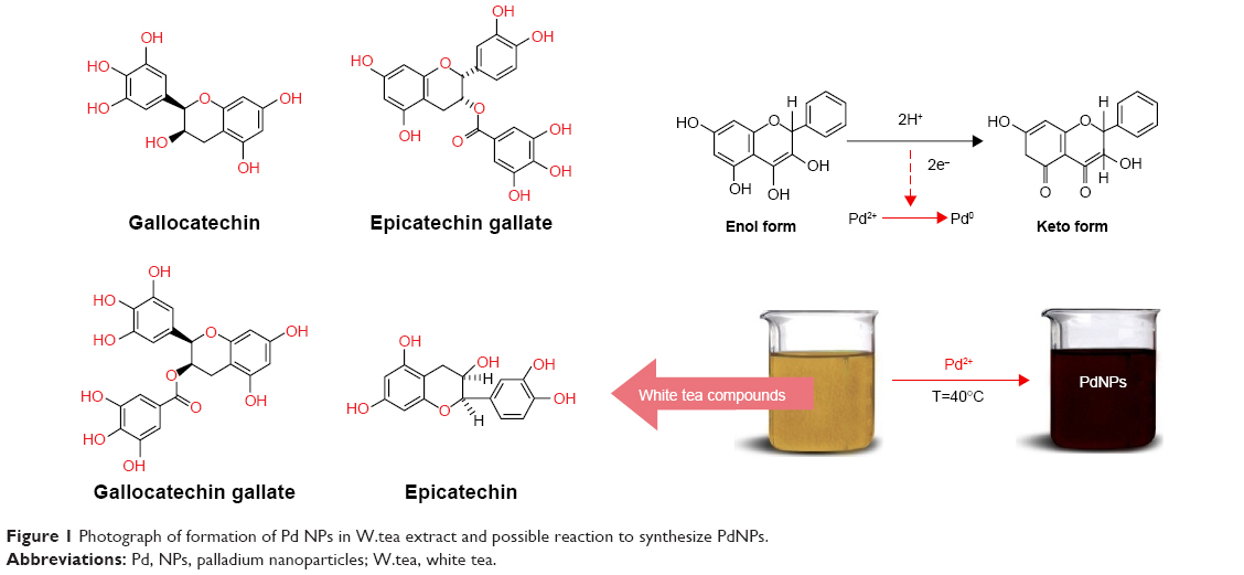 | Figure 1 Photograph of formation of Pd NPs in W.tea extract and possible reaction to synthesize PdNPs. |
To our knowledge, there is no report on the effect of white tea plant extract-derived PdNPs on leukemic cells. Among treatment options for leukemia are chemotherapy, radiation therapy, stem cell transplantation, biological or immune therapy, and targeted therapy. Chemotherapy and radiation are plagued by side effects that include anemia, hair loss, fatigue, gastrointestinal disorders, and susceptibility to bleeding and infections, whereas other treatments are effective in some receptive patients only. For these reasons, more effective alternative cancer therapeutic compounds with minimal side effects are desperately being sought.
Materials and methods
Materials
White tea plant was purchased from a local herbal store in Shiraz, Iran, and washed several times using distilled water to remove impurities. The leaves were sun-dried and then crushed into powder. PdCl2 (99.98%) was used as a palladium precursor and it was supplied from Merck (Darmstadt, Germany). All solutions were prepared with deionized water. The plant was authenticated by Department of Botany, Shahid Chamran University, Iran, and the voucher has been deposited.
Extract preparation
White tea powder sample (1.0 g) was dispersed in 100 mL distilled water with magnetic stirring and heated at 100°C for 20 min. The extract was cooled to room temperature and filtered through a muslin cloth to collect a clear extract.
Synthesis of palladium nanoparticles
An Erlenmeyer flask containing 50 mL of 1 mM PdCl2 solution was made to react with 50 mL of the aqueous white tea extract at 40°C with continuous stirring. The color of the reaction mixture gradually turned to dark brown from transparent yellow after 30 minutes, indicating the formation of PdNPs. The synthetic reaction was completed in 2 h. The initial pH of the solution was approximately 7.5, but changed to 5.6 by the end of the reaction. The product sample was collected through centrifugation at 6,000 rpm for 10 min and, after several washings with distilled water, dried in an oven at 60°C. The dried sample, palladium nanoparticles using white tea ([email protected]) NPs, was crushed into powder and stored in an airtight container for further analysis.
Characterization of synthesized [email protected] NPs
The Pd@W.tea NPs was quantitated by UV-Vis spectrophotometry (Lambda 25-Perkin Elmer, Waltham, MA, USA) over wavelength range of 200–800 nm, and the chemical composition was characterized by Fourier-transform infrared (FTIR) spectrometry (Perkin-Elmer 1725X) in the range of 400–4,000 cm−1. The phase purity and particle size of Pd@W.tea NPs were determined using the X-ray diffractometer (XRD-6000; Shimadzu) at 40 kV with nickel-filtered Cu (λ=1.542 Å) in the range of 10° to 80°.21 Morphological analysis of Pd@W.tea NPs was conducted by using transmission electron microscopy (TEM; HITACHI H-7650, Tokyo, Japan) at voltage 120 kV. The sample suspension was drop-casted on a carbon-coated copper grid and allowed to air-dry at room temperature overnight. The powdered sample was put on the carbon stub using carbon tape and then gold-coated using a sputter coater for ultrastructural examination via scanning electron microscopy (Philips XL-30).8
Quantification of phenolic and flavonoid content
The phenolic and flavonoid contents of Pd@W.tea NPs and crude white tea extract were quantified.
Total phenolic content
Phenolic content was determined by the Folin–Ciocalteu assay as described by Singleton and Rossi,22 with slight modifications. Briefly, 10 μL sample solution and 500 μL Folin-Ciocalteu reagents were placed in each well of 96-well plates. Then, 350 μL of 10% of Na2CO3 was added to the wells, and the plate was incubated in the dark at room temperature for 2 h. The absorbance was then recorded spectrophometerically (Agilent 8453 Spectrophotometer, USA) at 765 nm against 10% DMSO as the negative control. Phenolic content was estimated using the gallic acid calibration curve (R2=0.97), and was expressed as gallic acid equivalent (μg GAE).
Total flavonoid content
Total flavonoid content were determined by an aluminum chloride colorimetric method as described previously.23 In this test, the reaction mixture was prepared by mixing 100 μL Pd@W.tea NPs or white tea extract, 10 μL 10% aluminum chloride, 10 μL 5% (CH3CO2)K, and 30 μL distilled water. The mixture was incubated at room temperature for 30 min, and absorbance was read at 415 nm. The calibration curve (R2=0.99) was obtained using the quercetin solutions at concentrations of 0.0–10 μg/mL and the flavonoid content was determined from the curve and expressed as quercetin equivalent (μg QE).
Antioxidant activity
The antioxidant potential of the Pd@W.tea NPs was determined through 1-diphenyl-2-picrylhydrazyl (DPPH) and radical (−OH and −NO) scavenging activities.
DPPH scavenging activity
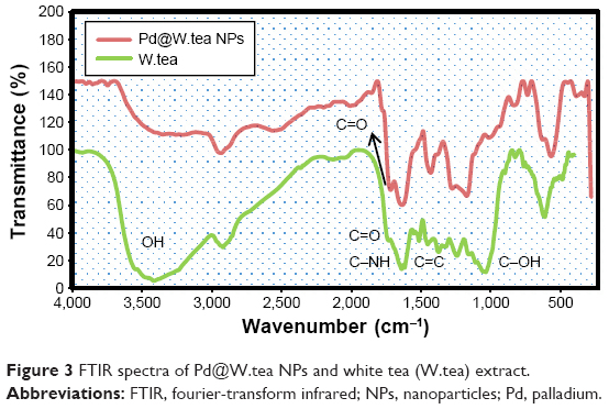
The DPPH scavenging activity of Pd@W.tea NPs was determined using the method described by Blois.24 In brief, approximately 20 μL each of Pd@W.tea NPs and white tea extract at concentrations ranging from 0.156 to 10 μM were added to 100 μL 0.1 mM methanolic DPPH solution. The mixture was incubated for 30 min at room temperature with constant shaking, and the absorbance was recorded at 517 nm. Butyl hydroxyl toluene was used as the reference. The DPPH radical scavenging activity (RSA) was calculated as percent inhibition using the following equation:
|
where Acontrol and Asample are the absorbance of the control and sample, respectively.
OH scavenging activity
Hydroxyl radical scavenging activity was determined by the degradation of 2-deoxyribose after condensation with thiobarbituric acid to produce OH radicals.25 The final reaction mixture containing 100 μM FeCl3, 100 μM ascorbic acid, 1 mM H2O2, 20 mM KOH buffer (pH 7.4), 100 μM EDTA, and 2.8 mM 2-deoxyribose was mixed with different concentrations (0.156–10 μM) of the Pd@W.tea NPs or control and incubated at 37°C for 1 h. Then, 0.5 mL reaction mixtures were added to 1 mL of 0.5% thiobarbituric acid diluted with NaOH (0.025 M) and 1 mL of 2.8% trichloroacetic acid. The mixtures were again incubated at 100°C for 30 min to obtain the chromogenic adduct. After cooling to room temperature, the concentration of chromogen was quantified at 532 nm. Gallic acid was used as the reference, and the percent inhibition of radical scavenging activity calculated using Eqn 1.
NO scavenging activity
The NO scavenging activity was determined spectrophotometrically (Agilent 8453 Spectrophotometer, Golden Valley, MN, USA).25 Approximately 50 μL of 5 mM sodium nitroprusside was added to various concentrations (0.156–10 μM) of Pd@W.tea NPs or control, and the mixture was incubated at 25°C for 1 h. Then, 1.5 mL of this mixture was obtained and treated with 1.5 mL Griess’ reagent (Sigma-Aldrich Co., St Louis, MO, USA) to diazotize and form chromophore. Gallic acid was used as the reference. The concentration of the chromophore was determined at 546 nm, and percent inhibition of scavenging activity was calculated using Eqn 1.
Antibacterial activity
The antibacterial potential of Pd@W.tea NPs was determined by the disc-diffusion method and minimum inhibitory concentration (MIC).3
Zone of inhibition
The antibacterial potential of Pd@W.tea NPs was determined using the disc-diffusion method on the Gram-positive Staphylococcus epidermidis S273 (S. epidermidis) and Gram-negative Escherichia coli E266 (E. coli) strains. After growing the bacteria overnight in nutrient broth, each bacterial inoculum standardized to 0.5 MF units (~108 cfu) was inoculated onto the Muller–Hilton agar (MHA) plate. Discs (6 mm) impregnated with samples at concentrations ranging from 0.156 to 10 μM were placed on the MHA plates and incubated at 37°C for 24 h. White tea extract was used for comparison, with streptomycin as a standard reference. The zone of inhibition was recorded.
Minimum inhibitory concentration
Serial tube dilution method was applied to determine the MIC of the Pd@W.tea NPs on S. epidermidis and E. coli. Test samples were prepared by mixing Pd@W.tea NPs, white tea, or streptomycin (0.156–10 μM) with 100 μL 10% DMSO. Then, 20 μL of the suspension was mixed with 450 mL LB broth (Sigma-Aldrich Co.) and 50 mL of fresh microbial inoculum (~108 cfu) and incubated at 37°C for 24 h to grow bacteria. The turbidity of the suspension was determined visually before and after incubation.
Antiproliferative activity
The antiproliferative effects of Pd@W.tea NPs and white tea extract was determined on the acute human T-lymphoblastic leukemia (MOLT-4) cell line (American Type Culture Collection [ATCC], Manassas, VA, USA), with cisplatin and doxorubicin as reference drugs.26 The analyses used the 3-(4,5-dimethylthiazol-2-yl)-2,5 diphenyl tetrazolium bromide (MTT) assay kit (Sigma-Aldrich Co.). Briefly, cells were allowed to grow in a 75 m2 cell culture flask until 95% confluent before seeding at 1×10⁵ cells/mL into each well of a 96-well plate. These cells were then treated with various concentrations (0.001–3.5 μM) of Pd@W.tea NPs, white tea extract, cisplatin, and doxorubicin. Normal adult human fibroblast (HDF-a) cell line was used as the control. After incubation for 48 h at 37°C at pH 7.5 and under 5% CO2, 25 μL 5.5 mg/mL MTT solution was added to each well and the plate was covered with aluminum foil and incubated for a further 3 h in the dark. The medium was immediately aspirated and the purple formazan lysed with MTT solution. The assay was conducted in triplicates. The absorbance was determined at 570 nm using the Spectra Max plus 384 UV–Vis plate reader. The half maximum inhibitory concentration (IC50) was determined by non-regression analysis using Graph Pad Prism V. 5.03 (Graph Pad Software Inc., CA, USA).
Annexin-V/PI double-staining assay
Phosphatidylserine translocation in MOLT-4 cells treated with Pd@W.tea NPs was determined by the Annexin-V-FITC/propidium iodide (PI) apoptosis detection kit (Sigma-Aldrich Co.).21 In brief, cells treated with 0.006 μM Pd@W.tea NPs for 12, 24, or 48 h were harvested, washed with PBS twice, resuspended in binding buffer, and incubated with Annexin V-FITC and propidium iodide solutions at room temperature in the dark for 30 min. Apoptosis was determined by flow cytometry using FACS Calibur (BD Biosciences, San Jose, CA, USA) with 15,000 ungated cells.
Cell-cycle analysis
MOLT-4 cell-cycle distribution after treatment with Pd@W.tea NPs was determined by flow cytometry according to the method described previously.27 Briefly, MOLT-4 cells after incubation for 12, 24, or 48 h with 0.006 μM Pd@W.tea NPs were collected and centrifuged at 200× g for 5 min before washing with a mixture of PBS and sodium azide. The harvested cells were fixed with chilled 70% ethanol at −20°C for 5 days and then centrifuged at 200× g for 5 min. The supernatant was discarded and the cells were again washed twice with PBS and sodium azide; stained with PBS-staining buffer containing 0.1% triton X-100, 10 mM ethylenediamine tetra-acetic acid, 50 μg/mL RNAase A, and 3 μg/mL PI; and incubated on ice in the dark for 30 min. Flow cytometric analysis was conducted using the BD FACS Calibur flow cytometer (BD), and data analysis was conducted using the Cell Quest Pro software with a DNA histogram to express the proportion of cells in cell-cycle phases. Apoptotic cells with hypodiploid DNA content were detected by quantifying the sub-G1 peak.
Protease activity of caspases 3 and 9
The caspase-3 and -9 activities were determined by a colorimetric assay kit (Gene script kit, code: L00289, GenScript, Piscataway, NJ, USA) in MOLT-4 cells.28 Cells treated with 0.006 μM Pd@W.tea NPs for 12, 24, or 48 h were washed twice with ice-cold PBS, centrifuged at 200× g for 5 min, and the medium was discarded before harvesting cells. The cell pellet was suspended in cells lysis buffer and incubated on ice for 1 h. Untreated MOLT-4 cells incubated for 12, 24, or 48 h served as controls. The protein concentrations were determined using the Bradford method. Approximately 1×10⁸ MOLT-4 treated cells were transferred to the 96-well plate and 5 μL caspase substrate was added to the cells; the plate was wrapped with aluminum foil and incubated at 37°C for 4 h. After incubation, the plate was read at 405 nm in a microplate reader (Universal Microplate reader, Biotech, Inc., Winooski, VT, USA) to determine caspase activity, and the results are presented as optical density (OD).
Results and discussion
Characterization of PdNPs
White tea contains several polyphenols and flavonoids. The [email protected] NPs suspension prepared from white tea was dark brown (Figure 1), which is due the PdNPs29 formed through a reduction process. Because hydroxyl groups are abundant in flavonoids, the reduction of palladium from Pd(II) to Pd(0) involved oxidation of the hydroxyl groups, with concomitant decrease in pH of the reaction suspension.30
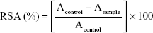
UV–Vis spectroscopy is a useful method to validate the presence of metal NPs. Using this technique, aqueous white tea solution showed an intense absorption peak at 250 nm, which is associated with the benzoyl π→π* transitions – an indication of the presence of flavonoids10 (Figure 2). The white tea extract with Pd(II) solution exhibited another discrete absorption peak at 410 nm, which is attributed to the Pd2+ ion content. [email protected] NPs produced a wide, continuous, absorption spectrum without the typical peaks of white tea extract or white tea with Pd(II) solution, indicating formation of PdNPs.
 | Figure 2 UV–vis spectra of [email protected] NPs, white tea (W.tea) plant extract, and white tea with Pd(II). |
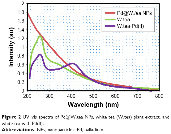
An FTIR analysis was used to identify molecules responsible for the reduction of Pd ions and coating of the PdNPs. The FTIR spectrum of white tea extract showed peaks at 3,345, 2,860, 1,712, 1,667, 1,520, 1,389, 1,124, and 720 cm−1, representing free OH, and stretching of CH group of aldehydes and alkanes, carbonyl group (C=O), (NH)−C=O group, C=C aromatic ring, C−OH, and phenyl (C−H), respectively (Figure 3). These peaks showed that flavonols and other phenolics molecules are present in the white tea extract. With reduction of Pd2+ ions, the position and intensity of peaks changed, especially by the decrease in the hydroxyl peak and appearance of the carbonyl peak, indicating the involvement of phytochemical sites in synthesis and binding of PdNPs. Thus, the results confirmed that reduction of Pd(II) to Pd(0) occurs through oxidation of hydroxyl to carbonyl groups.
 | Figure 3 FTIR spectra of [email protected] NPs and white tea (W.tea) extract. |
The phenolic content of Pd@W.tea NPs was significantly (P<0.05) low, and the flavonoid content lower in Pd@W.tea NPs than in crude white tea extract (Table 1). Differences in these chemical contents are responsible for some of the variability in biological activities between Pd@W.tea NPs and crude white tea extract.
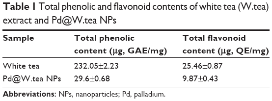 | Table 1 Total phenolic and flavonoid contents of white tea (W.tea) extract and [email protected] NPs |
The Pd@W.tea NPs were examined under X-ray diffraction to determine crystalline structure (Figure 4). There are five well-defined and characteristic diffraction peaks at 40.1°, 46.3°, 68.5°, 82.1°, and 86.2° representing reflections from (111), (200), (220), (311), and (222) planes, respectively, of face-centered cubic crystal structure of Pd(0). The presence of a broad and intense diffraction peak at 2θ of 13.5° may be assigned to the chemical contents of the white tea extract. Using the Scherrer equation, the average size of Pd@W.tea NPs was calculated at 15 nm.
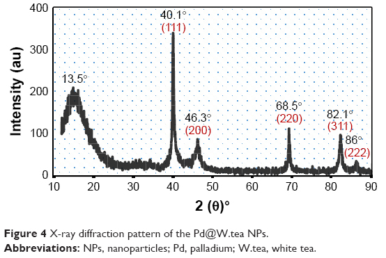 | Figure 4 X-ray diffraction pattern of the [email protected] NPs. |
Under scanning electron microscopy (SEM) and transmission electron microscopy (TEM), Pd@W.tea NPs were shown to be spherical with narrow size distribution (Figure 5). The particles did not appear to aggregate, although some were present in clumps that suggested passive contact. The ultrastructure studies showed that Pd@W.tea NPs are stable and had little tendency to aggregate, which is attributed to the presence of phenols and flavonoids in NPs. It is proposed that, while inhibiting particle aggregation, phenols and flavonoids play a major role in the chelation of NPs to ligands. The size distribution of Pd@W.tea NPS is in the range of 6–18 nm, averaging at 11 nm, which is marginally smaller than the value obtained by X-ray diffraction determination.
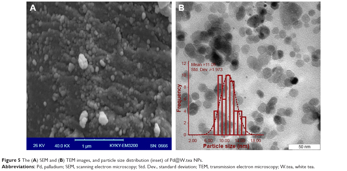 | Figure 5 The (A) SEM and (B) TEM images, and particle size distribution (inset) of [email protected] NPs. |
Radical scavenging
To assess the antioxidant activity of white tea extract and Pd@W.tea NPs, DPPH radical scavenging activity was determined in MOLT-4 cells (Figure 6). It is assumed that the content of phenolic and flavonoid compounds of the extract and Pd@W.tea NPs would afford them promising radical scavenging activity because these compounds are known to be scavengers of DPPH, OH, and NO.25,31,32 The radical scavenging activities of white tea extract for DPPH was high even at low doses and was not dose-dependent (Figure 7). On the other hand, the DPPH scavenging activity of Pd@W.tea NPs was low at low concentrations and only began to approach antioxidation efficacy of white tea extract only after reaching high doses.
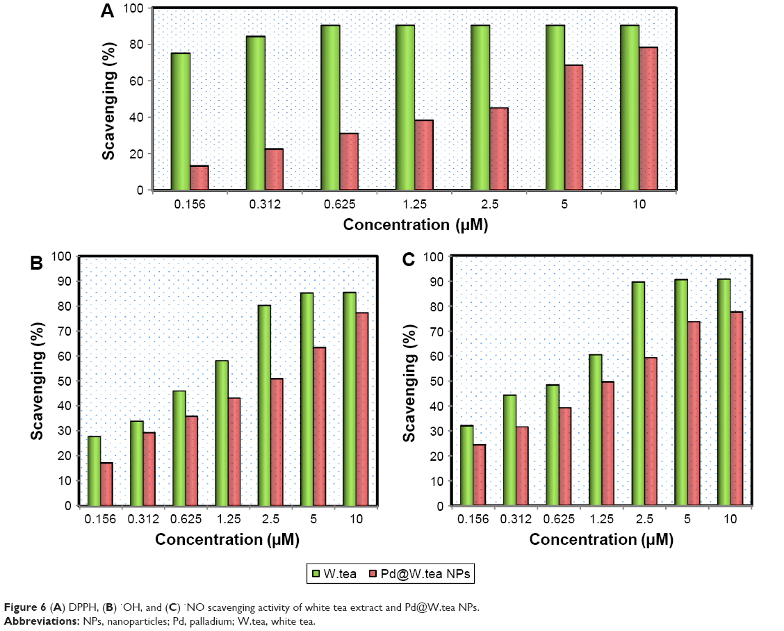 | Figure 6 (A) DPPH, (B) ·OH, and (C) ·NO scavenging activity of white tea extract and [email protected] NPs. |
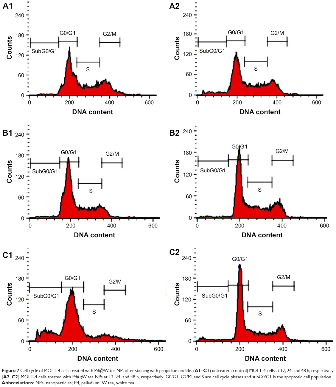 | Figure 7 Cell cycle of MOLT-4 cells treated with [email protected] NPs after staining with propidium iodide. (A1–C1) untreated (control) MOLT-4 cells at 12, 24, and 48 h, respectively. (A2–C2) MOLT-4 cells treated with [email protected] NPs at 12, 24, and 48 h, respectively. G0/G1, G2/M, and S are cell cycle phases and subG0/G1 is the apoptotic cell population. |
Both white extract and Pd@W.tea NPs showed dose-dependent scavenging activities of OH and NO. However, overall, white tea extract is still more efficacious than Pd@W.tea NPs at OH and NO scavenging. This phenomenon can be attributed to the concentration of phenolic and flavonoid compounds, which were much higher in the crude extract than Pd@W.tea NPs.
Antibacterial activity
The fact that white tea extract and Pd@W.tea NPs showed antioxidant activities makes them good candidates as antibacterial agents. By the disc diffusion assay, Pd@W.tea NPs and white tea extract variably inhibits growth of S. epidermidis and E. coli, with the effect being more potent on the S. epidermidis than E. coli (Table 2 and Figure 8). E. coli, a Gram-negative bacterium, possesses an external lipopolysaccharide membrane that protects the peptidoglycan layer and allows the bacteria to survive in harsh environments.33 Presumably, this protective layer is one of the contributing factors toward the greater ability of E. coli than S. epidermidis to survive the toxic effects of white tea extract and Pd@W.tea NPs.
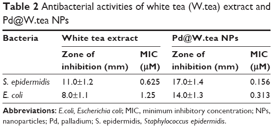 | Table 2 Antibacterial activities of white tea (W.tea) extract and [email protected] NPs |
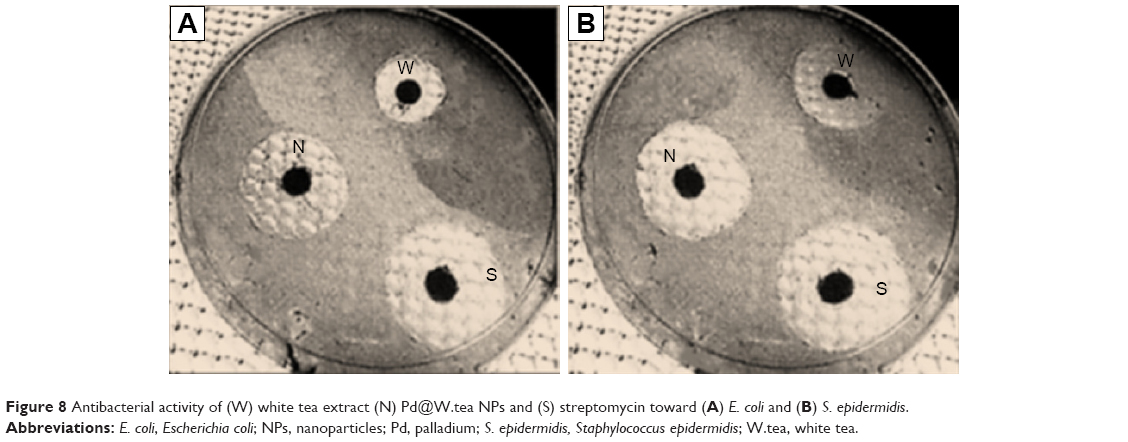 | Figure 8 Antibacterial activity of (W) white tea extract (N) [email protected] NPs and (S) streptomycin toward (A) E. coli and (B) S. epidermidis. |
The antibacterial efficacy of white tea extract and Pd@W.tea NPs was further verified via MIC. Pd@W.tea NPs showed higher MIC values toward S. epidermidis and E. coli than white tea extract (Table 3). These findings indicate that Pd@W.tea NP is a better antibacterial agent than white tea extract, possibly because of the greater ability of Pd@W.tea to specifically interact with the bacteria. Interaction of compounds with the bacterial membrane is dependent on physical and physicochemical characteristics of the ligands and surface of the bacterial membrane. In fact, for nanoparticulated drug delivery systems, particle size and surface area play significant roles in their antibacterial activities.34 The unique physicochemical properties of the NPs also facilitate interactions of the Pd@W.tea NPs with the bacterial cell membrane.35 White tea extract lacks formed structures with appropriate size and characteristics to aid specific interactions with bacteria surface effectively, and these account for its lesser antibacterial effects compared to Pd@W.tea NPs.
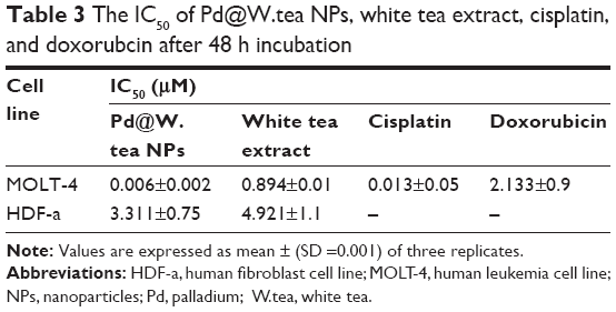 | Table 3 The IC50 of [email protected] NPs, white tea extract, cisplatin, and doxorubcin after 48 h incubation |
In vitro cytotoxicity
The antiproliferative activity of the white tea extract and Pd@W.tea NPs was determined on the MOLT-4 cell line with cisplatin and doxorubicin for comparison (Figure 9). [email protected] NPs were highly toxic to MOLT-4 cells, with effect increasing with increase in concentration. In fact, based on IC50, Pd@W.tea NPs are >2, >100, and >3,000 times more toxic to MOLT-4 cells than ciplastin, crude white tea extract, and doxorubicin, respectively (Table 3). These findings show that Pd@W.tea NPs have potential as an anticancer compound. Both Pd@W.tea NPs and crude white extract showed low toxicity to the normal HDF-a cell line. It is proposed that the high cytotoxicity of Pd@W.tea NPs to the cancer cell line is due to the presence of Pd, which interacts physicochemically with the functional groups of cellular proteins, nitrogen bases, and phosphate groups of the DNA,36 thus causing cell death. Moreover, previous studies showed that Pd causes production of free radical,37 lactate dehydrogenase leakage,15 and cell-cycle disturbances38 that could be among mechanisms of the anticancer effects of Pd@W.tea NPs.
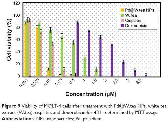 | Figure 9 Viability of MOLT-4 cells after treatment with [email protected] NPs, white tea extract (W.tea), cisplatin, and doxorubicin for 48 h, determined by MTT assay. |
Flavonoid compounds can control metabolic activities of cancer cells.39 The anticancer effects of flavonoids occur through oxidative destruction, inhibition of proliferation, inactivation of carcinogen, promotion of differentiation, induction of cell-cycle arrest and apoptosis, impairment of tumor angiogenesis, and suppression of metastasis.40–42 Flavonoids can interact with xenobiotic metabolizing enzymes and inhibit involvement of kinases signal transduction, interact with estrogen type II binding sites, and alter gene expression patterns,43,44 with resultant promotion of antiproliferative activity of Pd@W.tea NPs.
The size of NPs is an important factor in their cytotoxic effects. Small-sized NPs can exert greater cytotoxicity than large ones.45,46 At an average of 11 or 15 nm, Pd@W.tea NPs can evade the mononuclear phagocytic system whereas easily crossing cell membranes47 to exert anticancer effects. The antiproliferative effect of Pd@W.tea NPs on the MOLT-4 is selective because these NPs are relatively innocuous to normal fibroblast cells. The IC50 values of white tea extract and Pd@W.tea NPs on the HDF-a cells were considerably higher than on the MOLT-4 cells. It is suggested that surface modifications of the PdNPs by the flavonoids acquired from white tea extract are responsible for the toxic effect of Pd@W.tea NPs on cancers without adversely affecting normal cells.44,48
Phosphatidylserine translocation
Annexin-V/PI double-staining assay showed that MOLT-4 cells treated with Pd@W.tea NPs became apoptotic gradually, with concomitant decrease in viable cells in a time-dependent manner. After 12-h treatment, MOLT-4 cells were primarily in the early phase of apoptosis and began to enter the late phase of apoptosis from 24 h onward (Figure 10 and Table 4). This effect is similar to that seen with other green-synthesized NPs, such as Sargassum muticum gold nanoparticles on the human leukemia (K562) cell line induced,28 egg white-mediated silver NPs on human breast adenocarcinoma (MDA-MB231) cell line,49 hyaluronan/zinc oxide nanocomposite on acute promyelocytic leukemia cells (HL-60) cells,27 and selenium NPs on human (MCF-7) breast-cancer cells.50
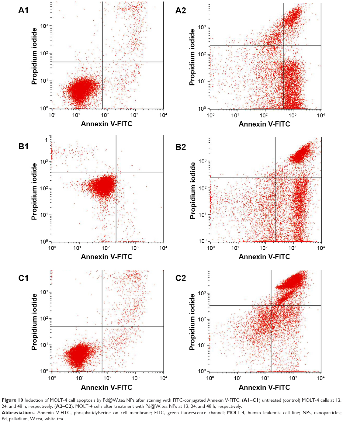 | Figure 10 Induction of MOLT-4 cell apoptosis by Pd@W.tea NPs after staining with FITC-conjugated Annexin V-FITC. (A1–C1) untreated (control) MOLT-4 cells at 12, 24, and 48 h, respectively. (A2–C2): MOLT-4 cells after treatment with Pd@W.tea NPs at 12, 24, and 48 h, respectively. |
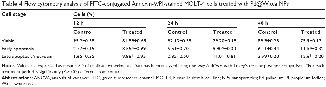 | Table 4 Flow cytometry analysis of FITC-conjugated Annexin-V/PI-stained MOLT-4 cells treated with [email protected] NPs |
MOLT-4 cell-cycle arrest
DNA fragmentation is an important marker of cell death that is reflected as cell-cycle arrest. We used PI-flow cytometric analysis to confirm that the mode of MOLT-4 cell death induced by Pd@W.tea NPs is, in fact, through apoptosis. MOLT-4 cells treated with Pd@W.tea NPs significantly entered the G2/M apoptotic phase after 48 h (Figure 7 and Table 5). The sub-G0/G1 population of Pd@W.tea NPs-treated leukemia cells decreased after 48 h. Thus, the study shows that Pd@W.tea NPs are antiproliferative toward leukemia cells by inducing DNA damage and G2/M arrest. This antiproliferative effect on cancer cell lines is typical of many NPs.27,28,49 Untreated MOLT-4 cells showed typical normal DNA content and cell-cycle distribution of viable cells.
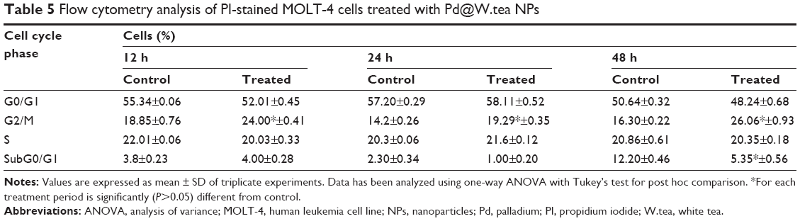 | Table 5 Flow cytometry analysis of PI-stained MOLT-4 cells treated with [email protected] NPs |
Caspases
Caspase-3 and -9 are the primary executioners of apoptosis. In MOLT-4 cells, treatment with Pd@W.tea NPs significantly increased the activities of caspase-3 and -9 (Table 6). Caspase pathway-mediated apoptosis seems to be the common mode of cell death instituted by NPs; for example, with zerumbone-loaded nanostructured lipid carriers and magnetic iron oxide NPs on Jurkat cells,51 amine-modified polystyrene NPs on astrocytoma cells,52 and nickel-zinc ferrite NPs on the HepG2, HT29, and MCF-7 cell lines.53
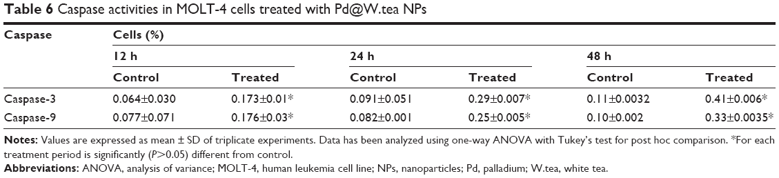 | Table 6 Caspase activities in MOLT-4 cells treated with [email protected] NPs |
Conclusion
We developed an ecofriendly and efficient process for the synthesis of PdNPs with white tea extract as the medium. The mild reaction condition and easy workup procedure as well as the scope of pharmacological and medicinal applications are making our methodology a valuable contribution to the processes for green synthesis of NPs. The Pd@W.tea NPs with size range between 6 and 18 nm are ideal as a drug carrier system, because it avoids clearance by the monocyte–phagocytic system while easily penetrating cell membranes. The Pd@W.tea NPs has potent radical scavenging capability, antibacterial properties, and are antiproliferative to the human leukemia cells without adversely affecting the normal human fibroblast cells. The antiproliferative of Pd@W.tea NPs is via apoptosis and G2/M cell-cycle arrest. Thus, Pd@W.tea NPs has the potential to be developed into an antibacterial and anticancer agent.
Acknowledgments
The authors are grateful to the Department of Bioprocess Technology, Faculty of Biotechnology and Bimolecular Sciences for providing the laboratory facilities, as well as the Universiti Putra Malaysia and University of Sulaimani, Iraq. Additionally, the authors would like to appreciate the Institute of Bioscience (IBS), Universiti Putra Malaysia for providing technical knowledge, assistance, and facilities for this study.
Disclosure
The authors report no conflicts of interest in this work.
References
Darroudi M, Zak AK, Muhamad MR, Huang NM, Hakimi M. Green synthesis of colloidal silver nanoparticles by sonochemical method. Mater Lett. 2012;66(1):117–120. | ||
Azizi S, Namvar F, Mahdavi M, Ahmad MB, Mohamad R. Biosynthesis of silver nanoparticles using brown marine macroalga, Sargassum muticum aqueous extract. Materials (Basel). 2013;6(12):5942–5950. | ||
Gopalakrishnan K, Ramesh C, Ragunathan V, Thamilselvan M. Antibacterial activity of Cu2O nanoparticles on E. coli synthesized from Tridax procumbens leaf extract and surface coating with polyaniline. Dig J Nanomater Bios. 2012;7(2):833–839. | ||
Shabestarian H, Homayouni-Tabrizi M, Soltani M, et al. Green synthesis of gold nanoparticles using Sumac aqueous extract and their antioxidant activity. Mater Res. 2017;20(1):264–270. | ||
Sathishkumar M, Sneha K, Kwak IS, Mao J, Tripathy SJ, Yun YS. Phyto-crystallization of palladium through reduction process using Cinnamom zeylanicum bark extract. J Hazard Mater. 2009;171(1–3):400–404. | ||
Song JY, Kwon EY, Kim BS. Biological synthesis of platinum nanoparticles using Diopyros kaki leaf extract. Bioproccess Biosyst Eng. 2010;33(1):159–164. | ||
Rosenberg B, VanCamp L, Trosko JE, Mansour VH. Platinum compounds: a new class of potent antitumour agents. Nature. 1969;222(5191):385–386. | ||
Chen H, Wei G, Ispas A, Hickey SG, Eychmüller A. Synthesis of palladium nanoparticles and their applications for surface-enhanced Raman scattering and electrocatalysis. J Phys Chem. 2010;114(50):21976–21981. | ||
Lebaschi S, Hekmati M, Veisi H. Green synthesis of palladium nanoparticles mediated by black tea leaves (Camellia sinensis) extract: catalytic activity in the reduction of 4-nitrophenol and Suzuki-Miyaura coupling reaction under ligand-free conditions. J Colloid Interface Sci. 2017;485:223–231. | ||
Nasrollahzadeh M, Sajadi SM, Maham M. Green synthesis of palladium nanoparticles using Hippophae rhamnoides Linn leaf extract and their catalytic activity for the Suzuki–Miyaura coupling in water. J Mol Catal A: Chem. 2015;396:297–303. | ||
Veisi H, Ghorbani-Vaghei R, Hemmati S, Aliani MH, Ozturk T. Green and effective route for the synthesis of monodispersed palladium nanoparticles using herbal tea extract (Stachys lavandulifolia) as reductant, stabilizer and capping agent, and their application as homogeneous and reusable catalyst in Suzuki coupling reactions in water. Appl Organomet Chem. 2015;29(1):26–32. | ||
Kora AJ, Rastogi L. Green synthesis of palladium nanoparticles using gum ghatti (Anogeissus latifolia) and its application as an antioxidant and catalyst. Arab J Chem. 2015;6(24):1–10. | ||
Tahir K, Nazir S, Ahmad A, et al. Biodirected synthesis of palladium nanoparticles using Phoenix dactylifera leaves extract and their size dependent biomedical and catalytic applications. RSC Adv. 2016;6(89):85903–85916. | ||
Manikandan V, Velmurugan P, Park JH, et al. Synthesis and antimicrobial activity of palladium nanoparticles from Prunus × yedoensis leaf extract. Mater Lett. 2016;185:335–338. | ||
Gurunathan S, Kim E, Han JW, Park JH, Kim JH. Green chemistry approach for synthesis of effective anticancer palladium nanoparticles. Molecules. 2015;20(12):22476–22498. | ||
Shanthi K, Sreevani V, Vimala K, Kannan S. Cytotoxic effect of palladium nanoparticles synthesized from Syzygium aromaticum aqueous extracts and induction of apoptosis in cervical carcinoma. Proc Natl Acad Sci India Sect B: Biol Sci. 2015:1–12. | ||
Hilal Y, Engelhardt U. Characterization of white tea comparison to green and black tea. J Verbrauch Lebensm. 2007;2:414–421. | ||
Almajano MP, Carbo R, Jiménez JAL, Gordon MH. Antioxidant and antimicrobial activities of tea infusions. Food Chem. 2008;108(1):55–63. | ||
Fassina G, Buffa A, Benelli R, Varnier OE, Noonan DM, Albini A. Polyphenolic antioxidant (–)-epigallocatechin-3-gallate from green tea as a candidate anti-HIV agent. AIDS. 2002;16(6):939–941. | ||
Yang CS, Wang X, Lu G, Picinich SC. Cancer prevention by tea: animal studies, molecular mechanisms and human relevance. Nat Rev Cancer. 2009;9(6):429–439. | ||
Ajdari Z, Rahman H, Shameli K, et al. Novel gold nanoparticles reduced by Sargassum glaucescens: preparation, characterization and anticancer activity. Molecules. 2016;21(3):123. | ||
Singleton VL, Rossi JA. Colorimetry of total phenolics with phosphomolybdic-phosphotungstic acid reagents. Am J Enol Vitic. 1965;16(3):144–158. | ||
Bibi G, Ullah N, Muazzam AG, Mannan A, Mirza B. Phytochemical evaluation of naturally growing Aster thomsonii plant species. J Pharm Harb Form. 2012;2:33–39. | ||
Blois MS. Antioxidant determinations by the use of a stable free radical. Nature. 1958;181(4617):1199–1200. | ||
Nimse SB, Pal D. Free radicals, natural antioxidant, and their reaction mechanisms. RSC Adv. 2015;5(35):27986–28006. | ||
Rahman HS, Rasedee A, Abdul AB, et al. Zerumbone-loaded nanostructured lipid carrier induces G2/M cell cycle arrest and apoptosis via mitochondrial pathway in a human lymphoblastic leukemia cell line. Int J Nanomedicine. 2014;9:527–538. | ||
Namvar F, Azizi S, Rahman HS, et al. Green synthesis, characterization, and anticancer activity of hyaluronan/zinc oxide nanocomposite. OncoTargets Ther. 2016;9:4549–4559. | ||
Namvar F, Rahman HS, Mohamad R, et al. Apoptosis induction in human leukemia cell lines by gold nanoparticles synthesized using the green biosynthetic approach. J Nanomater. 2015;2015:1–10. | ||
Rodríguez-León E, Iñiguez-Palomares R, Navarro RE, et al. Synthesis of silver nanoparticles using reducing agents obtained from natural sources (Rumex hymenosepalus extracts). Nanoscale Res Lett. 2013;8(1):318. | ||
Kuyucak N, Volesky B. Accumulation of gold by algal biosorbent. Biorecovery. 1989;1(3):189–204. | ||
Zhao HX, Zhang HS, Yang SF. Phenolic compounds and its antioxidant activities in ethanolic extracts from seven cultivars of Chinese jujube. Food Sci Human Wellness. 2014;3(3):183–190. | ||
Zhang H, Li X, Wu K, et al. Antioxidant activities and chemical constituents of flavonoids from the flower Paeonia ostii. Molecules. 2017;22(5):1–15. | ||
Azizi S, Ahmad M, Mahdavi M, Abdolmohammadi S. Preparation, characterization, and antimicrobial activities of ZnO nanoparticles/cellulose nanocrystal nanocomposites. BioResources. 2013;8(2):1841–1851. | ||
Uskoković V, Batarni SS, Schweicher J, King A, Desai TA. The effect of calcium phosphate particle shape and size on their antibacterial and osteogenic activity in the delivery of antibiotics in vivo. ACS Appl Mater Interfaces. 2013;5(7):2422–2431. | ||
Chen KL, Bothun GD. Nanoparticles meet cell membrane: probing nonspecific interactions using model membranes. Environ Sci Technol. 2014;48(2):873–880. | ||
Petrarca C, Clemente E, Di Giampaolo L, et al. Palladium nanoparticles induce disturbances in cell cycle entry and progression of peripheral blood mononuclear cells: paramount role of ions. J Immunol Res. 2014;2014:295092. | ||
Long R, Mao K, Ye X, et al. Surface facet of palladium nanocrystals: a key parameter to the activation of molecular oxygen for organic catalysis and cancer treatment. J Am Chem Soc. 2013;135(8):3200–3207. | ||
Yusop RM, Unciti-Broceta A, Johansson EM, Sánchez-Martín RM, Bradley M. Palladium-mediated intracellular chemistry. Nat Chem. 2011;3(3):239–243. | ||
Androutsopoulos VP, Ruparelia K, Arroo RJ, Tsatsakis AM, Spandidos DA. CYP1-mediated antiproliferative activity of dietary flavonoids in MDA-MB-468 breast cancer cells. Toxicology. 2009;264(3): 162–170. | ||
Bulzomi P, Bolli A, Galluzzo P, Acconcia F, Ascenzi P, Marino M. The naringenin-induced proapoptotic effect in breast cancer cell lines holds out against a high bisphenol a background. IUBMB Life. 2012;64(8):690–696. | ||
Kilani-Jaziri S, Frachet V, Bhouri W, Ghedira K, Chekir-Ghedira L, Ronot X. Flavones inhibit the proliferation of human tumor cancer cell lines by inducing apoptosis. Drug Chem Toxicol. 2012;35(1):1–10. | ||
Seelinger G, Merfort I, Wölfle U, Schempp CM. Anti-carcinogenic effects of the flavonoid luteolin. Molecules. 2008;13(10):2628–2651. | ||
Cai X, Ye T, Liu C, et al. Luteolin induced G2 phase cell cycle arrest and apoptosis on non-small cell lung cancer cells. Toxicol In Vitro. 2011;25(7):1385–1391. | ||
Ben Sghaier M, Skandrani I, Nasr N, Franca MG, Chekir-Ghedira L, Ghedira K. Flavonoids and sesquiterpenes from Tecurium ramosissimum promote antiproliferation of human cancer cells and enhance antioxidant activity: a structure–activity relationship study. Environ Toxicol Pharmacol. 2011;32(3):336–348. | ||
Azizi S, Mohamad R, Rahim RA, et al. ZnO-Ag core shell nanocomposite formed by green method using essential oil of wild ginger and their bactericidal and cytotoxic effects. Appl Surf Sci. 2016;384:517–524. | ||
Kittler S, Greulich C, Diendorf J, Köller M, Epple M. Toxicity of silver nanoparticles increases during storage because of slow dissolution under release of silver ions. Chem Mater. 2010;22(16):4548–4554. | ||
Li L, Sun J, Li X, et al. Controllable synthesis of monodispersed silver nanoparticles as standards for quantitative assessment of their cytotoxicity. Biomaterials. 2012;33(6):1714–1721. | ||
Plochmann K, Korte G, Koutsilieri E, et al. Structure–activity relationships of flavonoid-induced cytotoxicity on human leukemia cells. Arch Biochem Biophys. 2007;460(1):1–9. | ||
Lu R, Yang D, Cui D, Wang Z, Guo L. Egg white-mediated green synthesis of silver nanoparticles with excellent biocompatibility and enhanced radiation effects on cancer cells. Int J Nanomedicine. 2012;7:2101–2107. | ||
Ramamurthy CH, Sampath KS, Arunkumar P, et al. Green synthesis and characterization of selenium nanoparticles and its augmented cytotoxicity with doxorubicin on cancer cells. Bioprocess Biosyst Eng. 2013;36(8):1131–1139. | ||
Namvar F, Rahman HS, Mohamad R, et al. Cytotoxic effect of magnetic iron oxide nanoparticles synthesized via seaweed aqueous extract. Int J Nanomedicine. 2014;9:2479–2488. | ||
Bexiga MG, Varela JA, Wang F, et al. Cationic nanoparticles induce caspase 3-, 7- and 9-mediated cytotoxicity in a human astrocytoma cell line. Nanotoxicology. 2011;5(4):557–567. | ||
Al-Qubaisi MS, Rasedee A, Flaifel MH, et al. Cytotoxicity of nickel zinc ferrite nanoparticles on cancer cells of epithelial origin. Int J Nanomedicine. 2013;8:2497–2508. |
 © 2017 The Author(s). This work is published and licensed by Dove Medical Press Limited. The full terms of this license are available at https://www.dovepress.com/terms.php and incorporate the Creative Commons Attribution - Non Commercial (unported, v3.0) License.
By accessing the work you hereby accept the Terms. Non-commercial uses of the work are permitted without any further permission from Dove Medical Press Limited, provided the work is properly attributed. For permission for commercial use of this work, please see paragraphs 4.2 and 5 of our Terms.
© 2017 The Author(s). This work is published and licensed by Dove Medical Press Limited. The full terms of this license are available at https://www.dovepress.com/terms.php and incorporate the Creative Commons Attribution - Non Commercial (unported, v3.0) License.
By accessing the work you hereby accept the Terms. Non-commercial uses of the work are permitted without any further permission from Dove Medical Press Limited, provided the work is properly attributed. For permission for commercial use of this work, please see paragraphs 4.2 and 5 of our Terms.