")
Back to Journals » Drug Design, Development and Therapy » Volume 9
In vivo determination of the diclofenac skin reservoir: comparison between passive, occlusive, and iontophoretic application
Authors Clijsen R , Baeyens J, Barel AO, Clarys P
Received 17 October 2014
Accepted for publication 4 December 2014
Published 13 February 2015 Volume 2015:9 Pages 835—840
DOI https://doi.org/10.2147/DDDT.S76002
Checked for plagiarism Yes
Review by Single anonymous peer review
Peer reviewer comments 4
Editor who approved publication: Professor Shu-Feng Zhou
Ron Clijsen,1,2 Jean Pierre Baeyens,2 André Odilon Barel,2 Peter Clarys2
1Department of Health Sciences, University of Applied Sciences and Arts of Southern Switzerland, Landquart, Switzerland; 2Faculty of Physical Education and Physiotherapy, Vrije Universiteit Brussel, Brussels, Belgium
Aim: There is scarce information concerning the pharmacodynamic behavior of topical substances used in the physiotherapy setting. The aim of the present study was to estimate the formation and emptying of the diclofenac (DF) skin reservoir after passive, semiocclusive, and electrically assisted applications of DF.
Subjects and methods: Five different groups of healthy volunteers (ntotal=60, 23 male and 37 female), participated in this study. A 1% DF (Voltaren Emulgel) formulation (12 mg) was applied on the volar forearms on randomized defined circular skin areas of 7 cm2. DF was applied for 20 minutes under three different conditions at the same time. The presence of DF in the skin results in a reduction of the methyl nicotinate (MN) response. To estimate the bioavailability of DF in the skin, MN responses at different times following initial DF application (1.5, 6, 24, 32, 48, 72, 96, and 120 hours) were analyzed.
Results: At 1.5 hours after the initial DF application, a significant decrease in MN response was detected for the occluded and iontophoretic delivery. Passive application resulted in a decrease of the MN response from 6 hours post-DF application. The inhibition remained up to 32 hours post-DF application for the iontophoretic delivery, 48 hours for the occluded application, and 72 hours for the passive delivery. At 96 and 120 hours post-DF application none of the MN responses was inhibited.
Conclusion: The formation and emptying of a DF skin reservoir was found to be dependent on the DF-application mode. Penetration-enhanced delivery resulted in a faster emptying of the reservoir.
Keywords: transdermal drug delivery, passive diffusion, occlusion, iontophoresis, diclofenac
Introduction
Transdermal delivery of nonsteroidal anti-inflammatory drugs is a topical treatment routinely used in physiotherapy to reduce pain and inflammation in musculoskeletal disorders.1–4 In order to weaken the skin barrier and to achieve a clinically effective drug concentration in the target tissues, various transdermal enhancement techniques, such as sonophoresis, iontophoresis, and occlusion, are used.5–11 However, little information is available concerning the efficacy of the treatment protocol and the penetration profiles of the substances used in the physiotherapy setting. Indeed, most clinical studies have complex treatment protocols, and are unable to relate clinical outcome with drug penetration. A recent meta-analysis (2012) suggested a better clinical outcome for iontophoretically delivered substances.12 Despite methodological flaws (eg, combined therapies, lack of control for passive diffusion and occlusion), definite conclusions regarding the possible penetration enhancement of electrically assisted delivery cannot be inferred.12
The building up of a stratum corneum (SC) reservoir is an important component of the pharmacodynamics of topically applied substances. Rougier et al used the reservoir effect of the SC after 30 minutes of application to predict the total amount absorbed.13 Moreover, the human SC has the capacity to store topically applied substances through a prolonged time.14 The existence of a reservoir within the SC has been documented for several topically applied solutes such as corticosteroids,14–18 caffeine,19,20 nicotine,21 diclofenac (DF),22 and chemical ultraviolet filters.23 Roberts et al emphasized that reservoir properties can also be attributed to the viable epidermis and dermis. Specifically, DF-binding capacities in dermal tissue have been reported.24
Lambrecht et al estimated DF skin bioavailability by quantifying the reduction of methyl nicotinate (MN)-induced vasodilatation using chromametry and laser Doppler flowmetry. They detected a significant reduction in MN response 90 minutes after different application modalities of DF. Open application resulted in a 32% reduction in MN response, application under occlusion accounted for a reduction of 66%, and the iontophoretic application resulted in a 65% reduction.11 At that time, one could not discriminate between the occlusive and electrically assisted delivered DF. To obtain more insight into the pharmacodynamics and reservoir properties of DF, the aim of the present study was to evaluate the bioavailability in the skin of DF at longer time intervals after DF application using different application modalities. Therefore, three different forms of application (passive, occlusive, iontophoretic) were examined in order to detect the anti-inflammatory effect and thereby get a better understanding of the skin-penetrating and reservoir-building behavior of DF as a function of time. MN responses were quantified by skin colorimetry at different time points (1.5, 6, 24, 32, 48, 72, 96, and 120 hours) after initial DF application.
Subjects and methods
Subjects
Five different groups, (group 1, n=13 [1.5 hours]; group 2, n=12 [6 hours]; group 3, n=12 [24, 32 hours]; group 4, n=14 [48, 72 hours] and group 5, n=9 [96, 120 hours]) of healthy volunteers (n=60, 23 male and 37 female) not treated with any drugs participated in this study. The volunteers were Caucasians and had healthy skin. The mean age of the subjects was 21.4±4.1 years. Approval by the ethical committee of the Vrije Universiteit Brussel was obtained for this study (2002/136). Before testing, all subjects were informed of the research protocol and signed an informed consent. During the duration of the experiments, the volunteers were asked to maintain their daily activities, but to abstain from swimming and extensive showering.
The measurements were performed under standardized temperature (20°C±2°C) and relative humidity (45%±5%) conditions. Before every DF application and each measurement session, the volunteers participated in a 30-minute acclimatization period. For each time interval tested, five randomized circular skin areas (7 cm2) were demarcated on the subjects’ volar forearms, for the following treatment and/or control modes: 1) open application (without occlusion), 2) passive diffusion under a semiocclusive humid sponge, 3) iontophoretic application, 4) standard MN response, and 5) untreated skin side. This meant that either five skin areas for groups 1 and 2 or ten skin areas for groups 3, 4, and 5 (testing two time intervals) were randomly demarcated on both arms. This procedure enabled a single application of MN on every skin site at different time intervals, avoiding the occurrence of tachyphylaxis.
Bioavailability test
DF skin bioavailability was assessed by quantifying the reduction of the MN response, as proposed by Lambrecht et al.11 Topical application of MN provokes an increase of local cutaneous blood flow (erythema).25 When DF is present in the skin, the nicotinate response is depressed in a concentration-dependent way.26 Treffel and Gabard demonstrated a good correlation between the in vivo inhibition of the MN-induced inflammation and the in vitro determination (chromatographic analysis) of nonsteroidal anti-inflammatory drug-concentration levels in the SC.27 Lambrecht et al evaluated the inhibition of MN-induced erythema with both laser Doppler flowmetry and the colorimetric method, which resulted in equivalent conclusions. Therefore it was decided that the chromametric evaluation be used.11,28 In the present study, the inhibition of the MN response with sum of the a* parameter (Σa*) was evaluated up to 50 minutes post-MN application, making the protocol more feasible and bearable for the subjects.
Instruments
Skin-surface colorimetric measurements were performed with a Minolta CR 200 chromameter operating in the International Commission on Illumination L*a*b* color space. For the quantification of skin-surface color and erythema, the a* parameter is especially useful, as it represents the chromaticity between red/magenta and green (negative values indicate green, while positive values indicate magenta).29,30 Chromameter measurements were carried out before DF application, prior to MN application, and every 5 minutes until 50 minutes post-MN application for the different reservoir-estimation time points. Since different groups of subjects were involved, an untreated skin area was included in order to be able to correct for blank values.
Materials
For treatment modes 1, 2, and 3, a commercially available 1% DF (Voltaren® Emulgel®; Novartis Pharma, Bern, Switzerland) formulation (12 mg) was applied. The product was gently rubbed into the skin using a gloved finger. The MN test was applied using paper-filter disks that were saturated in a 0.005 M aqueous MN solution and applied on the circular demarcated skin areas for 30 seconds. After removal of the filter disk, excess solution was gently wiped away using a tissue paper.
Application modes
DF was applied for 20 minutes under three different conditions: 1) open application (without occlusion), 2) passive diffusion under a semiocclusive humid sponge, and 3) iontophoretic application (direct current, 0.2 mA/cm2, cathode). To reproduce physiotherapeutic conditions, skin occlusion was performed using electrode sponges as applied in the clinical therapeutic setting. Two marked skin areas were not treated with DF: the application side for the standard MN-induced response and the blank untreated skin side.
Reservoir estimation
The bioavailability of DF in the SC under the three conditions was assessed by quantification of MN induced erythema at 1.5, 6, 24, 48, 72, 96, and 120 hours post-DF application.
Calculations and statistics
To estimate the presence of DF in the skin, MN responses at the different time periods following initial DF application (1.5, 6, 24, 32, 48, 72, 96, and 120 hours) were compared. The Σa* corrected for blank values (untreated skin) up to 50 minutes post-MN application was used as an indicator for the magnitude of the MN response. Normality was evaluated using the Kolmogorov–Smirnov goodness-of-fit test. At the different time periods following initial DF application, the skin response to the different application modalities was compared using analysis of variance with Bonferroni correction for post hoc tests. Statistical significance between any application mode and the standard MN response was used as an indication for the presence of DF in the skin at that particular time point following initial DF application. The significance level was set at 5%.
Results
Analysis of the skin color (a* parameter) after MN application for the different application modes (open application, occlusion, and iontophoresis) and at the different reservoir-estimation times (1.5, 6, 24, 32, 48, 72, 96, and 120 hours) clearly showed changes in MN response as a function of the application mode and reservoir-estimation time. As an example, kinetics of the skin-color parameters are given in Figures 1 and 2. At 1.5 hours post-DF application, a clear inhibition was visible for application under occlusion and under iontophoresis, while open DF application did not provoke an inhibition in MN response (Figure 1). At 96 hours post-DF application, inhibition of the MN response was no longer perceived (Figure 2).
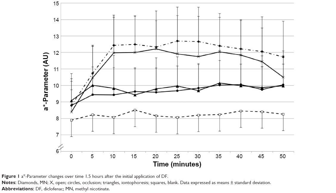 | Figure 1 a*-Parameter changes over time 1.5 hours after the initial application of DF. |
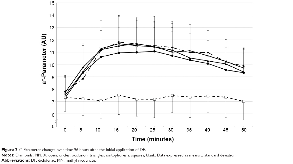 | Figure 2 a*-Parameter changes over time 96 hours after the initial application of DF. |
Differences between application modes at the different reservoir-estimation times were assessed using the Σa* values calculated from the different kinetics (Table 1). At 1.5 hours after the initial DF application, a significantly decreased response was detected for the occluded and iontophoretically delivered DF only. From 6 hours up to 32 hours post-DF application, a significantly reduced MN response was detected at all DF-pretreated skin sites. At 48 hours post-DF application, only the passive and occluded applications resulted in a significant reduction. At 72 hours post-DF application, only the response after the passively delivered DF remained significant. From 96 hours on, no differences between the MN responses were detected (see Table 1).
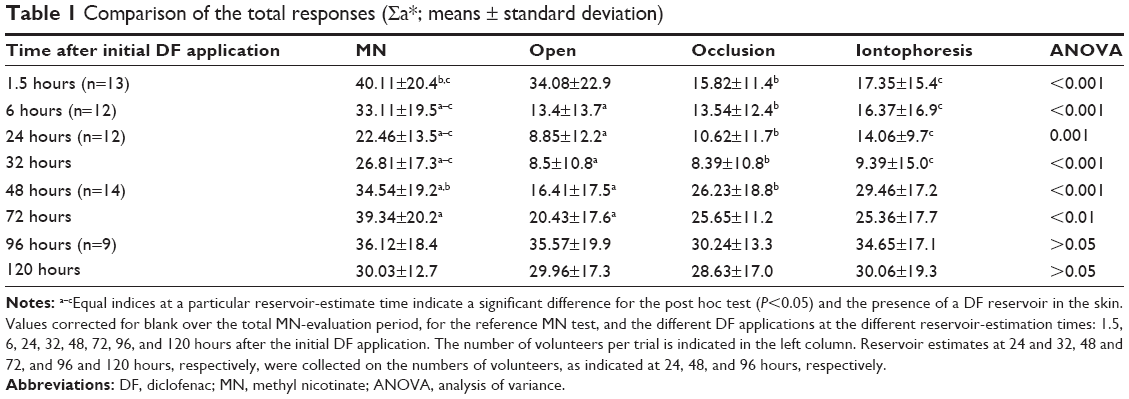 | Table 1 Comparison of the total responses (Σa*; means ± standard deviation) |
Discussion
The applied method – inhibition of a physiological reaction (MN) due to the presence of the inhibitor (DF) in the skin reservoir – enabled us to estimate penetration kinetics, DF skin-reservoir building, and emptying properties of three different application conditions up to 120 hours after initial DF application. In contrast with other reservoir protocols, an active substance-liberating action, such as occlusion or increased hydration, was not required.24 This may be a confirmation of the presence of DF in the viable tissue (dermal compound), since MN-induced vasodilatation is inhibited immediately.24
The application modality influenced the formation and the emptying of the reservoir. Penetration-enhancing factors, such as occlusion and current, induced faster formation and emptying of the reservoir compared to an open (passive) DF application. The presence of the reservoir for the passive DF application after 6 hours only is in contrast with earlier findings from our laboratory.11 Lambrecht et al11 presented the data of the MN response as calculated areas under the curve, relating the inhibition of MN response throughout a 65-minute time period. They found a 32% reduction of the MN response 1.5 hours after application, reaching borderline significance (P=0.04). The 25% reduction in MN response in the present study did not reach significance. Consequently, the weaker inhibition potential at 1.5 hours post-DF application noticed in both studies indicates lower bioavailability at that moment, due to a slower percutaneous penetration compared to the other application modes. According to the literature, the formation of a skin reservoir for topically applied substances is determined by a variety of factors (eg, lipid/water-solubility, protein-binding capacity, percutaneous absorption, compound concentration, clearance, application time, and application mode).11,14–16,23,24 Our results support the previous findings of Vickers, showing that increased drug diffusivity in the SC provoked by penetration enhancers results in faster formation of the reservoir.14 More specifically, Takahashi et al suggested increased diffusivity of DF into the SC from vehicles containing urea compared to vehicles without urea. The latter was explained by the hydration-enhancement effect of urea on the SC.31
The results concerning the penetration-enhancement effect of iontophoresis are in line with the results of Curdy et al on the in vivo uptake of piroxicam after passive, occlusive, and iontophoretic administration. Only after iontophoresis were enhanced drug uptakes found at 30, 60, and 125 minutes following the initial application at different depths in the SC. In contrast with our results, Curdy et al found no significant difference between passive delivery and the application under occlusion. A possible explanation could be the low lipophilicity of piroxicam resulting in a low passive uptake into the SC.32 Fang et al postulated that the route and mechanism for the iontophoretic delivery of DF through the skin might be different compared to passive delivery. Therefore, the importance of the SC as a rate-limiting barrier is reduced for iontophoretic delivery of DF.33
Our results indicate that the emptying of the reservoir is influenced by the application mode. After iontophoretic delivery, the DF reservoir was present up to 32 hours following initial application. However, after occlusive and passive delivery of DF, the reservoir was present up to 48 and 72 hours, respectively. It is well established that diffusivity influences clearance from the reservoir, with faster emptying as a function of increasing diffusivity. Increasing SC hydration as well as iontophoresis have been shown to be effective methods for enhancing the percutaneous penetration of DF.33 The present data fit in the model proposed by Roberts et al with a shorter lag time resulting in a faster reservoir emptying.24 Equally, based on the results of Rougier et al13 one can assume that the application with the fastest reservoir-buildup conditions may result in a greater delivery of active substances to the viable tissues, which may have an effect on the clinical outcomes of the physiotherapeutic treatment.
Within the limitations of our experimental design, we can hypothesize that the faster emptying of the reservoir after iontophoretic delivery compared to application under occlusion may be an indication for a higher diffusivity during the current-assisted application, leading to a superior delivery under iontophoretic circumstances.
Limitations of the present study are the lack of information on absolute DF quantities entering the viable skin and the fact that the methods used did not allow differentiation between an epidermal reservoir and a dermal reservoir. Further research estimating in vivo tissue concentrations after different modes of application are required for further elaboration of the pharmacodynamics of topical applied substances in physiotherapeutic practice.
Conclusion
This study measured the penetration kinetics and reservoir properties of DF after a single topical passive, occlusive, and electrical assisted application in a realistic physiotherapeutic setting. The results indicate that the contribution of occlusive and passive penetration in the iontophoretic delivery can be substantial. The prompt inhibition of the vasoactive reaction may be an indication for a dermal DF reservoir. The formation and emptying of the reservoir was found to be dependent on the application mode.
Disclosure
The authors report no conflicts of interest in this work.
References
Kaelin DL, Oh TH, Lim PA, Brander VA, Biundo JJ Jr. Rehabilitation of orthopedic and rheumatologic disorders. 4. Musculoskeletal disorders. Arch Phys Med Rehabil. 2000;81(3 Suppl 1):S73–S77; quiz S78–S86. | ||
Rovenský J, Miceková D, Gubzová Z, et al. Treatment of knee osteoarthritis with a topical non-steroidal antiinflammatory drug. Results of a randomized, double-blind, placebo-controlled study on the efficacy and safety of a 5% ibuprofen cream. Drugs Exp Clin Res. 2001;27(5–6):209–221. | ||
Machen J, Whitefield M. Efficacy of a proprietary ibuprofen gel in soft tissue injuries: a randomised, double-blind, placebo-controlled study. Int J Clin Pract. 2002;56(2):102–106. | ||
Başkurt F, Ozcan A, Algun C. Comparison of effects of phonophoresis and iontophoresis of naproxen in the treatment of lateral epicondylitis. Clin Rehabil. 2003;17(1):96–100. | ||
Pierre MB, Dos Santos Miranda Costa I. Liposomal systems as drug delivery vehicles for dermal and transdermal applications. Arch Dermatol Res. 2011;303(9):607–621. | ||
Meshali M, Abdel-Aleem H, Sakr F, Nazzal S, El-Malah Y. Effect of gel composition and phonophoresis on the transdermal delivery of ibuprofen: in vitro and in vivo evaluation. Pharm Dev Technol. 2011;16(2):93–101. | ||
Clijsen R, Baeyens JP, Barel AO, Clarys P. Influence of the timing of ultrasound application on the penetration of corticosteroids. Skin Res Technol. 2013;19(1):e279–e282. | ||
Aggarwal G, Dhawan S, Harikumar SL. Formulation, in vitro, and in vivo evaluation of matrix-type transdermal patches containing olanzapine. Pharm Dev Technol. 2013;18(4):916–925. | ||
Turner NG, Kalia YN, Guy RH. The effect of current on skin barrier function in vivo: recovery kinetics post-iontophoresis. Pharm Res. 1997;14(9):1252–1257. | ||
Luksurapan W, Boonhong J. Effects of phonophoresis of piroxicam and ultrasound on symptomatic knee osteoarthritis. Arch Phys Med Rehabil. 2013;94(2):250–255. | ||
Lambrecht R, Clarys P, Clijsen R, Barel AO. Determination of the in vivo bioavailability of iontophoretically delivered diclofenac using a methyl nicotinate skin inflammation assay. Skin Res Technol. 2006;12(3):211–216. | ||
Clijsen R, Taeymans J, Baeyens J, Barel A, Clarys P. The effects of iontophoresis in the treatment of musculoskeletal disorders – a systematic review and meta-analysis. Drug Deliv Lett. 2012;2(3):180–194. | ||
Rougier A, Dupuis D, Lotte C, Roguet R. The measurement of the stratum corneum reservoir. A predictive method for in vivo percutaneous absorption studies: influence of application time. J Invest Dermatol. 1985;84(1):66–68. | ||
Vickers CF. Stratum corneum reservoir for drugs. Adv Biol Skin. 1972;12:177–189. | ||
Pelchrzim R, Weigmann HJ, Schaefer H, et al. Determination of the formation of the stratum corneum reservoir for two different corticosteroid formulations using tape stripping combined with UV/VIS spectroscopy. J Dtsch Dermatol Ges. 2004;2(11):914–919. | ||
Clarys P, Gabard B, Barel AO. A qualitative estimate of the influence of halcinonide concentration and urea on the reservoir formation in the stratum corneum. Skin Pharmacol Appl Skin Physiol. 1999;12(1–2):85–89. | ||
Pershing LK, Bakhtian S, Poncelet CE, Corlett JL, Shah VP. Comparison of skin stripping, in vitro release, and skin blanching response methods to measure dose response and similarity of triamcinolone acetonide cream strengths from two manufactured sources. J Pharm Sci. 2002;91(5):1312–1323. | ||
Pellanda C, Ottiker E, Strub C, et al. Topical bioavailability of triamcinolone acetonide: effect of dose and application frequency. Arch Dermatol Res. 2006;298(5):221–230. | ||
Chambin-Remoussenard O, Treffel P, Bechtel Y, Agache P. Surface recovery and stripping methods to quantify percutaneous absorption of caffeine in humans. J Pharm Sci. 1993;82(11):1099–1101. | ||
Zesch A, Schaefer H, Stüttgen G. The quantitative distribution of percutaneously applied caffeine in the human skin. Arch Dermatol Res. 1979;266(3):277–283. | ||
Benowitz NL, LakeT, Keller KH, Lee BL. Prolonged absorption with development of tolerance to toxic effects after cutaneous exposure to nicotine. Clin Pharmacol Ther. 1987;42(1):119–220. | ||
Roberts MS, Cross SE. A physiological pharmacokinetic model for solute disposition in tissues below a topical application site. Pharm Res. 1999;16(9):1392–1398. | ||
Teichmann A, Jacobi U, Weigmann HJ, Sterry W, Lademann J. Reservoir function of the stratum corneum: development of an in vivo method to quantitatively determine the stratum corneum reservoir for topically applied substances. Skin Pharmacol Physiol. 2005;18(2):75–80. | ||
Roberts MS, Cross SE, Anissimov YG. Factors affecting the formation of a skin reservoir for topically applied solutes. Skin Pharmacol Physiol. 2004;17(1):3–16. | ||
Wilkin JK, Fortner G, Reinhardt LA, Flowers OV, Kilpatrick SJ, Streeter WC. Prostaglandins and nicotinate-provoked increase in cutaneous blood flow. Clin Pharmacol Ther. 1985;38(3):273–277. | ||
Treffel P, Gabard B. Feasibility of measuring the bioavailability of topical ibuprofen in commercial formulations using drug content in epidermis and a methyl nicotinate skin inflammation assay. Skin Pharmacol. 1993;6(4):268–275. | ||
Treffel P, Gabard B. Ibuprofen epidermal levels after topical application in vitro: effect of formulation, application time, dose variation and occlusion. Br J Dermatol. 1993;129(3):286–291. | ||
Clarys P, Alewaeters K, Lambrecht R, Barel AO. Skin color measurements: comparison between three instruments: the Chromameter(R), the DermaSpectrometer(R) and the Mexameter(R). Skin Res Technol. 2000;6(4):230–238. | ||
Weatherall IL, Coombs BD. Skin color measurements in terms of CIELAB color space values. J Invest Dermatol. 1992;99(4):468–473. | ||
Ale SI, Laugier JP, Maibach HI. Spacial variability of basal skin chromametry on the ventral forearm of healthy volunteers. Arch Dermatol Res. 1996;288(12):774–777. | ||
Takahashi K, Suzuki T, Sakano H, Mizuno N. Effect of vehicles on diclofenac permeation across excised rat skin. Biol Pharm Bull. 1995;18(4):571–575. | ||
Curdy C, Kalia YN, Naik A, Guy RH. Piroxicam delivery into human stratum corneum in vivo: iontophoresis versus passive diffusion. J Control Release. 2001;76(1–2):73–79. | ||
Fang J, Wang R, Huang Y, Wu PC, Tsai Y. Passive and iontophoretic delivery of three diclofenac salts across various skin types. Biol Pharm Bull. 2000;23(11):1357–1362. |
 © 2015 The Author(s). This work is published and licensed by Dove Medical Press Limited. The full terms of this license are available at https://www.dovepress.com/terms.php and incorporate the Creative Commons Attribution - Non Commercial (unported, v3.0) License.
By accessing the work you hereby accept the Terms. Non-commercial uses of the work are permitted without any further permission from Dove Medical Press Limited, provided the work is properly attributed. For permission for commercial use of this work, please see paragraphs 4.2 and 5 of our Terms.
© 2015 The Author(s). This work is published and licensed by Dove Medical Press Limited. The full terms of this license are available at https://www.dovepress.com/terms.php and incorporate the Creative Commons Attribution - Non Commercial (unported, v3.0) License.
By accessing the work you hereby accept the Terms. Non-commercial uses of the work are permitted without any further permission from Dove Medical Press Limited, provided the work is properly attributed. For permission for commercial use of this work, please see paragraphs 4.2 and 5 of our Terms.