")
Back to Journals » Journal of Experimental Pharmacology » Volume 11
Evaluation of hypoglycemic, antihyperglycemic and antihyperlipidemic activities of 80% methanolic seed extract of Calpurnia aurea (Ait.) Benth. (Fabaceae) in mice
Authors Belayneh YM , Birru EM , Ambikar D
Received 14 April 2019
Accepted for publication 2 July 2019
Published 25 July 2019 Volume 2019:11 Pages 73—83
DOI https://doi.org/10.2147/JEP.S212206
Checked for plagiarism Yes
Review by Single anonymous peer review
Peer reviewer comments 2
Editor who approved publication: Professor Bal Lokeshwar
Yaschilal Muche Belayneh,1 Eshetie Melese Birru,2 Digambar Ambikar2
1Department of Pharmacy, College of Medicine and Health Sciences, Wollo University, Dessie, Ethiopia; 2Department of Pharmacology, College of Medicine and Health Sciences, University of Gondar, Gondar, Ethiopia
Background: Diabetes mellitus is one of the most common chronic health problems in the world. As currently available antidiabetic medications have limitations in terms of safety, efficacy, and cost, it is an important research area to investigate medicinal plants for new antidiabetic compounds that can lead to effective, safe and less costly pharmacotherapy. The present study was done to evaluate the antidiabetic and antidyslipidemic activities of 80% methanolic seed extract of Calpurnia aurea (Ait.) Benth. (Fabaceae) in mice.
Methods: Blood glucose lowering activity of three doses (2.75 mg/kg, 5.5 mg/kg and 11 mg/kg) of the hydromethanolic seed extract of Calpurnia aurea was studied in three animal models: normoglycemic mice, oral glucose-loaded mice, and streptozotocin-induced diabetic mice. Additionally, the effect of the seed extract on body weight and serum lipid profile was studied in the streptozotocin-induced diabetic mice. Glibenclamide (5 mg/kg) was used as a standard drug in all animal models of the study. Blood glucose level was measured using a glucose meter, whereas serum lipid level was measured using an automated chemistry analyzer. Data were analyzed using one-way analysis of variance followed by Tukey’s post hoc multiple comparison test.
Results: Hydromethanolic extract of C. aurea seeds showed blood glucose lowering activity in all animal models, and it improved body weight loss and diabetic dyslipidemia in diabetic mice after 14 days of treatment.
Conclusion: This study revealed that hydromethanolic extract of Calpurnia aurea seeds has significant hypoglycemic, antihyperglycemic and antihyperlipidemic activities.
Keywords: diabetes mellitus, calpurnia aurea, streptozotocin, seed, mice
Background
Diabetes mellitus (DM) is one of four priority non-communicable public health problems targeted for action by WHO.1 Diabetes directly affects lipid levels in the blood, resulting in diabetic dyslipidemia.2 Diabetic patients are likely to have lower serum levels of high-density lipoprotein cholesterol (HDL-C), higher serum levels of triglyceride (TG), and similar serum values for low-density lipoprotein cholesterol (LDL-C) but with higher levels of small dense LDL when compared with non-diabetic patients.3,4
Currently available medications for DM are often limited in efficacy, carry the risk of adverse effects, and are often too costly, especially for the developing world.5 Therefore, searching for plant-derived antidiabetic compounds which are accessible and do not require intensive and costly pharmaceutical processing is an attractive research area. It was estimated that more than 1,000 plant species are traditionally being used for the treatment of DM.5 The families of plants with the most potent antidiabetic activities include Leguminosae (Fabaceae), Asteraceae, Moraceae, Lamiaceae, Liliaceae, Cucurbitaceae, Rosaceae, Euphorbiaceae, and Araliaceae.6
Calpurnia aurea (Ait.) Benth. (Fabaceae) is a yellow-flowered, multi-stemmed, 3–4 m tall small tree or shrub widely distributed in Africa, ranging from the Cape Province to Eritrea, and it also occurs in southern India.7,8 There are two subspecies of C. aurea, subsp. aureu which is found in Ethiopia and other parts of Africa and subsp. indica which occurs in India.9
C. aurea (Ait.) Benth. (Fabaceae) is traditionally used for the treatment of DM in parts of Ethiopia. An ethnobotanical survey of Shenasha, Agew-awi, and Amhara peoples of northwest Ethiopia reported that the seed, as well as the leaf of the plant, is used orally for the treatment of DM.10 Another survey in Nekemtae town (east Wollega, Ethiopia) reported that a leaf decoction of the plant is taken orally to treat DM.11 However, the antidiabetic activity of this medicinal plant has not been scientifically studied.
There is considerable evidence that induction of oxidative stress is a key process in the pathogenesis of DM and diabetic complications.12–14 The role of antioxidants in treating diabetes and its complications through prevention of oxidative stress has been explained.13–15 C. aurea (Ait.) Benth. leaves and seeds have strong in vitro antioxidant activities.7,16
The antidiabetic activity of medicinal plants is mainly due to the presence of alkaloids, phenolic compounds, flavonoids, and terpenoids.5,6,17–19 A previous preliminary phytochemical study has shown that the hydromethanolic extract of C. aurea seeds also contains these secondary metabolites, which are known to have blood glucose lowering activity.20
The present study was therefore undertaken to investigate the antidiabetic and antidyslipidemic effects of an 80% methanolic seed extract of C. aurea (Ait.) Benth. (Fabaceae) using in vivo models. The findings of this study may serve as baseline information for the scientific community to further investigate the plant C. aurea by initiating advanced studies on molecular mechanisms with identification of the active phytochemicals which may serve as lead compounds for the development of new antidiabetic drugs.
Materials and methods
Collection of plant materials
Fresh matured seeds of C. aurea were collected from the compound of the University of Gondar, Gondar town (located in the Central Gondar zone of Amhara region, northwest Ethiopia) in January 2017. Taxonomic identification of the plant was done by a botanist, and a specimen of the plant material was preserved in the Herbarium of the Biology Department, University of Gondar with a voucher number YM001 for future reference.
Drugs, chemicals, and instruments
The following drugs, chemicals, and instruments were used during the study. Streptozotocin (STZ; Sigma Aldrich Co., St Louis, MO, USA), glibenclamide (GLC; Julphar Pharmaceuticals, Ras Al Khaimah, United Arab Emirates), citric acid monohydrate (Lab Tech Chemicals, India), tri-sodium citrate dihydrate (Blulux Laboratories, Faridabad, India), methanol absolute (Nice Chemicals Private Limited, Ernakulam, India), 40% glucose solution (Reyoung Pharmaceuticals, Shandong, China), sterilized water for injections (Nirma Ltd, Ahmedabad, India), analytical balance, pH meter, i-QARE DS-W® blood glucose meter and strips (Alliance International, Taiwan), distilled water (DW), automated chemistry analyzer (Shenzhen Mindray Bio-medical Electronics Co., Ltd, Shenzhen, China). All chemicals used were of analytical grade.
Preparation of plant crude extracts
The seeds of the plant were thoroughly washed with DW to remove dirt and then dried under shade with optimal ventilation. Then, the dried seeds were pulverized. The coarse-powdered seeds (1 kg) were macerated in 80% methanol for 72 h and then the extracts were filtered using Whatman filter paper No.1. The marc was remacerated two times with fresh solvent, each for 72 h, and the filtrates obtained from the successive maceration were dried in an oven at 40°C.
Experimental animals
Healthy male Swiss albino mice (weighing 25–30 g and aged 8–12 weeks) were used in all experiments except for the acute oral toxicity tests. The mice were obtained from the Ethiopian Public Health Institute (EPHI) and they were kept in the animal house of the Department of Pharmacology, University of Gondar. The animals were fed with a standard pellet diet and water ad libitum. Animals were acclimatized to the laboratory conditions for a week before the initiation of the experiment. Fasting animals were kept in raised mesh bottom cages to prevent coprophagy.
Acute toxicity study
The acute oral toxicity test of the hydromethanolic seed extract was done based on the limit and main test recommendations of OECD No 425 Guideline.21 On the first day of the test, one female Swiss albino mouse fasted for 3 h was given 2,000 mg/kg of the extract orally. As mortality was observed in the first mouse that received 2,000 mg/kg of the seed extract, the main test was conducted in order to determine the LD50. In the main test single female mice were dosed in sequence at 48 h intervals using a starting dose of 175 mg/kg and a dose progression factor of 3.2. The dosing was stopped after testing six consecutive animals because five reversals (response, which is the death of the mouse, was observed at one dose, and a non-response is observed at the next dose tested) occurred. Animals were observed individually at least once during the first 30 min after dosing, periodically during the first 24 h with special attention given during the first 4 h, and daily thereafter, for a total of 14 days.
Grouping and dosing of animals
Male animals were used for normoglycemic, oral glucose loaded, and diabetic mice models because female mice are less sensitive to streptozotocin,22,23 and they are also less sensitive to insulin compared to male animals.24
In the normoglycemic, oral glucose loaded and single dose treated diabetic animal models, mice were randomly divided into five groups (each group containing six mice). In all three animal models, Group I (negative control) was treated with 10 mL/kg DW; Groups II, III, and IV were treated with 2.75 mg/kg, 5.5 mg/kg, and 11 mg/kg hydromethanolic seed extract, respectively; and Group V (positive control) was treated with the standard drug, 5 mg/kg GLC.
In the repeated dose treated diabetic mice, animals were randomly divided into six groups (five groups of diabetic mice and one additional group of normal mice, each group containing six mice). Group I was treated with 10 mL/kg DW and served as a diabetic control; Groups II, III, and IV were treated with 2.75 mg/kg, 5.5 mg/kg, and 11 mg/kg hydromethanolic seed extract, respectively; Group V was treated with 5 mg/kg GLC and served as a diabetic positive control; and Group VI (a group of normoglycemic mice) was treated with 10 mL/kg DW and served as a normal control.
The three different doses of hydromethanolic C. aurea seed extract (CASE) for this study were selected based on the result of the acute oral toxicity test. GLC (5 mg/kg) was selected as a standard drug for this study based on reports of earlier studies.25–27 The study was conducted using the oral route of administration because people traditionally use the plant via the oral route.10,11 All the doses were administered at a volume not greater than 10 mL/kg body weight of mice.21
Measurement of blood glucose level
In all cases, blood samples for blood glucose measurement were withdrawn from the tail vein of each animal by cutting the tip of the tail aseptically. Blood glucose level (BGL) was measured using a DS-W® blood glucose meter. Measurement of the BGL was done in triplicate and the average value was taken.
Induction of experimental diabetes
Experimental diabetes was induced using STZ. The drug was dissolved in 0.1 M cold citrate buffer (pH=4.5). The freshly prepared solution was then administered intraperitoneally at 150 mg/kg dose to mice28 which were fasted overnight for 16 h prior to administration. Thirty minutes after the administration of STZ, animals were allowed to have free access to food and water. Additionally, animals were allowed to drink 5% sucrose solution 6 h after the administration of STZ for the next 24 h to prevent death secondary to hypoglycemic shock. Then, animals were screened for the induction of diabetes four days after STZ injection. Mice which showed fasting BGL>200 mg/dL were included in the study as diabetic mice.26,29 Diabetic mice were randomly divided into different groups just after the screening to perform the experimental studies. The bedding of the cages was changed every day after STZ injection to maintain dryness of cages for polyuric diabetic animals.
Assessing hypoglycemic activity in normoglycemic mice
Mice, which were fasted overnight for 16 h, were randomly divided into five different groups (six animals per group). Then, the animals were treated according to their respective groupings as mentioned above. BGL of each mouse was measured just before treatment (at 0 h) as a baseline, and then at 1, 2, 4 and 6 h post-treatment.
Assessing the antihyperglycemic activity of the seed extract in oral glucose-loaded mice
Mice were used to evaluate the effect of the plant extract on oral glucose tolerance because overnight fasting increases their insulin sensitivity.30,31 Thus, the model can be more sensitive to screen anti-hyperglycemic activity of the plant extract. Overnight fasted (for 16 h) mice were randomly divided into five groups (six mice per group). Then, mice were treated with DW, hydromethanolic seed extract and GLC according to their respective groupings. Thirty minutes after the treatments,25,32 2.5 g/kg of glucose solution were administered to each animal orally.25 BGLs were measured for each animal just before treatment (at 0 min) as a baseline, and then at 30, 60 and 120 min following oral glucose administration.25,33
Assessing the effect of a single dose of the seed extract on blood glucose of diabetic mice
After overnight fasting for 16 h, STZ-induced diabetic mice were assigned randomly into five groups and treated with DW, plant extract and GLC according to their respective groupings as explained above. BGL was measured just before treatment (at 0 h) as a baseline, and then at 2, 4, 6 and 8 h post-treatment.
Assessing the effects of repeated doses of the seed extract on blood glucose, body weight and serum lipid level of diabetic mice
Overnight fasted (for 16 h) diabetic mice and normal mice were randomly divided into six groups and treated with DW, seed extract and GLC once daily in the morning for 14 days according to their respective groupings as explained above. BGL and body weight of diabetic mice were measured just before starting treatment on the first day as a baseline, and then on the seventh and 14th day of treatment following overnight fasting for 16 h.34
On the 15th day, overnight fasted mice were euthanized by sodium pentobarbitone (150 mg/kg IP) anesthesia and blood samples were collected via cardiac puncture from each mouse. The blood samples were kept at room temperature for 2 h to allow coagulation and then centrifuged at 2,000 rpm for 10 min. Then, serum samples were prepared from supernatant of the centrifuged blood samples to measure the level of serum TG, TC, and HDL-C using an automated chemistry analyzer.
Ethical considerations
The experiment was conducted in accordance with the Guide for the Care and Use of Laboratory Animals,35 and the proposal of the study was submitted and approved by the ethical review committee of the School of Pharmacy, University of Gondar before the commencement of the study.
Statistical analysis
Data were expressed as mean ± standard error of the mean. Means of all parameters among groups and within a group were compared using one-way ANOVA followed by Tukey’s post hoc multiple comparison test. Values of p<0.05 were considered statistically significant. SPSS Version 20 software (IBM Corporation, Armonk, NY, USA) was used for statistical analysis.
Result
Percentage yield of the hydromethanolic seed extract of C. aurea
A total of 136 g of dried yellowish brown gummy seed extract was harvested at the end of the extraction process from 1 kg of dried powdered seeds (percentage yield, 13.60% (w/w)).
Acute oral toxicity study
The acute oral toxicity study revealed that the median lethal dose (LD50) of CASE is between 55 mg/kg and 175 mg/kg based on the main test recommendations of OECD guideline no 425 (Table 1).
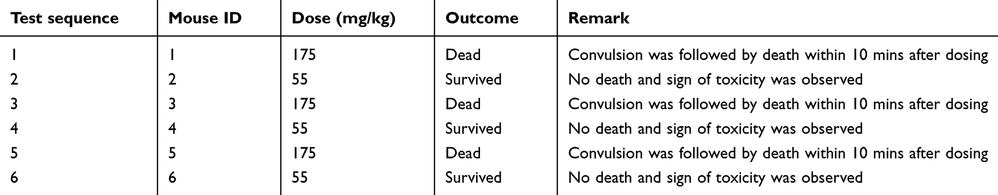 |
Table 1 Acute oral toxicity study of the 80% methanolic seed extract of Calpurnia aurea |
Hypoglycemic activity of 80% methanolic seed extract of C. aurea in normoglycemic mice
The effect of hydromethanolic seed extract of C. aurea on fasting BGL of normoglycemic mice is summarized in Table 2. Between-groups analysis revealed no significant difference in baseline fasting BGL across groups. CASE 11 mg/kg significantly reduced the BGL at the fourth and sixth hours (p<0.05) compared to the negative control. Similarly, BGL was significantly reduced by 5 mg/kg GLC at the second (p<0.05), fourth (p<0.01) and sixth (p<0.05) hour compared to the negative control. A statistically significant difference in BGL was not observed when groups treated with different doses of the seed extract compared with each other and compared with the positive control at all time points.
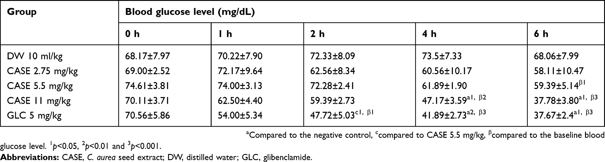 |
Table 2 Hypoglycemic activity of hydromethanolic Calpurnia aurea seed extract in normoglycemic mice |
Within-group analysis showed that treatment with 5.5 mg/kg CASE significantly reduced the BGL at the sixth hour (p<0.05) compared to the baseline level with a percentage reduction of 20.39%. Similarly, a significant reduction in BGL was induced with 11 mg/kg CASE at the fourth (p<0.01) and sixth (p<0.001) hour compared to the baseline level with percentage reduction in BGL, 32.72% and 46.11%, respectively. In addition, the standard drug (GLC) reduced the BGL significantly at the second, fourth and sixth hours compared to the baseline level with percentage reductions of 32.37%, 40.63%, and 46.61%, respectively.
Antihyperglycemic activity of the seed extract in oral glucose-loaded mice
There was no significant difference in baseline BGL across groups just before the administration of DW, plant extract and GLC (Table 3). Between-groups analysis showed that 5.5 mg/kg and 11 mg/kg CASE significantly reduced the hyperglycemia (p<0.05 in both cases) at the second hour compared to the negative control. Similarly, 5 mg/kg GLC reduced the BGL significantly at 60 and 120 min post oral glucose administration compared to the DW treated group. A statistically significant difference in BGL was not observed at all time points when the GLC treated group was compared with seed extract treated groups. Similarly, a statistically significant difference in BGL was not observed at all time points when all the seed extract treated groups were compared with one another.
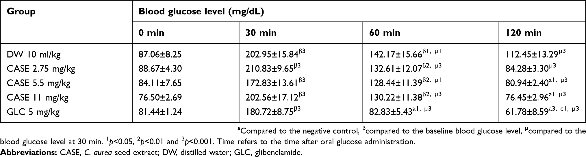 |
Table 3 Effect of the hydromethanolic Calpurnia aurea seed extract on oral glucose tolerance in normoglycemic mice |
Within-group analysis showed that oral glucose administration to mice caused a statistically significant (p<0.001) increment in BGL after 30 min in all groups regardless of the treatments given. Additionally, 60 min after oral glucose loading significant hyperglycemia was observed in all groups except the GLC treated group as compared to the respective baseline BGL. The BGL reduced to normal or the baseline level in all groups at the second hour post oral glucose load.
Antihyperglycemic activity of single dose of C. aurea seed extract in STZ-induced diabetic mice
A total of 69 male Swiss albino mice were injected with STZ and 52 of them were found to be diabetic (fasting BGL>200 mg/dL) four days after STZ injection, with a success rate of 75.36%. Among the 52 diabetic mice, one died before the administration of the test substances and all the remaining animals survived until the end of the experiment.
Antihyperglycemic activity of a single dose of the extracts was studied in STZ-induced diabetic mice. As shown in Table 4, between- and within-group comparisons were performed to analyze BGL differences across the various groups and time points, respectively. The between-group analysis indicated no significant difference in baseline fasting BGL across all groups. Similarly, a significant difference in BGL was not observed across all groups at the second hour post-treatment. Compared to the negative control, a significant BGL reduction was observed at sixth and eighth hour in 5.5 mg/kg and 11 mg/kg CASE treated groups; and at fourth, sixth, and eighth hours in the 5 mg/kg GLC treated group. There was no statistically significant difference in BGL at all time points when groups treated with the seed extract were compared to each other, and compared to the GLC treated group.
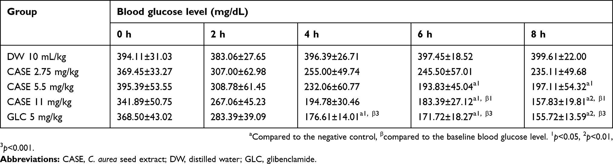 |
Table 4 Antihyperglycemic activity of single dose of Calpurnia aurea seed extract in streptozotocin-induced diabetic mice |
Within-group comparison indicated that significant BGL reduction was not observed in CASE 2.75 mg/kg, CASE 5.5 mg/kg and DW treated groups at all time points compared to the baseline fasting BGL. However, the percent reduction in BGL was recorded as 27.47% in the CASE 2.75 mg/kg treated group and 50.15% in the CASE 5.5 mg/kg treated group at the eighth hour compared to the respective baseline fasting BGL. CASE 11 mg/kg was able to decrease the BGL significantly at the sixth and eighth (p<0.05) hour compared to the initial value with percentage reductions of 46.36% and 51.47%, respectively. The standard drug, GLC, also produced a significant BGL reduction at the fourth, sixth and eighth (p<0.001) hour compared to the baseline level.
Antihyperglycemic activity of the repeated doses of C. aurea seed extract in STZ-induced diabetic mice
Between-group comparisons indicated that the baseline BGL of the diabetic groups was significantly higher than the baseline BGL of the normal control, but no statistically significant difference was observed in baseline BGL across diabetic groups (Table 5). Groups treated with 2.75 mg/kg, 5.5 mg/kg and 11 mg/kg CASE and 5 mg/kg GLC showed a significant reduction in BGL on the seventh and 14th day of treatment compared to the diabetic control. There was no statistically significant difference in BGL at all time points when groups treated with the different doses of CASE compared with each other. Similarly, the GLC treated group showed no significant difference in BGL at all time points when compared to CASE treated groups.
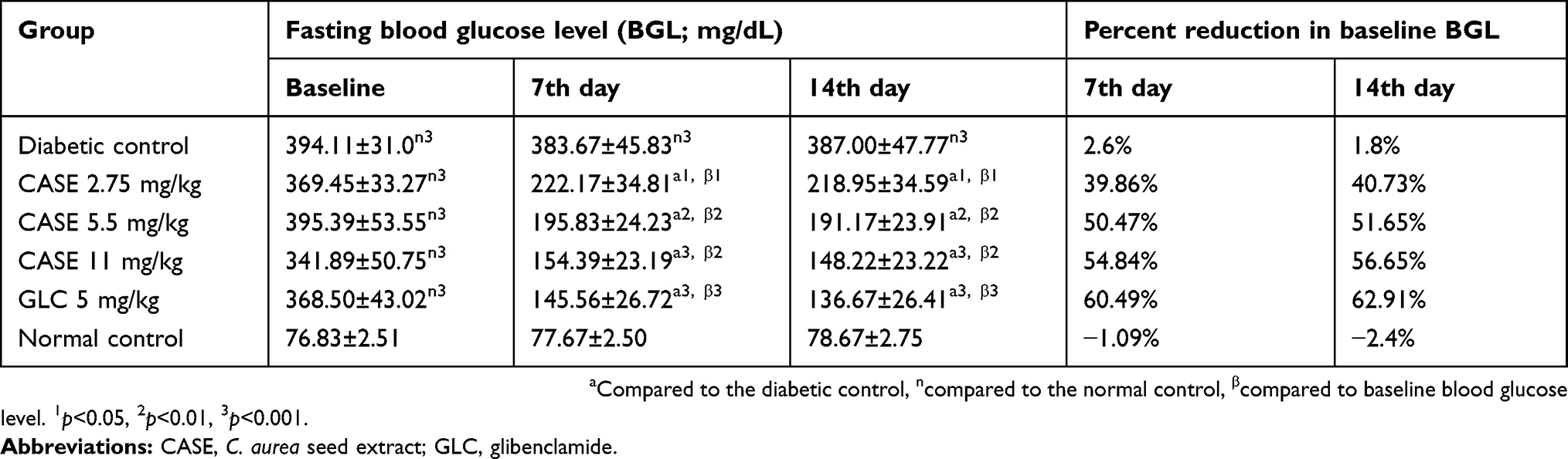 |
Table 5 Antihyperglycemic effect of repeated doses of Calpurnia aurea seed extract in Streptozotocin-induced diabetic mice |
Within-group analysis revealed that all the CASE treated groups and the GLC treated group showed a significant reduction in BGL at the seventh and 14th day of treatment compared to the baseline level. But a significant change in BGL was not observed in the diabetic and normal control groups at all time points compared to the baseline level (Table 5).
Effect of the repeated doses of C. aurea seed extract on body weight of STZ-induced diabetic mice
STZ-induced diabetes caused a statistically significant body weight loss in the diabetic control at the seventh and 14th day of treatment compared to the normal control group (Table 6). All the three doses of CASE (2.75, 5.5 and 11 mg/kg) and GLC significantly improved the body weight of diabetic mice at the 14th day of treatment compared to the DW treated diabetic control.
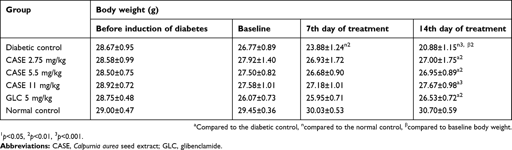 |
Table 6 Effect of the repeated doses of CASE on body weight of streptozotocin-induced diabetic mice |
Intra-group analysis revealed that the diabetic control showed significant (p<0.01) body weight loss at the 14th day of treatment compared to the baseline body weight, but the normal control, groups treated with the plant extract and the GLC treated group did not show a significant body weight change at all time points compared to the respective baseline body weight.
Effect of the repeated doses of C. aurea seed extract on serum lipid level of STZ-induced diabetic mice
There was a significant elevation (p<0.001) of serum total cholesterol, triglycerides, and a significant reduction (p<0.001) of HDL cholesterol in the diabetic control compared to the normal control (Table 7). The administration of 5.5 mg/kg and 11 mg/kg C. aurea seed extract for 14 days significantly reduced (p<0.05) the levels of serum total cholesterol while significantly increasing (p<0.05) the HDL cholesterol level. Similarly, all three doses of CASE significantly reduced the serum triglyceride level. The standard drug GLC also significantly reduced (p<0.001) TC and TG level while increasing (p<0.01) the HDL-C. Additionally, a significant difference in the level of serum TC, TG and HDL-C was not observed when groups treated with CASE compared with each other.
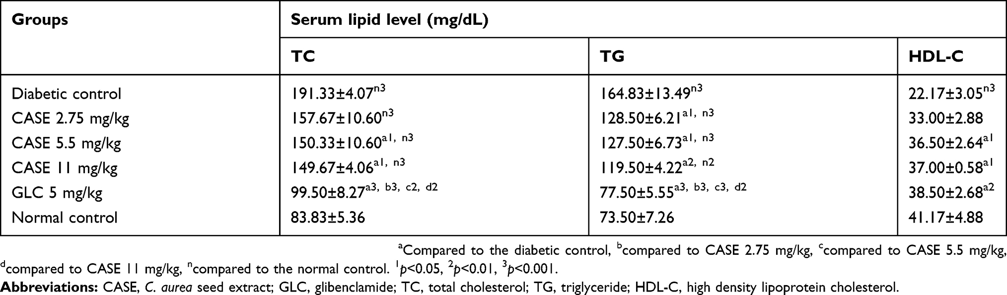 |
Table 7 Effect of repeated doses of Calpurnia aurea seed extract on serum lipid level of streptozotocin-induced diabetic mice |
Discussion
DM is one of the largest global health problems of the twenty-first century.36 There is a need for safer and more effective drug therapy because currently available medications for DM have definite limitations. Investigating plant-derived compounds, which are easily accessible and do not require intensive pharmaceutical processing, for the treatment of DM is an important research area.5,34
There was no previous acute oral toxicity study on the hydromethanolic seed extract of C. aurea. This study revealed that the median lethal dose of the hydromethanolic seed extract is between 55 mg/kg and 175 mg/kg, showing the toxic nature of the seeds.
In the present study, experimental diabetes in mice was induced using STZ [2-deoxy-2-(3-methyl-3-nitrosourea)-1-D-glucopyranose]. STZ-induced DM is well documented and a commonly used model of experimental diabetes in mice.37 Previous studies showed that single intraperitoneal injection of 150 mg/kg STZ can produce sustained hyperglycemia in mice at least for 8 weeks.38 Similarly, the present study revealed that STZ-induced persistent hyperglycemia without significant change in BGL during the study period of two weeks as observed in the diabetic control mice. STZ is a better diabetogenic agent than alloxan with wider species effectiveness and greater reproducibility, and this could be attributed to the fact that STZ is more stable in solution before and after injection in animals than alloxan.39 The three major mechanisms associated with pancreatic β cell death secondary to STZ exposure are DNA methylation, nitric oxide, and reactive oxygen species production.39 STZ toxicity to β cells is short-lived and further impairment of the surviving β cell function is due to hyperglycemic toxicity.40
In this study, there were no detectable differences in baseline BGL across groups in each animal model; additionally, the vehicle treated groups did not show significant reduction of BGL compared to the baseline level. However, significant reductions in BGL were observed in all models after the administration of the hydroalcoholic seed extract and standard drug, indicating changes induced on BGL were attributed to treatments received.
The study on normoglycemic mice revealed that the 80% methanolic seed extract of C. aurea at the dose of 5.5 mg/kg and 11 mg/kg showed significant hypoglycemic activity. Similarly, the extract at the dose of 5.5 mg/kg and 11 mg/kg showed significant antihyperglycemic activity after administration of single dose of the extract in oral glucose loaded mice as well as in STZ induced diabetic mice. Additionally, all three doses of CASE showed significant antihyperglycemic activity and improvement in body weight after administration of repeated doses of the extract in diabetic mice. The hypoglycemic and antihyperglycemic activities of CASE were dose-dependent. In all cases, a higher reduction in BGL was observed with 11mg/kg CASE.
The antidiabetic activity of medicinal plants is due to the presence of phytochemicals like alkaloids, phenolic compounds, flavonoids, and terpenoids.5,6,17–19 Flavonoids are known to have insulinogenic and pancreatic beta cell regenerating activities.6,17 Thus, the blood glucose lowering effect of the hydromethanolic extract may be due to the presence of these different secondary metabolites known to have antidiabetic activity with possible additive or synergistic effects.
The antidiabetic activity of CASE may be due to the induction of insulin secretion from beta cells of the pancreas or enhancement of glucose uptake in the peripheral tissue.41 However, detailed pharmacological and biochemical studies are required to identify the exact mechanism for the hypoglycemic and antihyperglycemic effects observed in the study.
The extract showed a relatively delayed onset of blood glucose lowering action compared to the standard drug. This might be due to the presence of compounds with a higher glycemic index that could lead to increased BGL following absorption. The presence of such an effect in the face of the blood glucose lowering actions by the active compounds could lead to a delay in the action of the plant extract.26
STZ-induced diabetes is associated with significant body weight loss.22,27 Mice with severe STZ-induced hyperglycemia tend to lose a large percentage of their body weight.22,38 Similarly, the present study revealed that STZ caused significant body weight loss in the diabetic control mice. STZ-induced diabetes leads to body weight loss due to increased wasting of fat stores,42 muscle and tissue proteins.43,44 Hence, the weight gain after repeated administration of the hydromethanolic C. aurea seed extract in STZ-induced diabetic mice suggests the antihyperglycemic activity of the extract.
One of the complications of DM is a disturbance in serum lipid level which is manifested mainly by high serum TG, TC, and low HDL-C.4,27 This lipid abnormality is due to activation of hormone-sensitive lipase that leads to increased lipolysis and increased secretion of VLDL from the liver.3,4 Insulin deficiency also causes decreased activity of lipoprotein lipase which leads to decreased clearance of VLDL and chylomicrons.45 Additionally, an increased triglyceride level can stimulate the enzymatic activity of cholesteryl ester transfer protein, resulting in increased triglyceride content of HDL and LDL. Triglyceride-enriched HDL particles are subjected to increased catabolism, whereas triglyceride-enriched LDL particles undergo subsequent hydrolysis via lipoprotein lipase or hepatic lipase, resulting in LDL particle size.4 In this study, the diabetic control showed significantly increased serum TG, TC, and decreased HDL-cholesterol as expected. Administration of CASE for 2 weeks significantly reduced serum TG, TC, and increased HDL-C in a dose-dependant manner, but it is not known whether the seed extract had a direct effect on lipid metabolism or the antidyslipidemic activity is achieved only due to the controlled hyperglycemia.
Conclusion
Methanolic extract of C. aurea seeds showed significant hypoglycemic, antihyperglycemic and antihyperlipidemic activities, justifying the traditional use of the plant for the treatment of DM. However, further phytochemical investigations are required to isolate and identify the active compounds responsible for the antidiabetic activity of the plant.
Abbreviations list
CASE, Calpurnia aurea seed extract; IP, intraperitoneal; LD50, Median lethal dose; OECD, Organization for Economic Cooperation and Development.
Availability of data and materials
All the datasets used and analyzed during the current study are available from the corresponding author on reasonable request.
Ethics approval
Ethical approval was obtained from the Ethical Review Committee of the School of Pharmacy, University of Gondar, before conducting the experiment.
Acknowledgments
We are grateful to the University of Gondar for funding this study.
An abstract of this paper was presented at the 29th Annual Conference of EPHA (Ethiopian Public Health Association) as a poster presentation and conference talk. The poster’s abstract was published in “Poster Abstracts” and is available at: http://www.etpha.org/conference/index.php/29thConference/29thConference/paper/view/1007.
Disclosure
The authors declare that they have no competing interests.
References
1. WHO. Global report on diabetes. Geneva, Switzerland; 2016.
2. David S, Paul Z. Diabetes and hyperlipidemia: a direct quantitative analysis—A direct analysis of the effects of insulin resistance on lipid levels in relation to atherosclerotic coronary artery disease. World J Cardiovasc Dis 2012;2(1):20–25. doi:10.4236/wjcd.2012.21004
3. Basak RC, Chatterjee M, Sarma P. An overview on management of diabetic dyslipidemia. J Diabetes Endocrinol. 2013;4(3):27–36.
4. Wu L, Parhofer KG. Diabetic dyslipidemia. Metabolism. 2014;63(12):1469–1479. doi:10.1016/j.metabol.2014.08.010
5. Rao MU, Sreenivasulu M, Chengaiah B, Reddy KJ, Chetty CM. Herbal medicines for diabetes mellitus: a review. Int J PharmTech Res. 2010;2(3):1883–1892.
6. Patel D, Prasad S, Kumar R, Hemalatha S. An overview on antidiabetic medicinal plants having insulin mimetic property. Asian Pac J Trop Biomed. 2012;2(4):320–330. doi:10.1016/S2221-1691(12)60032-X
7. Adedapo AA, Jimoh FO, Koduru S, Afolayan AJ, Masika PJ. Antibacterial and antioxidant properties of the methanol extracts of the leaves and stems of Calpurnia aurea. BMC Complement Altern Med. 2008;8(1):1. doi:10.1186/1472-6882-8-62
8. Zorloni A, Penzhorn BL, Eloff JN. Extracts of Calpurnia aurea leaves from southern Ethiopia attract and immobilise or kill ticks. Vet Parasitol. 2010;168(1):160–164. doi:10.1016/j.vetpar.2009.10.026
9. Beaumont A, Beckett R, Edwards T, Stiron C. Revision of the genus Calpurnia (Sophoreae: leguminosae). Bothalia. 1999;29(1):5–23. doi:10.4102/abc.v29i1.568
10. Giday M, Teklehaymanot T, Animut A, Mekonnen Y. Medicinal plants of the Shinasha, Agew-awi and Amhara peoples in northwest Ethiopia. J Ethnopharmacol. 2007;110(3):516–525. doi:10.1016/j.jep.2006.10.011
11. Suleman S, Alemu T. A survey on utilization of ethnomedicinal plants in Nekemte Town, East Wellega (Oromia), Ethiopia. J Herbs Spices Med Plants. 2012;18(1):34–57. doi:10.1080/10496475.2011.645188
12. Matough FA, Budin SB, Hamid ZA, Alwahaibi N, Mohamed J. The role of oxidative stress and antioxidants in diabetic complications. Sultan Qaboos Univ Med J. 2012;12(1):5. doi:10.12816/0003082
13. Asmat U, Abad K, Ismail K. Diabetes mellitus and oxidative stress - A concise review. Saudi Pharm J. 2016;24(5):547–553.
14. Pérez-Matute P, Zulet MA, Martínez JA. Reactive species and diabetes: counteracting oxidative stress to improve health. Curr Opin Pharmacol. 2009;9(6):771–779. doi:10.1016/j.coph.2009.08.005
15. Robertson RP. Chronic oxidative stress as a central mechanism for glucose toxicity in pancreatic islet beta cells in diabetes. J Biol Chem. 2004;279(41):42351–42354. doi:10.1074/jbc.R400019200
16. Mulata H, Gnanasekaran N, Melaku U, Daniel S. Phytochemical screening and assessment of in vitro antioxidant activities of Calpurnia aurea seeds and leaves. Ijppr Human. 2015;2(2):1–12.
17. Jung M, Park M, Lee HC, Kang Y-H, Kang ES, Kim SK. Antidiabetic agents from medicinal plants. Curr Med Chem. 2006;13(10):1203–1218. doi:10.2174/092986706776360860
18. Subramanian SP, Prasath GS. Antidiabetic and antidyslipidemic nature of trigonelline, a major alkaloid of fenugreek seeds studied in high-fat-fed and low-dose streptozotocin-induced experimental diabetic rats. Biomed Preventive Nutr. 2014;4(4):475–480. doi:10.1016/j.bionut.2014.07.001
19. Verma AK, Singh H, Satyanarayana M, et al. Flavone-based novel antidiabetic and antidyslipidemic agents. J Med Chem. 2012;55(10):4551–4567. doi:10.1021/jm201107g
20. Getiye Y, Tolessa T, Engidawork E. Antihypertensive activity of 80% methanol seed extract of Calpurnia aurea (Ait.) Benth. subsp. aurea (Fabaceae) is mediated through calcium antagonism induced vasodilation. J Ethnopharmacol. 2016;189(189):99–106. doi:10.1016/j.jep.2016.04.056
21. OECD/OCDE. OECD guideline for the testing of chemichals: acute oral toxicity; Up-and-Down Procedure (UDP). OECD, No 4252008.
22. Deeds M, Anderson J, Armstrong A, et al. Single dose streptozotocin-induced diabetes: considerations for study design in islet transplantation models. Lab Anim. 2011;45(3):131–140. doi:10.1258/la.2010.010090
23. Furman BL. Streptozotocin-induced diabetic models in mice and rats. Current Protocols Pharmacol. 2015;5(47):1–5.
24. Vital P, Larrieta E, Hiriart M. Sexual dimorphism in insulin sensitivity and susceptibility to develop diabetes in rats. J Endocrinol. 2006;190(2):425–432. doi:10.1677/joe.1.06596
25. Tesfaye A, Makonnen E, Gedamu S. Hypoglycemic and antihyperglycemic activity of aqueous extract of Justicia Schimperiana leaves in normal and streptozotocin-induced diabetic mice. IntJ Pharma Sci Res. 2016;7(02):110–113.
26. Tamiru W, Engidawork E, Asres K. Evaluation of the effects of 80% methanolic leaf extract of Caylusea abyssinica (fresen.) fisch. & Mey. on glucose handling in normal, glucose loaded and diabetic rodents. BMC Complement Altern Med. 2012;12(1):1. doi:10.1186/1472-6882-12-151
27. Yanyan Z, Fu F, Ting C, Zhongwen L, Qingwu WS. Antidiabetic and antihyperlipidemic activities of Forsythia suspensa (Thunb.) Vahl (fruit) in streptozotocin-induced diabetic mice. J Ethnopharmacol. 2016;192:256–263. doi:10.1016/j.jep.2016.07.002
28. Etuk E. Animal models for studying diabetes mellitus. Agric Biol JN Am. 2010;1(2):130–134.
29. Baquer NZ, Kumar P, Taha A, Kale R, Cowsik S, McLean P. Metabolic and molecular action of Trigonella foenum-graecum (fenugreek) and trace metals in experimental diabetic tissues. J Biosci. 2011;36(2):383–396.
30. Bowe JE, Franklin ZJ, Hauge-Evans AC, King AJ, Persaud SJ, Jones PM. Metabolic phenotyping guidelines: assessing glucose homeostasis in rodent models. J Endocrinol. 2014;222(3):G13–G25. doi:10.1530/JOE-14-0182
31. Ayala JE, Samuel VT, Morton GJ, et al. Standard operating procedures for describing and performing metabolic tests of glucose homeostasis in mice. Dis Model Mech. 2010;3(9–10):525–534. doi:10.1242/dmm.006239
32. Chika A, Bello SO. Antihyperglycaemic activity of aqueous leaf extract of Combretum micranthum (Combretaceae) in normal and alloxan-induced diabetic rats. J Ethnopharmacol. 2010;129(1):34–37. doi:10.1016/j.jep.2010.02.008
33. Rajurkar B. Phyto-pharmacological investigations of Clerodendrum infortunatum Gartn. Int Res J Pharm. 2011;2(11):130–132.
34. Toma A, Makonnen E, Mekonnen Y, Debella A, Adisakwattana S. Antidiabetic activities of aqueous ethanol and n-butanol fraction of Moringa stenopetala leaves in streptozotocin-induced diabetic rats. BMC Complement Altern Med. 2015;15(1):1. doi:10.1186/s12906-015-0779-0
35. Clark J, Baldwin R, Bayne K, et al. Guide for the Care and Use of Laboratory Animals. Washington, DC: Institute of Laboratory Animal Resources, National Research Council; 1996:125.
36. IDF. Diabetes Atlas.
37. Arora S, Ojha SK, Vohora D. Characterisation of streptozotocin induced diabetes mellitus in swiss albino mice. Global J Pharmacol. 2009;3(2):81–84.
38. He-Lin T, Li-Shun W, Zhong-Xin X, Ru-Tong Z, Dong-Ling J, Jin-Sheng G. Correlation between blood glucose level and diabetes signs in streptozotocin induced diabetic mice. Global J Pharmacol. 2010;4(3):111–116.
39. Eleazu CO, Eleazu KC, Chukwuma S, Essien UN. Review of the mechanism of cell death resulting from streptozotocin challenge in experimental animals, its practical use and potential risk to humans. J Diabetes Metab Disord. 2013;12(1):60. doi:10.1186/2251-6581-12-60
40. Wu J, Yan L-J. Streptozotocin-induced type 1 diabetes in rodents as a model for studying mitochondrial mechanisms of diabetic β cell glucotoxicity. Diabetes Metab Syndrome Obesity. 2015;8:181.
41. Sharma S, Choudhary M, Bhardwaj S, Choudhary N, Rana AC. Hypoglycemic potential of alcoholic root extract of Cassia occidentalis Linn. in streptozotocin induced diabetes in albino mice. Bulletin Faculty Pharm Cairo Univ. 2014;52(2):211–217. doi:10.1016/j.bfopcu.2014.09.003
42. Howarth F, Jacobson M, Shafiullah M, Adeghate E. Long-term effects of streptozotocin-induced diabetes on the electrocardiogram, physical activity and body temperature in rats. Exp Physiol. 2005;90(6):827–835. doi:10.1113/expphysiol.2004.028316
43. Chikhi I, Allali H, Dib MEA, Medjdoub H, Tabti B. Antidiabetic activity of aqueous leaf extract of Atriplex halimus L. (Chenopodiaceae) in streptozotocin–induced diabetic rats. Asian Pac J Trop Dis. 2014;4(3):181–184. doi:10.1016/S2222-1808(14)60501-6
44. Kumar S, Kumar V, Prakash O. Antidiabetic and hypolipidemic activities of Kigelia pinnata flowers extract in streptozotocin induced diabetic rats. Asian Pac J Trop Biomed. 2012;2(7):543–546. doi:10.1016/S2221-1691(12)60093-8
45. Goldberg IJ. Diabetic dyslipidemia: causes and consequences. J Clin Endocrinol Metab. 2001;86(3):965–971. doi:10.1210/jcem.86.3.7304
 © 2019 The Author(s). This work is published and licensed by Dove Medical Press Limited. The full terms of this license are available at https://www.dovepress.com/terms.php and incorporate the Creative Commons Attribution - Non Commercial (unported, v3.0) License.
By accessing the work you hereby accept the Terms. Non-commercial uses of the work are permitted without any further permission from Dove Medical Press Limited, provided the work is properly attributed. For permission for commercial use of this work, please see paragraphs 4.2 and 5 of our Terms.
© 2019 The Author(s). This work is published and licensed by Dove Medical Press Limited. The full terms of this license are available at https://www.dovepress.com/terms.php and incorporate the Creative Commons Attribution - Non Commercial (unported, v3.0) License.
By accessing the work you hereby accept the Terms. Non-commercial uses of the work are permitted without any further permission from Dove Medical Press Limited, provided the work is properly attributed. For permission for commercial use of this work, please see paragraphs 4.2 and 5 of our Terms.