")
Back to Journals » Diabetes, Metabolic Syndrome and Obesity » Volume 12
Ethnic differences and heterogeneity in genetic and metabolic makeup contributing to nonalcoholic fatty liver disease
Authors Szanto KB, Li J, Cordero P, Oben JA
Received 14 August 2018
Accepted for publication 20 October 2018
Published 19 March 2019 Volume 2019:12 Pages 357—367
DOI https://doi.org/10.2147/DMSO.S182331
Checked for plagiarism Yes
Review by Single anonymous peer review
Peer reviewer comments 2
Editor who approved publication: Dr Steven F. Abcouwer
Krisztina B Szanto,1,2 Jiawei Li,2,3 Paul Cordero,2 Jude A Oben2,4
1Faculty of Life Sciences and Medicine, School of Medicine, King’s College London, London, UK; 2Institute for Liver and Digestive Health, University College London, London, UK; 3Institute of Child Health, University College London, London, UK; 4Department of Gastroenterology and Hepatology, Guy’s and St Thomas’ Hospital, NHS Foundation Trust, London, UK
Abstract: Obesity is the most prevalent noncommunicable disease in the 21st century, associated with triglyceride deposition in hepatocytes leading to nonalcoholic fatty liver disease (NAFLD). NAFLD is now present in around a third of the world’s population. Epidemiological studies have concluded that ethnicity plays a role in complications and treatment response. However, definitive correlations of ethnicity with NAFLD are thoroughly under-reported. A comprehensive review was conducted on ethnic variation in NAFLD patients and its potential role as a crucial effector in complications and treatment response. The highest NAFLD prevalence is observed in Hispanic populations, exhibiting a worse disease progression. In contrast, African-Caribbeans exhibit the lowest risk, with less severe steatosis and inflammation, lower levels of triglycerides, and less metabolic derangement, but conversely higher prevalence of insulin resistance. The prevalence of NAFLD in Asian cohorts is under-reported, although reaching epidemic proportions in these populations. The most well-documented NAFLD patient population is that of Caucasian ethnicity, especially from the US. The relative paucity of available literature suggests there is a vital need for more large-scale multi-ethnic clinical cohort studies to determine the incidence of NAFLD within ethnic groups. This would improve therapy and drug development, as well as help identify candidate gene mutations which may differ within the population based on ethnic background.
Keywords: NAFLD, steatosis, obesity, ethnicity, steatohepatitis
Introduction
Nonalcoholic fatty liver disease (NAFLD) is an emerging public health concern in affluent economies and is defined by liver fat infiltration greater than 5%–10% of liver weight. It describes a spectrum from uncomplicated fatty liver (no liver injury), through nonalcoholic steatohepatitis (tissue inflammation) to liver cirrhosis.1 In the US, its prevalence in adults has risen from 18% in 1988–1991, to 29% in 1999–2000, and to 31% in 2011–2012.2 Mean age in this cohort was 48 years, and 45.8% of NAFLD patients according to the Third National Health and Nutrition Examination Survey were female. Currently, an estimated 1 billion people are now affected by NAFLD worldwide.3 NAFLD cannot be considered a disease only prevalent in affluent Western countries, as high rates of NAFLD are reported in the Middle East (32%) and South America (31%) followed by Asia (27%). Lower prevalence is observed in the US (24%) and Europe (23%), and rates are reported at 14% in Africa.4 Overall, NAFLD was most prevalent in 70–79 year-olds, where 33.99% of patients according to a meta-analysis were affected.4 This review focuses on exploring ethnic heterogeneity in NAFLD to uncover factors that may be contributing to the global variance of disease burden.
The only treatment currently recommended for patients diagnosed with NAFLD is lifestyle change such as exercise or diet-induced weight loss. Recent studies have shown reduced prevalence of liver damage of at-risk patients receiving statin treatment.5 Metformin, ursodeoxycholic acid, and orlistat have also been shown to be beneficial.6–8 Antioxidants seem to have some limited efficacy in treating NAFLD, and an increasing number of trials have demonstrated improvements in enzyme abnormalities in patients taking vitamins A, B, D, and E.9 In practice, some clinicians recommend these patients to take vitamin E as a therapeutic option. Further emerging management options include, among others, angiotensin receptor blockers and α1 adrenoceptors antagonists.10
Without any reliable test, the presence of NAFLD is largely determined by histological and radiological confirmation of hepatic steatosis with the exclusion of excessive alcohol intake. Biopsies show principal features of NAFLD: peri-sinusoidal fibrosis, microvesicular steatosis, lobular inflammation, hepatocellular ballooning, and the absence of lipogranulomas.11
The metabolic syndrome seems to also be strongly correlated with NAFLD. This is comprised of glucose intolerance, central obesity, hypertriglyceridemia, low levels of high-density lipoprotein (HDL), and hypertension. Most individuals with NAFLD will exhibit some of these characteristics, with 65–71% of patients being obese, 57–68% having disturbed lipid profiles, 36–70% suffering from hypertension, and 12–37% with impaired fasting glucose tolerance. As a result of this association, NAFLD is widely considered a further feature of the metabolic syndrome.12,13 As many as 70–75% of type two diabetes mellitus patients, and as high as 95% of obese patients have clinical signs of NAFLD.14
An important factor driving the development of NAFLD is lifestyle changes of populations across the globe. Economic success, access to media, travel, and modernization in personal as well as professional lifestyles have led to an overall more sedentary lifestyle. Readily available calorie sources have contributed to excessive consumption in many countries, for example, India and China have been greatly affected by these trends.3 The perception of food and calories as well as cultural influence and resource availability accounts for greatly differentiated epidemiology of NAFLD across the countries.
Although many environmental risk factors are implicated in ethnic variations of NAFLD, more research focusing on the genetic background of NAFLD is required. Multivariable models adjusted for sex, age, and ethnicity have shown 52% heritability rates of NAFLD, but evidence for specific genetic mutations is sparse.3 The patatin-like phospholipase domain-containing protein 3 (PNPLA3) gene, otherwise known as adiponutrin, has been identified to be responsible for increased hepatic triglyceride levels, fibrosis, and inflammation. This is currently regarded as the only confirmed gene heterogeneity underpinning ethnic differences in NAFLD.15 Homozygote patients have a two-fold increase in hepatic fat content compared to those not carrying the allele. Hispanic populations seem to have the highest frequency of this polymorphism, with almost 49% reported as carriers, compared to 23% in Caucasian and 17% in black patients in studies.16,17 Mutations in the Hemochromatosis (HFE) gene have also been commonly associated with developing NAFLD. The HFE gene is responsible for iron uptake and transferrin plasma concentration, and the association with NAFLD may be due to iron deposition in the liver, though the mechanisms of this remain unclear.18,19
Several other factors have been identified in the development and subsequent outcomes of NAFLD. Epigenetic alterations, maternal nutrition during pregnancy, gut microbiota, and reactive oxygen species have been suggested to be of importance.20–25 However, results for racial and ethnic differences in oxidative stress levels are inconsistent. Ito et al26 found higher lipid peroxidation among Japanese in the US compared to Caucasians, and Block et al27 found African American individuals to have a significantly lower level of lipid peroxidation. Additionally, changes to innate immunity are implicated in patients with NAFLD. Studies have shown an increased number of Kupffer cells (KC); however, these KC showed defective function.22 Some studies also report a decreased number of natural killer T cells.28
It is crucial to examine disease-progression and factors that modify this in NAFLD. While fibrosis seems to be associated with the natural progression of NAFLD over time, patients may remain stable for several years, providing a potential window for intervention.29,30 It is important to note that increased BMI and insulin resistance have been associated with a more rapid progression to fibrosis, which may eventually result in the patient requiring liver transplantation.31,32 The UK National Health Service Blood and Transplant agency has reported an increase in the proportion of patients undergoing surgery due to nonalcoholic steatohepatitis, where the liver has become inflamed following fat accumulation (12% in 2013 compared to 4% in 1995).33 Furthermore, the death of those patients with cirrhotic stage nonalcoholic steatohepatitis, where the liver has become inflamed following fat accumulation is most likely related to end-stage liver disease within 4–10 years of follow-up.34 A US community-based study of 103 NAFLD patients reported that 37% of patients progressed to liver fibrosis within a mean of 3.2±3.0 years.35 A meta-analysis found an increased mortality within NAFLD patients (OR=1.57) attributed to liver-related or cardiovascular causes, and a two-fold risk of developing diabetes.36 A Swedish study by Söderberg et al37 followed 256 patients with positive biopsies for approximately 28 years, and reported an overall increased mortality ratio of 1.69. They concluded that patients with mild steatosis had a 55% higher risk of mortality, and patients with moderate-to-advanced fibrosis had an 86% higher risk compared to the general population. Finally, in a meta-analysis of five observational studies of 1,495 biopsy-proven NAFLD patients, an exponentially higher risk of mortality was observed, and the mortality risk progressively increases with fibrosis stage.38
There is evidence suggesting significant variation in the risk of developing NAFLD as well as differences in disease progression due to ethnic background. This has been argued to act as a potential independent risk factor for disease severity, along with advanced age and male sex.39 However, there is a need for an up-to-date analysis, as these associations could potentially serve as an important and novel clinical viewpoint, and may aid the refinement of management options.
This review summarizes available evidence to support, and suggests potential causes for the significant variation in prevalence and severity of NAFLD observed in different ethnic groups. We interrogated literature on NAFLD in African-Caribbean, Caucasian, Hispanic, East Asian, and South Asian ethnic groups (see Table 1), and uncovered that different ethnic groups in NAFLD patients may contribute to an improved understanding of pathogenesis and management of the disease, and thus improve long-term outcomes.
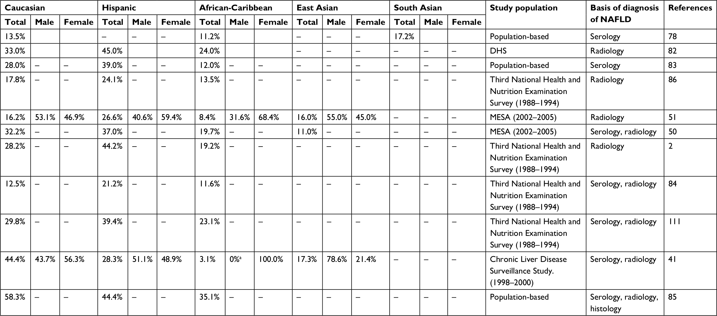 | Table 1 Epidemiology of NAFLD based on selected multi-ethnic studies Notes: aAll NAFLD patients identified in this study were female. Female, % identified patients female, where data available. Male, % of identified patients male, where data available; Total = total number of NAFLD patients identified in the study cohort. Abbreviations: DHS, Dallas Heart Study; MESA, Multi-Ethnic Study of Atherosclerosis; NAFLD, nonalcoholic fatty liver disease. |
Afro-Caribbean ethnic group
For this review, Afro-Caribbean ethnicity is defined as an individual of African descent, living in or coming from Africa or the Caribbean region. Contrary to observed prevalence of metabolic disease and obesity in Afro-Caribbean populations, several studies have shown a relative sparsity of NAFLD cases among these groups compared to Caucasian and Hispanic populations.40,41 However, as Caldwell et al42 argues, it is hard to tell if the discrepancy might simply arise from “under-recognition, under-referral or a truly lower prevalence” in these ethnic groups, as Afro-Caribbean patients are empirically less likely to be referred to health services.43
Evidence suggests that, in Afro-Caribbean populations of NAFLD patients, liver biopsies show less steatosis, and decreased inflammation when compared with biopsies from Caucasian patients.44 Additionally, Afro-Caribbean patients with NAFLD were shown to be affected by hypertension more commonly compared to Caucasian or Hispanic patients.45 For metabolic profiles, Afro-Caribbean patients were found to have lower levels of triglycerides and significantly lower serum HDL compared to other ethnic groups.46 This correlates to less metabolic derangement in these cohorts. Afro-Caribbean patients also have less visceral fat, which appears to contribute to a lower risk of developing advanced liver pathology after the diagnosis of NAFLD.47,48 However, the literature showed that, despite lower grade hepatosteatosis, Afro-Caribbean individuals had a similar prevalence of insulin resistance compared to Hispanics, who were found to have the highest level of hepatic fat content.32
Some specific transcriptomic profile differences have been isolated between Afro-Caribbean and Caucasian populations, which may contribute to the different metabolic makeup. In a study of 94 patients undergoing bariatric surgery, there was over-expression of CYP/CYP450 (CYP3A), insulin-like growth factor (IGF2), acyl-CoA synthetase long chain family member 4 (ACSL4), fumarylacetoacetate hydrolase (FAH), fucosyltransferase 4 (FUT4), erythrocyte membrane protein band 4.1 (EPB41L1), glutathione transferase 2 (GSTM2), 4 (GSTM4), and 5 (GSTM5).40 These genes are responsible for the coding of monooxydases, peroxidases, catalysis of tyrosine, glycolysis, and detoxification of electroliphic compounds. Differences in the coding of these genes thus may contribute to pathologies associated with the metabolic syndrome, such as NAFLD. Indeed, GSTM2, GSTM4, and GSTM5 have previously been implicated in the pathogenesis of the condition.40 Heritable missense variants in PNPLA3 and glucokinase regulator gene (GCKR) have been identified by another study, which was shown to be associated with a predisposition to hepatic steatosis across ancestries in both African and Hispanic Americans.15
East Asian ethnic group
In this review, East Asian ethnic groups were defined as: those with Chinese, Japanese, Taiwanese, Indonesian, and Korean ethnic backgrounds. The management of obesity-associated conditions is swiftly becoming a public health issue in this cohort. There are several potential factors contributing to this phenomenon; increased diagnosing or the wider availability of “Western” diets with more energy-dense food consumption are among the most often-cited culprits.49 NAFLD among East Asians has been widely studied in large scales. However, only a few studies compare this cohort with patients from a Caucasian, Hispanic, or African origin.44,50,51
Chinese individuals have been shown to have higher body fat percentages than Caucasian controls for any given BMI, and have also been reported to have a higher visceral adiposity and subcutaneous body fat overall. Conversely, patients affected by NAFLD have been shown to have lower BMI than patients from other ethnic backgrounds.52 This is consistent with the now generally recognized fact that obesity-related metabolic disorders begin at much lower levels of BMI in those of Asian descent. For this reason, some health care professionals now adapt a lower BMI threshold for clinical assessment and referral in these patients.53,54 A meta-analysis of 48 studies, conducted between 1997 and 2013, and including 356,367 subjects in China, found a pooled prevalence of NAFLD of approximately 20% in healthy cohorts (18–22%).55
In Japan, a study found that the prevalence of NAFLD was 25% in 2005, compared to figures of 12.6% in 1989, indicating a rapid rise in NAFLD prevalence.56 Some annual health checks show that as many as 9–30% of Japanese adults have evidence of NAFLD on ultrasonography.57 However, a research article using a model to predict changes in the prevalence of NAFLD found that the number of cases in Japan will show a 2.6% decrease by 2030.58 Nevertheless, the rising prevalence of NAFLD in Japan is not negligible.
In Korean adults, the study of NAFLD prevalence compared to other ethnic groups is even less. One study stated a 18.7% prevalence (23% in men and 13.7% in women), investigating 6,648 subjects by ultrasound scanning.59 Within East-Asia ethnic groups, Taiwanese individuals were found to have the lowest prevalence: a study enrolling 3,245 Taiwanese adults from a rural village found that the prevalence of NAFLD was 11.5%, based on blood tests and ultrasonography. Other studies reported ranges as high as 11–41%, which is similar to that of other Asian countries.60 Indonesian individuals were found to have the highest prevalence (30%) of NAFLD among East-Asian ethnic groups.61
East Asian NAFLD patients seem to develop more significant pathological changes on liver biopsies. On histological data, East Asian individuals were almost three times as likely (OR=2.67) to develop high grades of ballooning of hepatocytes, with higher grades of inflammation.44 One possible explanation for this observation may be Apolipoprotein C3 (APOC3) gene polymorphisms, which have been identified to correlate with hepatic fat accumulation in Asian populations. Studies conducted in other ethnic groups did not replicate this genotype-phenotype association. It is, therefore, plausible that APOC3 genotypes are solely linked with NAFLD within Asian patient populations, contributing to the explanation of ethnic variations of NAFLD.62 Ethnic differences have not been thoroughly investigated, but a study of 2,410 male Korean individuals has shown that serum ferritin levels predicted incidental findings of hepatic steatosis, and, therefore, may also be significant in these populations.63
NAFLD in these populations developed within a much shorter period and within a younger patient population and lower BMI.49 Different rates of obesity have been recorded within subgroups of East-Asia, suggesting that different stages of urbanization are a strong confounding factor, as well as genetic differences. The effect of the Asian diet needs to be explored in greater depth, as the study of the influence of nutritional factors may be of importance in these cohorts.64 There has been a rapid economic growth, especially in Japan, and this could bring about a shift in lifestyle and diet preferences. This may partially explain the “obesity epidemics” observed in these countries, as these populations are traditionally adapted to a different diet compared to the West. Historically, Asian diets largely consist of low levels of fat and meat, and are mainly vegetable-based with rice as the staple carbohydrate. Thus, over-nutrition and the consumption of energy-dense food may contribute greatly to the accelerated and more significant development of obesity-associated liver changes in these cohorts.65
Identifying susceptibility factors in East-Asian individuals is important, and a personalized approach needs to be taken when managing these patients.
South Asian ethnic group
South Asian populations (Indian, Bangladeshi, Pakistani, Nepalese, Sri Lankan, and Malaysian) have high rates of diabetes, insulin resistance, and other obesity-associated pathology when compared with Western populations. Indeed, India has been ranked as the country with the highest number of individuals with diabetes worldwide.66 This ethnic group is investigated separately from other Asian countries because of the potential differences in diet, economic status, and growth, as well as other confounding factors.
There are at least 25 million NAFLD patients in India alone, which is a country with a population of more than 1.324 billion people.67 However, there are only limited data on South Asian NAFLD patients compared with other ethnic groups.68 This could be due to the fact that, in some developing parts of these countries, a large component of hepatobiliary disease burden is viral hepatitis, reducing the priority accorded to NAFLD.69
It has been shown that South Asians have a more adverse body fat distribution and are more likely to have dyslipidemia than Western patients.70,71 A study investigating samples from 100 Asian Indian NAFLD patients suggested a significant difference in the pathological profile, when compared with studies on Western patients.68 An overall lower average body weight was observed, and, contrary to Caucasian patients, diabetes mellitus was not strongly associated with the development of the disease. This is often referred to as the “Asia paradox”, describing the observation that South Asian patients with NAFLD have an overall lower BMI. The average BMI of these patients is only 26, and BMI has been shown to not represent the true risk of developing the condition.72 PNPLA3 single nucleotide polymorphisms were associated with increased hepatic fat content in South Asian ethnic groups, particularly in Asian Indians according to studies.73 Other contributing factors may include physical inactivity, reduced disease awareness, and health-seeking, delayed diagnosis, and religious and sociocultural factors between these and Western patient populations.74
Studies have shown that Indian NAFLD patients were younger and had lower prevalence of diabetes mellitus compared to Caucasian cohorts, with similar necro-inflammatory activity and levels of fibrosis.75 Several studies have concluded, however, that insulin resistance and the metabolic syndrome were similarly indicated in these cohorts as other ethnic groups.76 Iron abnormalities were not significantly observed in this population group.77 Several other epidemiological studies cited a prevalence of 16–30% in South-Asian populations.78
Pakistani patients had high odd ratios (OR=1.31) of developing NAFLD, but, surprisingly, Indian study subjects were found to be less likely to be diagnosed with the condition than those of other ethnic groups.78 In Indian men, insulin resistance seems to be 2–3-fold higher than other ethnic groups, leading to a 2-fold increase in hepatic triglyceride content compared to Caucasians.79 This may imply important ethnic differences in insulin resistance and its association with NAFLD. Bangladeshi patients were more frequently diagnosed with NAFLD, with one study reporting prevalence as high as 34.34%.80
In conclusion, South-Asian individuals seem to be at a much higher risk of developing NAFLD when compared to Caucasians individuals.81
Hispanic ethnic group
The most extensive evidence available on the ethnic variations of NAFLD is that of its prevalence in Hispanics, defined as “Spanish-speaking” individuals of Central and South American origin. Several studies have shown that NAFLD is disproportionately frequent in Hispanic minorities compared to Caucasian or Afro-Caribbean patients.82–86 This ethnic disparity seems to be independent of age, and studies have shown that children and adolescents of Hispanic heritage have similarly high incidence of NAFLD compared to their non-Hispanic peers.87 In fact, mortality rates in Hispanic populations from the US due to cirrhosis have been reported to be almost 2-fold greater than non-Hispanic white populations, however deaths from alcoholic liver disease were included in these studies.88,89 In another study, Hispanic patients were more than twice as likely to show higher grades of ballooning and Mallory bodies, respectively, than Caucasians and other ethnic groups combined. Thus, in these cohorts, the evidence shows that NAFLD is not only more prevalent, but may also have a worse diagnosis.44
There seems to be variation in prevalence among Hispanic patients of different heritage, although little is known of the distribution of NAFLD among Hispanic subgroups. Individuals with a Cuban, Puerto Rican, and Dominican heritage seem to have lower prevalence of suspected NAFLD than Central-, South American, and Mexican populations.90 Riquelme et al91 found that 23% of Chilean Hispanics had evidence of NAFLD on ultrasound in the absence of Hepatitis infection or alcohol consumption. Serum and radiological evidence from the multi-ethnic study of atherosclerosis (MESA) cohort study concluded that Mexican Hispanics had a 2-fold prevalence (33%) of NAFLD compared to individuals of Dominican (16%) and Puerto Rican origin (18%).92 The reasons for these variations within the ethnic group are unclear, but may point to genetic background, different levels of insulin resistance, and lifestyle factors as main contributing factors.
One explanation for higher rates of NAFLD in Hispanics may be that these individuals seem to have a higher BMI overall compared to other ethnic groups.93 When compared to women with similar BMI and socioeconomic status, Hispanic women have also been found to have a higher level of adiposity.94 Likewise, socioeconomic and cultural risk factors have been indicated in these cohorts. Evidence shows that residence in high-poverty areas is correlated with higher prevalence of lower-cost sugar and fat-rich foods and drinks, factors especially prevalent in Latino communities. According to 2015 census data, one in four Latino adults lived at or below the poverty line (22.6%), a figure more than twice as high as non-Latino ethnic groups (10.4%).95
Some genetic influences have been uncovered, and a genome-wide association study (GWAS) study by including samples from 248 Hispanic children has found associations of several genetic loci with the diagnosis of NAFLD on chromosomes 8, 14, and 20.96 A further GWAS study showed a positive correlation of a PNPLA3 gene variant with hepatic fat content, a polymorphism known to be more common in Latinos compared with other ethnic groups.97 A GWAS study conducted in the US investigating genetic markers in Hispanic women attained strong correlations with farnesyl-diphosphate farnesyltransferase one within these cohorts, a gene that regulates cholesterol homeostasis, which could be a contributing factor to the ethnic disparity.98 However, more focused research is required to interrogate genetic as well as metabolic differences within these cohorts, with a focus on Hispanics of varying heritage.
Caucasian ethnic group
Although incidence of NAFLD in Caucasians in the US is well-documented and well-researched, few studies investigate discrepancies within Caucasian cohorts of different countries. Notably, within Europe, most forms of hepatitis have been demarcating a gradient of increasing prevalence; however, this does not seem to be the case for NAFLD.99
The DIONYSOS study found NAFLD prevalence to be 20–25% within Italian cohorts, and a Greek study found NAFLD to be 31% in autopsy reports.100,101 NAFLD is estimated to be present in 20% of Romanians.102 In obese patients, NAFLD was diagnosed in as high as 20–30% of Caucasian cohorts.103
Poor dietary habits, especially a typical Western diet (incorporating high intakes of red or processed meat, full-fat products, fried food, refined cereals, sugar, and soft drinks) has been correlated with and believed to be a cornerstone in the development of NAFLD in Caucasian cohorts, as well as other ethnic groups. However, the role of specific dietary patterns has not been formally investigated. One study compared 995 adolescents following healthy and Western diets, respectively. Participants were interviewed at 14 years, and then followed up with a liver ultrasound at 17 years. This study found that a Western dietary pattern at 14 years was associated with the development of NAFLD at 17 years (OR=1.59), and found a healthy diet protective.104 Recently, significant association was found by a meta-analysis between sugar-sweetened soda consumption and developing NAFLD, a staple in economically advanced Western countries.105 A meta-analysis reviewing 21 population-based surveys has found that the prevalence of NAFLD was positively correlated with gross national income, thus Europe exhibited a much higher prevalence in all studies (28% compared to 13% in Middle East and 19% in East-Asia). In this study, urban living was also contributing to these prevalence rates, signifying a strong link between economic success and NAFLD.106
Some studies have interrogated genetic discordance in Caucasian-only cohorts. A large GWAS study comparing results from three distinct European ancestry studies previously reported associations with PNPLA3 and GCKR, but also uncovered two novel gene loci associations near PPP1R3B and NCAN.107 Feldman et al108 compared Caucasian lean individuals with NAFLD with those without, and found significant differences in the subjects with the disease. Whilst their fasting insulin concentrations were similar to the healthy cohort, they had marked glucose tolerance and higher rates of PNPLA3 allele mutations. Certain lysophosphatidylcholines were also indicated to be of importance in the study, as well as lysine, tyrosine, and valine. Uncovering protective factors of Caucasian ethnic descent compared to Asian and Hispanic patients may greatly contribute to decreasing NAFLD disease burden in the Western countries.
Conclusion
NAFLD is a spectrum of liver disease, which is set to become the most prevalent cause of liver transplant globally by 2020.109 There is a greater risk of the disease in Asian and Hispanic ethnic groups, and this can be directly linked to metabolic factors, genetic predisposition, as well as the environment. Table 2 summarizes the differences our study has concluded within these cohorts. However, specific effectors and their individual contributions remain unclear. It is important to note that the majority of studies defined and diagnosed NAFLD using surrogate measures, such as liver enzymes and imaging. Few of the included studies have used biopsies as an indicator, which remains the gold standard for diagnosis. The specificity of liver enzymes is limited, as is the use of ultrasound, and this may result in uncertain prevalence estimation of NAFLD.110 Most of the studies reviewed utilized primary data collection done by the Third National Health and Nutrition Examination Survey (1988–1994), or MESA (2002–2005), which may produce outdated results.2,50,51,84,86,111 Prevalence of NAFLD, thus, in current, prospective population-based studies, may be higher.
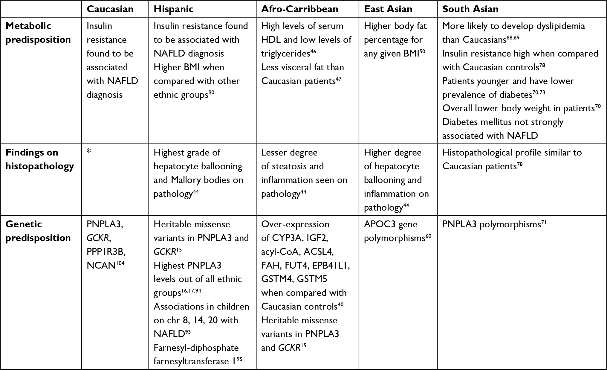 | Table 2 Identified variations in genetic and metabolic makeup contributing to NAFLD Note: *Histopathology for Caucasian ethnicity was not usually defined as it was used as baseline or controls for comparison with other ethnicities. Abbreviations: APOC3, apolipoprotein C3; BMI, body mass index; GCKR, glucokinase regulator gene; HDL, high-density lipoprotein; NCAN, neurocan; PNPLA3, Patatin-like phospholipase domain-containing protein 3; PP1R3B, protein phosphatase 1, regulatory subunit 3B; chr, chromosome; NAFLD, nonalcoholic fatty liver disease. |
Specific ethnic groups that have different heritages should be compared in large cohort studies, using biopsies for measure of disease. This would aid the identification of contributors to susceptibility of developing NAFLD, variability in gene mutations, as well as provide a larger patient sample for shared biomarker discovery. Furthermore, with increasing rates of NAFLD in children and adolescents, studies should further focus on the interplay between developmental, genetic, and environmental factors.
This review summarized the literature available investigating ethnic variations in the development of this highly prevalent but widely unrecognized noncommunicable disease. Although there are several population-based cohort studies investigating ethnic variations in the pathogenesis of NAFLD, large-scale multi-ethnic clinical studies are vital for targeted therapy and drug development.112 Identifying and outlining high-risk populations would aid clinicians to successfully adapt screening and prevention programmes. A more targeted approach also needs to be taken to interrogate genetic crosstalk within the genome, to uncover more generalizable patterns within our genetic makeup, and their effect on susceptibility to obesity-related illnesses. This would help develop strategies for the implementation of public health policies in different geographical areas.
Acknowledgment
We appreciate the support of the Welcome Trust and the Obesity Action Campaign.
Disclosure
The authors report no conflicts of interest in this work.
References
Salt WB. Nonalcoholic fatty liver disease (NAFLD): a comprehensive review. J Insur Med. 2004;36(1):27–41. | ||
Ruhl CE, Everhart JE. Fatty liver indices in the multiethnic united states national health and nutrition examination survey. Aliment Pharmacol Ther. 2015;41(1):65–76. | ||
Loomba R, Sanyal AJ. The global NAFLD epidemic. Nat Rev Gastroenterol Hepatol. 2013;10(11):686–690. | ||
Younossi ZM, Koenig AB, Abdelatif D, Fazel Y, Henry L, Wymer M. Global epidemiology of nonalcoholic fatty liver disease-Meta-analytic assessment of prevalence, incidence, and outcomes. Hepatology. 2016;64(1):73–84. | ||
Athyros VG, Alexandrides TK, Bilianou H, et al. The use of statins alone, or in combination with pioglitazone and other drugs, for the treatment of non-alcoholic fatty liver disease/non-alcoholic steatohepatitis and related cardiovascular risk. an expert panel statement. Metabolism. 2017;71:17–32. | ||
Doycheva I, Loomba R. Effect of metformin on ballooning degeneration in nonalcoholic steatohepatitis (NASH): when to use metformin in nonalcoholic fatty liver disease (NAFLD). Adv Ther. 2014;31(1):30–43. | ||
Steinacher D, Claudel T, Trauner M. Therapeutic mechanisms of bile acids and Nor-Ursodeoxycholic acid in non-alcoholic fatty liver disease. Dig Dis. 2017;35(3):282–287. | ||
Ali Khan R, Kapur P, Jain A, Farah F, Bhandari U. Effect of orlistat on periostin, adiponectin, inflammatory markers and ultrasound grades of fatty liver in obese NAFLD patients. Ther Clin Risk Manag. 2017;13:139–149. | ||
Li J, Cordero P, Nguyen V, Oben JA. The role of vitamins in the pathogenesis of non-alcoholic fatty liver disease. Integr Med Insights. 2016;11:19–25. | ||
Mouralidarane A, Lin CI, Suleyman N, Soeda J, Oben JA. Practical management of the increasing burden of non-alcoholic fatty liver disease. Frontline Gastroenterol. 2010;1(3):149–155. | ||
Kleiner DE, Brunt EM, van Natta M, et al. Design and validation of a histological scoring system for nonalcoholic fatty liver disease. Hepatology. 2005;41(6):1313–1321. | ||
Ahmed MH, Abu EO, Byrne CD. Non-alcoholic fatty liver disease (NAFLD): new challenge for general practitioners and important burden for health authorities? Prim Care Diabetes. 2010;4(3):129–137. | ||
Ballestri S, Zona S, Targher G, et al. Nonalcoholic fatty liver disease is associated with an almost twofold increased risk of incident type 2 diabetes and metabolic syndrome. Evidence from a systematic review and meta-analysis. J Gastroenterol Hepatol. 2016;31(5):936–944. | ||
Portillo-Sanchez P, Bril F, Maximos M, et al. High prevalence of nonalcoholic fatty liver disease in patients with type 2 diabetes mellitus and normal plasma aminotransferase levels. J Clin Endocrinol Metab. 2015;100(6):2231–2238. | ||
Palmer ND, Musani SK, Yerges-Armstrong LM, et al. Characterization of European ancestry nonalcoholic fatty liver disease-associated variants in individuals of African and Hispanic descent. Hepatology. 2013;58(3):966–975. | ||
Romeo S, Huang-Doran I, Baroni MG, Kotronen A. Unravelling the pathogenesis of fatty liver disease: patatin-like phospholipase domain-containing 3 protein. Curr Opin Lipidol. 2010;21(3):247–252. | ||
Rotman Y, Koh C, Zmuda JM, Kleiner DE, Liang TJ; NASH CRN. The association of genetic variability in patatin-like phospholipase domain-containing protein 3 (PNPLA3) with histological severity of nonalcoholic fatty liver disease. Hepatology. 2010;52(3):894–903. | ||
Britton LJ, Subramaniam VN, Crawford DH. Iron and non-alcoholic fatty liver disease. World J Gastroenterol. 2016;22(36):8112. | ||
Miyake T, Kumagi T, Furukawa S, et al. Non-alcoholic fatty liver disease: factors associated with its presence and onset. J Gastroenterol Hepatol. 2013;28(Suppl 1):71–78. | ||
Cordero P, Campion J, Milagro FI, Martinez JA. Transcriptomic and epigenetic changes in early liver steatosis associated to obesity: effect of dietary methyl donor supplementation. Mol Genet Metab. 2013;110(3):388–395. | ||
Mouralidarane A, Soeda J, Sugden D, et al. Maternal obesity programs offspring non-alcoholic fatty liver disease through disruption of 24-h rhythms in mice. Int J Obes. 2015;39(9):1339–1348. | ||
Oben JA, Mouralidarane A, Samuelsson AM, et al. Maternal obesity during pregnancy and lactation programs the development of offspring non-alcoholic fatty liver disease in mice. J Hepatol. 2010;52(6):913–920. | ||
Aron-Wisnewsky J, Gaborit B, Dutour A, Clement K. Gut microbiota and non-alcoholic fatty liver disease: new insights. Clin Microbiol Infect. 2013;19(4):338–348. | ||
Henao-Mejia J, Elinav E, Jin C, et al. Inflammasome-mediated dysbiosis regulates progression of NAFLD and obesity. Nature. 2012;482(7384):179–185. | ||
Gambino R, Musso G, Cassader M. Redox balance in the pathogenesis of nonalcoholic fatty liver disease: mechanisms and therapeutic opportunities. Antioxid Redox Signal. 2011;15(5):1325–1365. | ||
Ito Y, Shimizu H, Yoshimura T, et al. Serum concentrations of carotenoids, alpha-tocopherol, fatty acids, and lipid peroxides among Japanese in Japan, and Japanese and Caucasians in the US. Int J Vitam Nutr Res. 1999;69(6):385–395. | ||
Block G, Dietrich M, Norkus EP, et al. Factors associated with oxidative stress in human populations. Am J Epidemiol. 2002;156(3):274–285. | ||
Kremer M, Thomas E, Milton RJ, et al. Kupffer cell and interleukin-12-dependent loss of natural killer T cells in hepatosteatosis. Hepatology. 2010;51(1):130–141. | ||
European Association for the Study of the Liver (EASL)European Association for the Study of Diabetes (EASD)European Association for the Study of Obesity (EASO). EASL-EASD-EASO clinical practice guidelines for the management of non-alcoholic fatty liver disease. J Hepatol. 2016;64(6):1388–1402. | ||
Harrison SA, Torgerson S, Hayashi PH. The natural history of nonalcoholic fatty liver disease: a clinical histopathological study. Am J Gastroenterol. 2003;98(9):2042–2047. | ||
Ekstedt M, Franzén LE, Mathiesen UL, et al. Long-term follow-up of patients with NAFLD and elevated liver enzymes. Hepatology. 2006;44(4):865–873. | ||
Guerrero R, Vega GL, Grundy SM, Browning JD. Ethnic differences in hepatic steatosis: an insulin resistance paradox? Hepatology. 2009;49(3):791–801. | ||
Williams R, Aspinall R, Bellis M, et al. Addressing liver disease in the UK: A blueprint for attaining excellence in health care and reducing premature mortality from lifestyle issues of excess consumption of alcohol, obesity, and viral hepatitis. Lancet. 2014;384(9958):1953–1997. | ||
Angulo P. Gi epidemiology: nonalcoholic fatty liver disease. Aliment Pharmacol Ther. 2007;25(8):883–889. | ||
Adams LA, Sanderson S, Lindor KD, Angulo P. The histological course of nonalcoholic fatty liver disease: a longitudinal study of 103 patients with sequential liver biopsies. J Hepatol. 2005;42(1):132–138. | ||
Musso G, Gambino R, Cassader M, Pagano G. Meta-analysis: natural history of non-alcoholic fatty liver disease (NAFLD) and diagnostic accuracy of non-invasive tests for liver disease severity. Ann Med. 2011;43(8):617–649. | ||
Söderberg C, Stål P, Askling J, et al. Decreased survival of subjects with elevated liver function tests during a 28-year follow-up. Hepatology. 2010;51(2):595–602. | ||
Dulai PS, Singh S, Patel J, et al. Increased risk of mortality by fibrosis stage in nonalcoholic fatty liver disease: systematic review and meta-analysis. Hepatology. 2017;65(5):1557–1565. | ||
Non-alcoholic Fatty Liver Disease Study Group, Lonardo A, Bellentani S, et al. Epidemiological modifiers of non-alcoholic fatty liver disease: focus on high-risk groups. Dig Liver Dis. 2015;47(12):997–1006. | ||
Stepanova M, Hossain N, Afendy A, et al. Hepatic gene expression of Caucasian and African-American patients with obesity-related non-alcoholic fatty liver disease. Obes Surg. 2010;20(5):640–650. | ||
Weston SR, Leyden W, Murphy R, et al. Racial and ethnic distribution of nonalcoholic fatty liver in persons with newly diagnosed chronic liver disease. Hepatology. 2005;41(2):372–379. | ||
Caldwell SH, Harris DM, Patrie JT, Hespenheide EE. Is NASH underdiagnosed among African Americans? Am J Gastroenterol. 2002;97(6):1496–1500. | ||
Morgan C, Mallett R, Hutchinson G, et al. Pathways to care and ethnicity. 2: source of referral and help-seeking. Report from the AESOP study. Br J Psychiatry. 2005;186(04):290–296. | ||
Mohanty SR, Troy TN, Huo D, O’Brien BL, Jensen DM, Hart J. Influence of ethnicity on histological differences in non-alcoholic fatty liver disease. J Hepatol. 2009;50(4):797–804. | ||
Foster T, Anania FA, Li D, Katz R, Budoff M. The prevalence and clinical correlates of nonalcoholic fatty liver disease (NAFLD) in African Americans: the multiethnic study of atherosclerosis (MESA). Dig Dis Sci. 2013;58(8):2392–2398. | ||
Solga SF, Clark JM, Alkhuraishi AR, et al. Race and comorbid factors predict nonalcoholic fatty liver disease histopathology in severely obese patients. Surg Obes Relat Dis. 2005;1(1):6–11. | ||
Karelis AD, St-Pierre DH, Conus F, Rabasa-Lhoret R, Poehlman ET. Metabolic and body composition factors in subgroups of obesity: what do we know? J Clin Endocrinol Metab. 2004;89(6):2569–2575. | ||
Perry AC, Applegate EB, Jackson ML, et al. Racial differences in visceral adipose tissue but not anthropometric markers of health-related variables. J Appl Physiol. 2000;89(2):636–643. | ||
Yoon K-H, Lee J-H, Kim J-W, et al. Epidemic obesity and type 2 diabetes in Asia. Lancet. 2006;368(9548):1681–1688. | ||
Al Rifai M, Silverman MG, Nasir K, et al. The association of nonalcoholic fatty liver disease, obesity, and metabolic syndrome, with systemic inflammation and subclinical atherosclerosis: the multi-ethnic study of atherosclerosis (MESA). Atherosclerosis. 2015;239(2):629–633. | ||
Remigio-Baker RA, Allison MA, Forbang NI, et al. Race/ethnic and sex disparities in the non-alcoholic fatty liver disease-abdominal aortic calcification association: the multi-ethnic study of atherosclerosis. Atherosclerosis. 2017;258:89–96. | ||
Wong RJ, Ahmed A. Obesity and non-alcoholic fatty liver disease: disparate associations among Asian populations. World J Hepatol. 2014;6(5):263. | ||
Deurenberg P, Deurenberg-Yap M, Guricci S. Asians are different from Caucasians and from each other in their body mass index/body fat per cent relationship. Obesity Reviews. 2002;3(3):141–146. | ||
Fan JG, Saibara T, Chitturi S, et al. What are the risk factors and settings for non-alcoholic fatty liver disease in Asia-Pacific? J Gastroenterol Hepatol. 2007;22(6):794–800. | ||
Li Z, Xue J, Chen P, Chen L, Yan S, Liu L. Prevalence of nonalcoholic fatty liver disease in mainland of China: a meta-analysis of published studies. J Gastroenterol Hepatol. 2014;29(1):42–51. | ||
Kojima S, Watanabe N, Numata M, Ogawa T, Matsuzaki S. Increase in the prevalence of fatty liver in Japan over the past 12 years: analysis of clinical background. J Gastroenterol. 2003;38(10):954–961. | ||
Hashimoto E, Tokushige K, Prevalence TK. Prevalence, gender, ethnic variations, and prognosis of NASH. J Gastroenterol. 2011;46(Suppl 1):63–69. | ||
Estes C, Anstee QM, Arias-Loste MT, et al. Modeling NAFLD disease burden in China, France, Germany, Italy, Japan, Spain, United Kingdom, and united states for the period 2016-2030. J Hepatol. 2018;69(4):896–904. | ||
Park SH, Jeon WK, Kim SH, et al. Prevalence and risk factors of non-alcoholic fatty liver disease among Korean adults. J Gastroenterol Hepatol. 2006;21(1 Pt 1):138–143. | ||
Hsu CS, Kao JH. Non-alcoholic fatty liver disease: an emerging liver disease in Taiwan. J Formos Med Assoc. 2012;111(10):527–535. | ||
Agrawal S, Duseja AK. Non-alcoholic fatty liver disease: East versus West. J Clin Exp Hepatol. 2012;2(2):122–134. | ||
Petersen KF, Dufour S, Hariri A, et al. Apolipoprotein C3 gene variants in nonalcoholic fatty liver disease. N Engl J Med. 2010;362(12):1082–1089. | ||
Kim CW, Chang Y, Sung E, Shin H, Ryu S. Serum ferritin levels predict incident non-alcoholic fatty liver disease in healthy Korean men. Metabolism. 2012;61(8):1182–1188. | ||
Chitturi S, Wong VW, Farrell G. Nonalcoholic fatty liver in Asia: firmly entrenched and rapidly gaining ground. J Gastroenterol Hepatol. 2011;26(Suppl 1):163–172. | ||
Enjoji M, Nakamuta M. Is the control of dietary cholesterol intake sufficiently effective to ameliorate nonalcoholic fatty liver disease? World J Gastroenterol. 2010;16(7):800–803. | ||
Kaveeshwar SA, Cornwall J. The current state of diabetes mellitus in India. Australas Med J. 2014;7(1):45–48. | ||
United Nations, Department of Economic and Social Affairs, Population Division; 2017. World Population Prospects: The 2017 Revision; Key Findings and Advance Tables. Working Paper No. ESA/P/WP/248. Available from: https://population.un.org/wpp/Publications/. Accessed March 6, 2019. | ||
Duseja A, Das A, Das R, et al. The clinicopathological profile of Indian patients with nonalcoholic fatty liver disease (NAFLD) is different from that in the West. Dig Dis Sci. 2007;52(9):2368–2374. | ||
Duseja A. Nonalcoholic fatty liver disease in India - a lot done, yet more required! Indian J Gastroenterol. 2010;29(6):217–225. | ||
Banerji MA, Faridi N, Atluri R, Chaiken RL, Lebovitz HE. Body composition, visceral fat, leptin, and insulin resistance in Asian Indian men. J Clin Endocrinol Metab. 1999;84(1):137–144. | ||
Wulan SN, Westerterp KR, Plasqui G. Ethnic differences in body composition and the associated metabolic profile: a comparative study between Asians and Caucasians. Maturitas. 2010;65(4):315–319. | ||
Singh S, Kuftinec GN, Sarkar S. Non-alcoholic fatty liver disease in South Asians: a review of the literature. J Clin Transl Hepatol. 2017;5(1):76–81. | ||
Bhatt SP, Nigam P, Misra A, Guleria R, Pandey RM, Pasha MA. Genetic variation in the patatin-like phospholipase domain-containing protein-3 (PNPLA-3) gene in Asian Indians with nonalcoholic fatty liver disease. Metab Syndr Relat Disord. 2013;11(5):329–335. | ||
Misra A, Khurana L. Obesity-related non-communicable diseases: South Asians vs white Caucasians. Int J Obes. 2011;35(2):167–187. | ||
Singh SP, Kar SK, Panigrahi MK, et al. Profile of patients with incidentally detected nonalcoholic fatty liver disease (IDNAFLD) in coastal eastern India. Trop Gastroenterol. 2013;34(3):144–152. | ||
Mohan V, Farooq S, Deepa M, Ravikumar R, Pitchumoni CS. Prevalence of non-alcoholic fatty liver disease in urban South Indians in relation to different grades of glucose intolerance and metabolic syndrome. Diabetes Res Clin Pract. 2009;84(1):84–91. | ||
Duseja A, das R, Nanda M, das A, Garewal G, Chawla Y. Nonalcoholic steatohepatitis in Asian Indians is neither associated with iron overload nor with HFE gene mutations. WJG. 2005;11(3):393. | ||
Alazawi W, Mathur R, Abeysekera K, et al. PTU-138 population-based study of ethnicity and the diagnosis gap in liver disease. Gut. 2014;63(Suppl 1):A99.1–A99. | ||
Petersen KF, Dufour S, Feng J, et al. Increased prevalence of insulin resistance and nonalcoholic fatty liver disease in Asian-Indian men. Proc Natl Acad Sci. 2006;103(48):18273–18277. | ||
Alam S, Fahim SM, Chowdhury MAB, et al. Prevalence and risk factors of non-alcoholic fatty liver disease in Bangladesh. JGH Open. 2018;2(2):39–46. | ||
Pati GK, Singh SP. Nonalcoholic fatty liver disease in South Asia. Euroasian J Hepatogastroenterol. 2016;6(2):154–162. | ||
Browning JD, Szczepaniak LS, Dobbins R, et al. Prevalence of hepatic steatosis in an urban population in the United States: impact of ethnicity. Hepatology. 2004;40(6):1387–1395. | ||
Kallwitz ER, Kumar M, Aggarwal R, et al. Ethnicity and nonalcoholic fatty liver disease in an obesity clinic: the impact of triglycerides. Dig Dis Sci. 2008;53(5):1358–1363. | ||
Schneider AL, Lazo M, Selvin E, Clark JM. Racial differences in nonalcoholic fatty liver disease in the U.S. population. Obesity. 2014;22(1):292–299. | ||
Williams CD, Stengel J, Asike MI, et al. Prevalence of nonalcoholic fatty liver disease and nonalcoholic steatohepatitis among a largely middle-aged population utilizing ultrasound and liver biopsy: a prospective study. Gastroenterology. 2011;140(1):124–131. | ||
Lazo M, Hernaez R, Eberhardt MS, et al. Prevalence of nonalcoholic fatty liver disease in the United States: the third National Health and Nutrition Examination Survey, 1988-1994. Am J Epidemiol. 2013;178(1):38–45. | ||
Marzuillo P, Miraglia del Giudice E, Santoro N. Pediatric fatty liver disease: role of ethnicity and genetics. World J Gastroenterol. 2014;20(23):7347. | ||
Kung H, Hoyert D, Xu J, et al. Deaths: final data for 2005. Natl Vital Stat Rep. 2008;56(10):1–120. | ||
Stinson FS, Grant BF, Dufour MC. The critical dimension of ethnicity in liver cirrhosis mortality statistics. Alcohol Clin Exp Res. 2001;25(8):1181–1187. | ||
Kallwitz ER, Daviglus ML, Allison MA, et al. Prevalence of suspected nonalcoholic fatty liver disease in Hispanic/Latino individuals differs by heritage. Clin Gastroenterol Hepatol. 2015;13(3):569–576. | ||
Riquelme A, Arrese M, Soza A, et al. Non-alcoholic fatty liver disease and its association with obesity, insulin resistance and increased serum levels of C-reactive protein in Hispanics. Liver Int. 2009;29(1):82–88. | ||
Fleischman MW, Budoff M, Zeb I, Li D, Foster T. NAFLD prevalence differs among Hispanic subgroups: the multi-ethnic study of atherosclerosis. World J Gastroenterol. 2014;20(17):4987. | ||
Wang J, Thornton JC, Burastero S, et al. Comparisons for body mass index and body fat percent among Puerto Ricans, blacks, whites and Asians living in the New York City area. Obes Res. 1996;4(4):377–384. | ||
Casas YG, Schiller BC, Desouza CA, Seals DR. Total and regional body composition across age in healthy Hispanic and white women of similar socioeconomic status. Am J Clin Nutr. 2001;73(1):13–18. | ||
Proctor BD, Semega JL, Kollar MA. US Census Bureau. Income and Poverty in the United States: 2015. Washington DC: US Department of Commerce; 2015:13. | ||
Saab S, Manne V, Nieto J, Schwimmer JB, Chalasani NP. Nonalcoholic fatty liver disease in Latinos. Clin Gastroenterol Hepatol. 2016;14(1):5–12. | ||
Romeo S, Kozlitina J, Xing C, et al. Genetic variation in PNPLA3 confers susceptibility to nonalcoholic fatty liver disease. Nat Genet. 2008;40(12):1461–1465. | ||
Chalasani N, Guo X, Loomba R, et al. Genome-wide association study identifies variants associated with histologic features of nonalcoholic fatty liver disease. Gastroenterology. 2010;139(5):1567–1576. | ||
Younossi Z, Anstee QM, Marietti M, et al. Global burden of NAFLD and NASH: trends, predictions, risk factors and prevention. Nat Rev Gastroenterol Hepatol. 2018;15(1):11–20. | ||
Bedogni G, Miglioli L, Masutti F, Tiribelli C, Marchesini G, Bellentani S. Prevalence of and risk factors for nonalcoholic fatty liver disease: the Dionysos nutrition and liver study. Hepatology. 2005;42(1):44–52. | ||
Zois CD, Baltayiannis GH, Bekiari A, et al. Steatosis and steatohepatitis in postmortem material from northwestern Greece. WJG. 2010;16(31):3944. | ||
Radu C, Grigorescu M, Crisan D, et al. Prevalence and associated risk factors of non-alcoholic fatty liver disease in hospitalized patients. J Gastrointestin Liver Dis. 2008;17(3):255–260. | ||
Milić S, Stimac D. Nonalcoholic fatty liver disease/steatohepatitis: epidemiology, pathogenesis, clinical presentation and treatment. Dig Dis. 2012;30(2):158–162. | ||
Oddy WH, Herbison CE, Jacoby P, et al. The Western dietary pattern is prospectively associated with nonalcoholic fatty liver disease in adolescence. Am J Gastroenterol. 2013;108(5):778–785. | ||
Wijarnpreecha K, Thongprayoon C, Edmonds PJ, Cheungpasitporn W. Associations of sugar- and artificially sweetened soda with nonalcoholic fatty liver disease: a systematic review and meta-analysis. QJM. 2016;109(7):461–466. | ||
Zhu JZ, Dai YN, Wang YM, Zhou QY, Yu CH, Li YM. Prevalence of nonalcoholic fatty liver disease and economy. Dig Dis Sci. 2015;60(11):3194–3202. | ||
Speliotes EK, Yerges-Armstrong LM, Wu J, et al. Genome-wide association analysis identifies variants associated with nonalcoholic fatty liver disease that have distinct effects on metabolic traits. PLoS Genet. 2011;7(3):e1001324. | ||
Feldman A, Eder SK, Felder TK, et al. Clinical and metabolic characterization of lean Caucasian subjects with non-alcoholic fatty liver. Am J Gastroenterol. 2017;112(1):102–110. | ||
Xu R, Tao A, Zhang S, Deng Y, Chen G. Association between patatin-like phospholipase domain containing 3 gene (PNPLA3) polymorphisms and nonalcoholic fatty liver disease: a huge review and meta-analysis. Sci Rep. 2015;5(1):9284. | ||
Lazo M, Clark JM. The epidemiology of nonalcoholic fatty liver disease: a global perspective. Semin Liver Dis. 2008;28(4):339–350. | ||
Smits MM, Ioannou GN, Boyko EJ, Utzschneider KM. Non-alcoholic fatty liver disease as an independent manifestation of the metabolic syndrome: results of a US national survey in three ethnic groups. J Gastroenterol Hepatol. 2013;28(4):664–670. | ||
Rich NE, Oji S, Mufti AR, et al. Racial and ethnic disparities in nonalcoholic fatty liver disease prevalence, severity, and outcomes in the United States: a systematic review and meta-analysis. Clin Gastroenterol Hepatol. 2018;16(2):198–210. |
 © 2019 The Author(s). This work is published and licensed by Dove Medical Press Limited. The full terms of this license are available at https://www.dovepress.com/terms.php and incorporate the Creative Commons Attribution - Non Commercial (unported, v3.0) License.
By accessing the work you hereby accept the Terms. Non-commercial uses of the work are permitted without any further permission from Dove Medical Press Limited, provided the work is properly attributed. For permission for commercial use of this work, please see paragraphs 4.2 and 5 of our Terms.
© 2019 The Author(s). This work is published and licensed by Dove Medical Press Limited. The full terms of this license are available at https://www.dovepress.com/terms.php and incorporate the Creative Commons Attribution - Non Commercial (unported, v3.0) License.
By accessing the work you hereby accept the Terms. Non-commercial uses of the work are permitted without any further permission from Dove Medical Press Limited, provided the work is properly attributed. For permission for commercial use of this work, please see paragraphs 4.2 and 5 of our Terms.