")
Back to Journals » Infection and Drug Resistance » Volume 13
Emergence of Terbinafine Resistant Trichophyton mentagrophytes in Iran, Harboring Mutations in the Squalene Epoxidase (SQLE) Gene
Authors Taghipour S, Shamsizadeh F , Pchelin IM , Rezaei-Matehhkolaei A , Zarei Mahmoudabadi A , Valadan R, Ansari S, Katiraee F , Pakshir K, Zomorodian K , Abastabar M
Received 15 January 2020
Accepted for publication 2 March 2020
Published 13 March 2020 Volume 2020:13 Pages 845—850
DOI https://doi.org/10.2147/IDR.S246025
Checked for plagiarism Yes
Review by Single anonymous peer review
Peer reviewer comments 3
Editor who approved publication: Dr Eric Nulens
Simin Taghipour,1 Forough Shamsizadeh,2 Ivan M Pchelin,3 Ali Rezaei-Matehhkolaei,4 Ali Zarei Mahmoudabadi,4 Reza Valadan,5 Saham Ansari,6 Farzad Katiraee,7 Keyvan Pakshir,8 Kamiar Zomorodian,8 Mahdi Abastabar9
1Department of Medical Parasitology and Mycology, Faculty of Medicine, Shahrekord University of Medical Sciences, Shahrekord, Iran; 2Student Research Committee, Ahvaz Jundishapur University of Medical Sciences, Ahvaz, Iran; 3Kashkin Research Institute of Medical Mycology, North‐Western State Medical University Named After I.I. Mechnikov, Saint Petersburg, Russia; 4Infectious and Tropical Diseases Research Center, Health Research Institute, Ahvaz Jundishapur University of Medical Sciences, Ahvaz, Iran; 5Molecular and Cell Biology Research Center, School of Medicine, Mazandaran University of Medical Sciences, Sari, Iran; 6Department of Medical Parasitology and Mycology, School of Medicine, Shahid Beheshti University of Medical Sciences, Tehran, Iran; 7Department of Pathobiology, Faculty of Veterinary Medicine, University of Tabriz, Tabriz, Iran; 8Basic Sciences in Infectious Diseases Research Center, Parasitology & Mycology Department, School of Medicine, Shiraz University of Medical Sciences, Shiraz, Iran; 9Invasive Fungi Research Center, Department of Medical Mycology and Parasitology, School of Medicine, Mazandaran University of Medical Sciences, Sari, Iran
Correspondence: Ali Rezaei-Matehhkolaei
Infectious and Tropical Diseases Research Center, Health Research Institute, Ahvaz Jundishapur University of Medical Sciences, Ahvaz, Iran
Tel +98-912-7112573
Fax +98-61-33332036
Email [email protected]
Mahdi Abastabar
Invasive Fungi Research Center, Department of Medical Mycology and Parasitology, School of Medicine, Mazandaran University of Medical Sciences, Sari, Iran
Tel +98-911-2111347
Email [email protected]
Introduction: Trichophyton mentagrophytes and T. interdigitale are important causative agents of superficial mycoses, demonstrating emergent antifungal drug resistance. We studied the antifungal susceptibility profiles in Iranian isolates of these two species.
Methods: A total of 96 T. interdigitale and 45 T. mentagrophytes isolates were subjected to molecular typing by ribosomal ITS region. Antifungal susceptibility profiles for terbinafine, griseofulvin, clotrimazole, efinaconazole, luliconazole, amorolfine and ciclopirox were obtained by CLSI broth microdilution method. The squalene epoxidase (SQLE) gene was subjected to sequencing for mutations, if any, in isolates exhibiting elevated MICs for terbinafine.
Results: Luliconazole and efinaconazole showed the lowest MIC values against T. mentagrophytes and T. interdigitale isolates. There were five isolates with terbinafine MICs ≥ 32 μg/mL in our sample. They belonged to T. mentagrophytes type VIII and harbored two alternative SQLE gene sequence variants, leading to Phe397Leu and Ala448Thr or Leu393Ser and Ala448Thr substitutions in the enzyme. All terbinafine resistant strains could be inhibited by luliconazole and efinaconazole.
Conclusion: This study documented a step in the global spread of resistance mechanisms in T. mentagrophytes. However, treatment alternatives for resistant isolates were available.
Keywords: Trichophyton mentagrophytes, SQLE, terbinafine, antifungal drug resistance, Iran
Introduction
Dermatophytosis or tinea is known as the most common superficial mycosis in dermatological practice. It involves skin, nails and hair. Dermatophytosis is caused by a group of fungi, called dermatophytes. The dermatophytes are cosmopolitan and encompass more than 50 species from the genera Trichophyton, Microsporum, Epidermophyton, Arthroderma, Nannizzia, Lophophyton, and Paraphyton.1 Most skin infections by dermatophytes, especially by Trichophyton spp., are successfully treated by using terbinafine (TRB), an allylamine compound which is the first-line oral medication for the treatment of such infections.2 The drug blocks the formation of ergosterol, the major component of fungal membrane, by inhibiting squalene epoxidase enzyme (SQLE) and subsequently inhibits the fungal growth.3 However, an increasing number of difficultly treated cases is being documented.4,5 This phenomenon can be connected in part with relapses because of poor adherence to antifungal treatment regime.6 However, the most important part is emergence of recalcitrant dermatophytosis due to verified in vivo/in vitro resistance to TRB.7,8 In the first decade of the 21st century, terbinafine resistance in dermatophytes was found to be rare and primarily limited to T. rubrum isolates.9,10
Nonetheless, recent reports from India and some other Asian and European countries indicate that clinical/microbial TRB resistance now involves T. interdigitale and T. mentagrophytes.5,11-19 Since these fungi are essentially conspecific,20 they can be treated together as the T. mentagrophytes/T. interdigitale species group (TMTISG).21 In most mentioned reports, clinical therapeutic failures have been correlated with nonsynonymous point mutations in the SQLE gene. But, there has been no report on TRB resistance and molecular mechanisms underlying reduced susceptibility to this antifungal agent in Iranian TMTISG isolates.
Our study aimed to assess antifungal susceptibility of Iranian TMTISG isolates. We described five T. mentagrophytes strains, harboring some known point mutations in SQLE gene, which probably were responsible for high terbinafine MICs and proposed treatment alternatives for TRB resistant cases.
Materials and Methods
Clinical Isolates
A total of 141 clinical TMTISG isolates from different provinces of Iran were included in the study. These isolates were a part of a recent investigation on the epidemiological aspects of infections due to the TMTISG in Iran over a two-year study during 2016–2018.21 The study was approved by the Ethic Committee of Ahvaz Jundishapur University of Medical Sciences, Ahvaz, Iran (approval ID: IR.AJUMS.REC.1398.851).
Molecular Identification of the Isolates
Identification of all isolates was performed by sequencing of the internal transcribed spacer region of ribosomal DNA (ITS). Briefly, the isolates were sub-cultured on Sabouraud dextrose agar (SDA; BD Diagnostics, USA) and incubated at 28°C for one week. Genomic DNA was isolated from the mycelium of each strain by mechanical homogenization in lysis buffer, consisting of 200 mM Tris-HCl [pH 8.0], 0.5% sodium dodecyl sulfate, 250 mM NaCl, and 25 mM EDTA22 with the use of SpeedMill device (Analytik Jena, Germany), followed by purification with phenol-chloroform-isoamyl alcohol (25:24:1) and precipitation with ethanol. ITS region was amplified in each isolate by V9G (5′-TTACGTCCCTGCCCTTTGTA-3′) and LS266 (5′-GCATTCCCAAACAACTCGACTC-3′) primers.23 The amplification program could be schematically represented in the following way: 6 min 94°C; 35 × [30 s 94°C, 30 s 58°C, 1 min 72°C]; 10 min 72°C. The amplified products were sequenced with the use of mentioned primers and the BigDye Terminator Kit version 3.1 (Applied Biosystems, USA), in an ABI Prism 3130XL Genetic Analyzer (Applied Biosystems, USA). The obtained raw sequences were imported to MEGA software ver. 7.024 and quality-checked. Each isolate was identified down to the species level by BLAST search of respective ITS sequence against annotated sequences, deposited in the Westerdijk Fungal Biodiversity Institute (CBS) database (http://www.cbs.knaw.nl).
Antifungal Susceptibility Testing
In vitro AFST against TMTISG isolates was done by the Clinical and Laboratory Standards Institute broth microdilution method, according to CLSI M38-A2 document.25 The following drugs were tested: terbinafine (TRB; Combi-Blocks, USA), itraconazole (ITC; Sigma-Aldrich, USA), griseofulvin (GRE; Wako Pure Chemical, Japan), clotrimazole (CLT; Sigma-Aldrich, USA), efinaconazole (EFN; Nihon Nohyaku, Japan), luliconazole (LUZ; Funakoshi, Japan), amorolfine hydrochloride (AHC; LKT Laboratories, USA) and ciclopirox olamine (CPO; LKT Laboratories, USA). The final concentration of each antifungal agent was as follows: 0.001–0.5 µg/mL for TRB, 0.016–8 µg/mL for ITC, 0.015–8 µg/mL for GRE, 0.0625–32 µg/mL for CLT, 0.001 to 0.5 µg/mL for EFN, 0.00006–0.031 µg/mL for LUZ, 0.03–16 µg/mL for AHC and 0.004–2 µg/mL for CPO. To promote conidiation, we cultured the strains for a week at 28°C on Petri dishes containing modified Sabouraud Glucose agar, diluted 10-fold (peptone 0.2%, KH2PO4 0.1%, MgSO4·7H2O 0.1%, glucose 0.1%, agar 1.5%).26 The sterile saline containing 0.05% (w/v) Tween 80 was added to the surface of each mature colony and inoculum suspension was prepared by rubbing with a sterile scraper. Conidial suspension was harvested by a sterile syringe and transferred to a sterile filter (0.4 µm) to eliminate the hyphal masses. After gentle shaking conidial suspension was diluted to achieve 65–70% light transmission at wavelength of 530 nm. To obtain final density of 1 × 10³ to 3 × 10³ CFU/mL, the conidial suspension was diluted in RPMI 1640 medium, with the ratio of 1:50. For each isolate, the dermatophyte-free and antifungal-free controls were used and microplates were incubated at 30°C. To check the performance accuracy of AFST, the CLSI reference strains of Candida parapsilosis ATCC 22019 and C. krusei ATCC 6258 were used as control for every new batch of tested isolates. The minimal inhibitory concentrations (MICs) for all used antifungals were defined as the lowest concentration that led to complete inhibition of observable growth after 96 hrs.
Sequencing of Squalene Epoxidase (SQLE) Gene and Analysis
In order to determine whether a mutation in the SQLE gene could be potentially involved in elevated TRB MICs, the partial SQLE gene in five resistant isolates, and also in five susceptible isolates for comparison, was amplified and sequenced with the TrSQLE-F1 (5’-ATGGTTGTAGAGGCTCCTCCC-3’) and TrSQLE-R1 (5’-CTAGCTTTGAAGTTCGGCAAA-3’) primer pair.11 A PCR machine was run according to a program, modified from Singh et al: 5 min 95°C; 34 × [30 s 95°C, 30 s 60°C, 3 min 72°C]; 10 min 72°C.5 The predicted amino acid sequences of SQLE in all tested T. mentagrophytes isolates were compared with the reference sequence for T. mentagrophytes and T. interdigitale (GenBank accession number KU242352).
ITS Typing
Given that some recent reports correlated in vitro/in vivo TRB resistance in the TMTISG with a distinct ITS genotype (Type VIII), the nucleotide sequences for ribosomal ITS1‐5.8S‐ITS2 region in all isolates were used for typing by a script (https://github.com/Ivan-Pchelin/genotyping-by-sequencing). All point mutations and indel events strictly within the borders of ITS region were considered significant.21
Results
By ITS sequencing, 96 isolates were identified as T. interdigitale and 45 isolates as T. mentagrophytes. The T. interdigitale isolates originated from tinea pedis (n = 71), tinea unguium (n = 20) and tinea corporis (n = 5) infections, while the majority of T. mentagrophytes isolates were from tinea corporis (n = 43), followed by tinea capitis (n = 1) and nail infection (n = 1).
Table 1 summarizes MIC ranges, geometric means (GMs) of MICs, and the MIC50/MIC90 ratios of 8 antifungal drugs used against 141 TMTISG isolates. GM MIC values of LUZ and EFN against T. interdigitale isolates were 0.0016 and 0.0057 μg/mL while these values for T. mentagrophytes isolates were 0.0024 and 0.009 μg/mL, respectively. Whereas GRE had the highest GM MIC value (1.1 μg/mL) for T. interdigitale isolates, CLT showed the highest GM MIC value (3.25 μg/mL) against T. mentagrophytes isolates. In view of susceptibility to TRB, all T. interdigitale isolates were susceptible to this agent (MICs range = 0.003–0.25 µg/mL and MIC90 = 0.0125 µg/mL) while the MICs of TRB for T. mentagrophytes isolates were in the range 0.007-≥32 µg/mL. We found 5 T. mentagrophytes isolates with TRB MICs ≥32 µg/mL (resistant). From a total of 45 T. mentagrophytes isolates, 28 isolates harbored ITS type VIII and all five TRB resistant strains were from this genotype (5/28; 18%).
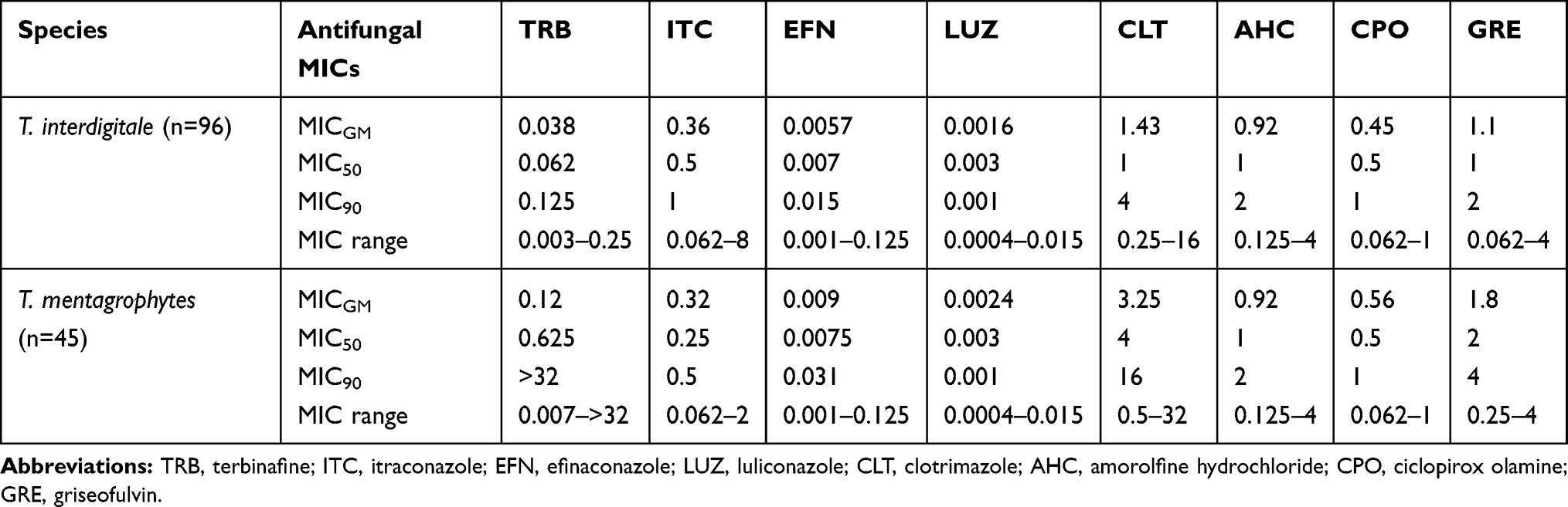 |
Table 1 MIC Values (μg/mL) of 8 Antifungal Agents Against 141 Trichophyton interdigitale and T. mentagrophytes Isolates |
The partial SQLE sequence was successfully amplified in all 5 sensitive and 5 resistant strains. The obtained sequences were deposited in GenBank under accession numbers MN893286 and MN901902–MN901905 for susceptible and MN901906–MN901910 for resistant strains. All T. mentagrophytes resistant isolates harbored missense mutations in SQLE, corresponding to amino acid substitution Ala448Thr in combination with Leu393Ser (isolate R1) or Phe397Leu (isolates R2 to R5). On the other hand, two susceptible isolates (MICs = 0.125–0.25 µg/mL) revealed wild type SQLE sequence, while the three other isolates (MICs = 0.0075–0.25 µg/mL) showed a single Ala448Thr substitution (Table 2).
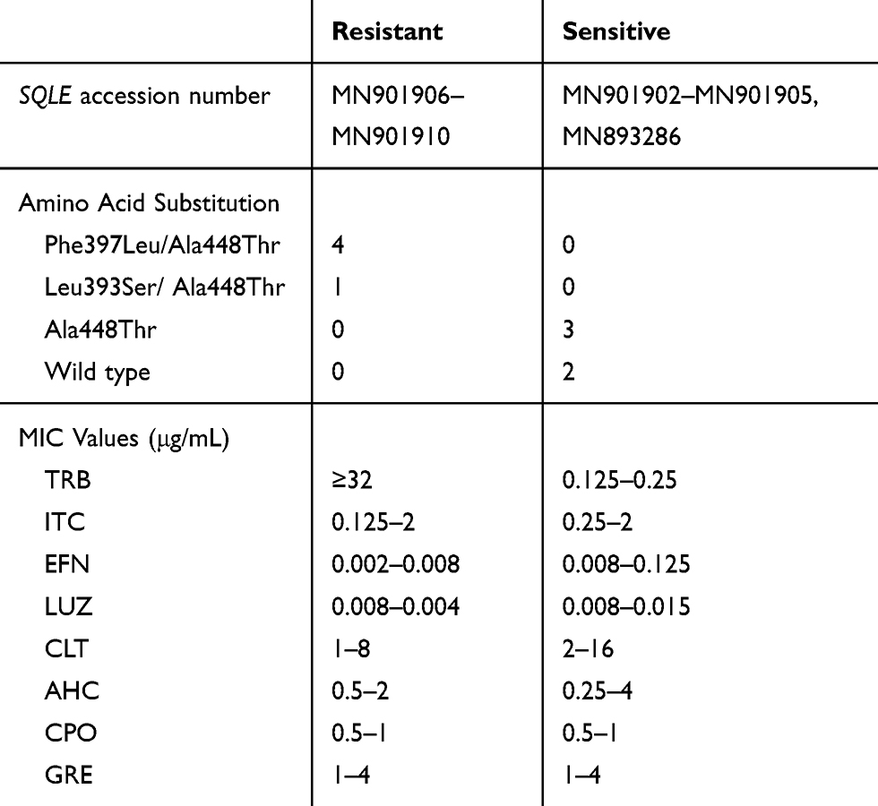 |
Table 2 MIC Values (μg/mL) of Used Antifungal Agents Against 10 Terbinafine Resistant/Susceptible T. mentagrophytes Isolates and Corresponding GenBank Accession with Consequential Amino Acid Substitutions in SQLE Gene |
Discussion
The antifungal susceptibility profile of Iranian TMTISG isolates, described in our results (Table 1), was comparable to that ones from other Iranian and foreign studies.12,27-30 The MIC values of EFN against TMTISG isolates from Japan, Canada, US and Iran in three studies all in all ranged from 0.001 to 0.03 µg/mL.27–29 They did not differ significantly from the MIC values for EFN in the current study (MICs = 0.001–0.125 µg/mL). Similarly, LUZ inhibitory concentrations against our TMTISG isolates ranged from 0.0004 to 0.015 µg/mL and were comparable with 0.0001–0.004 µg/mL for this imidazole against TMTISG strains in two previous reports from US and Iran.28,29 In two other investigations, LUZ has also had the most pronounced in vitro effect against TMTISG isolates from India and Iran, when compared with other antifungals, though the MIC values were higher (0.016–0.25 µg/mL) than in the current study.12,30 In our assessment, AHC and CPO in comparison with other novel antifungals, such as LUZ and EFN, demonstrated lower activities against TMTISG isolates (Table 1). The GM MIC value for CPO against T. interdigitale (0.45 µg/mL) in the present study, was similar to those found by Rudramurthy et al12 (0.25 µg/mL) and Magagnin et al (0.6 µg/mL).31 In the present study, the MIC90 for AHC against TMTISG isolates was 2 μg/mL, which was not compatible with those reported for T. interdigitale (0.02 μg/mL) and T. mentagrophytes (0.125 μg/mL) in India and US.12,28 In agreement with the reports by Singh et al and Baghi et al, TMTISG strains showed increased susceptibility to CLT and GRE.5,30 In our study, all T. mentagrophytes isolates with high MICs for terbinafine (≥32 µg/mL) and point mutations in the SQLE gene, were inhibited by 0.015 μg/mL of LUZ and 0.125 μg/mL of EFN. Given that there are no reports on dermatophyte resistance to newly FDA approved antifungals EFN and LUZ, these agents should be taken into consideration in the cases of TRB resistance as alternatives.
To the extent of our knowledge, TRB resistance in TMTISG isolates has already been reported from at least eight Asian and European countries, including India,5,12,14,15 Switzerland,11,16 Japan,13 Finland,17 Denmark,18 Bahrain,19 Russia,32 and Germany.19 The TRB MIC values in resistant TMTISG isolates from Iran and other countries varied in the range ≥1–≥32 μg/mL.12–14,16–18 In the current report, similar to some other studies,5,15,17,19 all TRB resistant isolates belonged to T. mentagrophytes species and ITS type VIII (5/28; 18%). Despite this, careful analysis of the literature revealed resistant Type II strains (Table 3). Hence, more data are needed to clarify whether in the TMTISG there is an association between genotypes and a potential to develop antifungal drug resistance. Among amino acid substitutions in SQLE, leading to TRB resistance, the most commonly encountered are Phe397Leu and Leu393Phe (Tables 2 and 3). The substitution Leu393Ser or other less common substitutions were also correlated with a high MIC value (≥32 μg/mL) of TRB. In some cases, in vitro resistance could not be explained by the presence of any mutation in SQLE (wild type).12,13 Then, mechanisms other than Phe397Leu and Leu393Phe substitution should still be considered as alternatives for TRB treatment failure. For example, Santos et al, recently showed that TRB resistance in T. rubrum can be mediated by multiplication of salicylate 1-monooxygenase (salA) gene.33
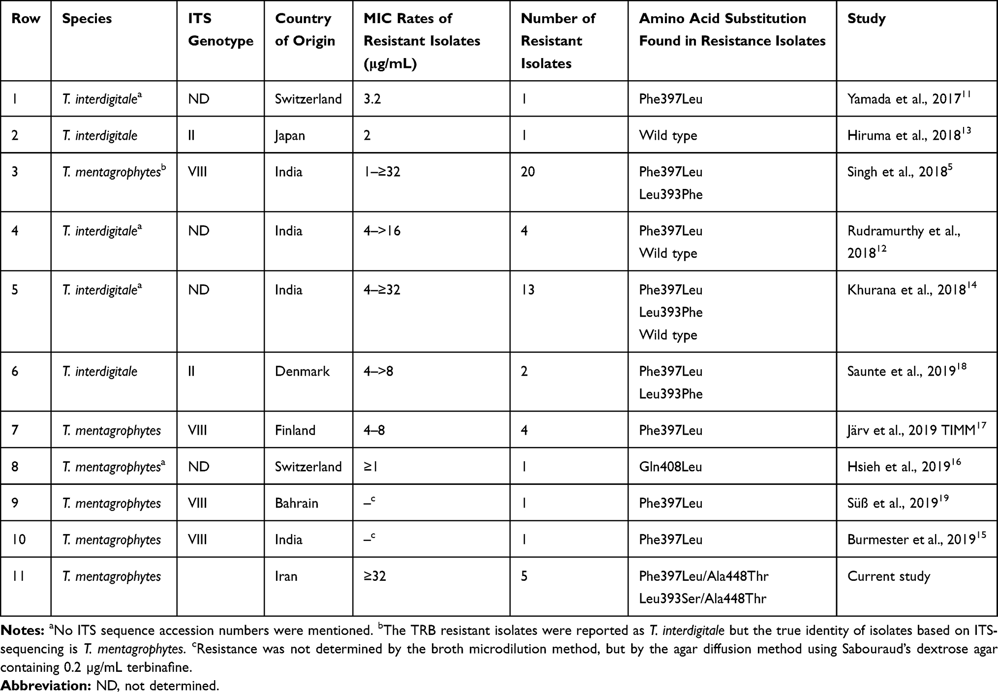 |
Table 3 Number of Terbinafine (TRB) Resistant Isolates Showing Mutations in the Squalene Epoxidase (SQLE) Gene in Regards to the ITS Genotypes and TRB MIC Values in Different Studies |
Conclusion
Overall, in both T. mentagrophytes and T. interdigitale, the potencies of LUZ and EFN against TMTISG isolates were apparently greater than those of other agents. Here, for the first time in Iran, we described TRB resistance in TMTISG isolates with its molecular mechanisms. The emergence of high level of in vitro TRB resistance with proven mutation in SQLE gene in Iranian T. mentagrophytes isolates is unpromising and warrants the genotyping of isolates primarily resistant to TRB.
Acknowledgments
This work was financially supported by Dr. Ali Rezaei-Matehkolaei from Ahvaz Jundishapur University of Medical Sciences, Ahvaz, Iran.
Disclosure
The authors declare that they have no competing interests in this work.
References
1. De Hoog GS, Dukik K, Monod M, et al. Toward a novel multilocus phylogenetic taxonomy for the dermatophytes. Mycopathologia. 2017;182:5–31. doi:10.1007/s11046-016-0073-9
2. Hay RJ. Dermatophytosis (ringworm) and other superficial mycoses. Mandell, Douglas, and Bennett’s Principles and Practice of Infectious Diseases.
3. Gupta AK, Foley KA, Versteeg SG. New antifungal agents and new formulations against dermatophytes. Mycopathologia. 2017;182(1–2):127–141. doi:10.1007/s11046-016-0045-0
4. Verma S, Madhu R. The great Indian epidemic of superficial dermatophytosis: an appraisal. Indian J Dermatol. 2017;62(3):227. doi:10.4103/ijd.IJD_206_17
5. Singh A, Masih A, Khurana A, et al. High terbinafine resistance in Trichophyton interdigitale isolates in Delhi, India harboring mutations in the squalene epoxidase gene. Mycoses. 2018;61(7):477–484. doi:10.1111/myc.12772
6. Majid I, Sheikh G, Kanth F, Hakak R. Relapse after oral terbinafine therapy in dermatophytosis: a clinical and mycological study. Indian J Dermatol. 2016;61(5):529–533. doi:10.4103/0019-5154.190120
7. Dogra S, Uprety S. The menace of chronic and recurrent dermatophytosis in India: is the problem deeper than we perceive? Indian Dermatol Online J. 2016;7(2):73–76. doi:10.4103/2229-5178.178100
8. Khurana A, Sardana K, Chowdhary A. Antifungal resistance in dermatophytes: recent trends and therapeutic implications. Fungal Genet Biol. 2019;132:103255. doi:10.1016/j.fgb.2019.103255
9. Mukherjee PK, Leidich SD, Isham N, Leitner I, Ryder NS, Ghannoum MA. Clinical Trichophyton rubrum strain exhibiting primary resistance to terbinafine. Antimicrob Agents Chemother. 2003;47(1):82–86. doi:10.1128/AAC.47.1.82-86.2003
10. Osborne CS, Leitner I, Hofbauer B, Fielding CA, Favre B, Ryder NS. Biological, biochemical, and molecular characterization of a new clinical Trichophyton rubrum isolate resistant to terbinafine. Antimicrob Agents Chemother. 2006;50(6):2234–2236. doi:10.1128/AAC.01600-05
11. Yamada T, Maeda M, Alshahni MM, et al. Terbinafine resistance of Trichophyton clinical isolates caused by specific point mutations in the squalene epoxidase gene. Antimicrob Agents Chemother. 2017;61(7):e00115–17. doi:10.1128/AAC.00115-17
12. Rudramurthy SM, Shankarnarayan SA, Dogra S, et al. Mutation in the squalene epoxidase gene of Trichophyton interdigitale and Trichophyton rubrum associated with allylamine resistance. Antimicrob Agents Chemother. 2018;62(5):e02522–17. doi:10.1128/AAC.02522-17
13. Hiruma J, Kitagawa H, Noguchi H, et al. Terbinafine‐resistant strain of Trichophyton interdigitale strain isolated from a tinea pedis patient. J Dermatol. 2019;46(4):351–353. doi:10.1111/1346-8138.14809
14. Khurana A, Masih A, Chowdhary A, et al. Correlation of in vitro susceptibility based on MICs and squalene epoxidase mutations with clinical response to terbinafine in patients with tinea corporis/cruris. Antimicrob Agents Chemother. 2018;62(12):e01038–18. doi:10.1128/AAC.01038-18
15. Burmester A, Hipler UC, Hensche R, Elsner P, Wiegand C. Point mutations in the squalene epoxidase gene of Indian ITS genotype VIII T. mentagrophytes identified after DNA isolation from infected scales. Med Mycol Case Rep. 2019;26:23–24. doi:10.1016/j.mmcr.2019.09.001
16. Hsieh A, Quenan S, Riat A, Toutous-trellu L, Fontao L. A new mutation in the SQLE gene of Trichophyton mentagrophytes associated to terbinafine resistance in a couple with disseminated tinea corporis. J Mycol Med. 2019;29(4):352–355. doi:10.1016/j.mycmed.2019.100903
17. Järv H, Uhrlass S, Simkin T, et al. Terbinafine resistant Trichophyton mentagrophytes genotype VIII, Indian type, isolated in Finland. J Fungi. 2019;5(95):P039.
18. Saunte DM, Hare RK, Jørgensen KM, et al. Emerging terbinafine resistance in Trichophyton: clinical characteristics, squalene epoxidase gene mutations, and a reliable EUCAST method for detection. Antimicrob Agents Chemother. 2019;63(10):e01126–e011219. doi:10.1128/AAC.01126-19
19. Süß A, Uhrlaß S, Ludes A, et al. Extensive tinea corporis due to a terbinafine-resistant Trichophyton mentagrophytes isolate of the Indian genotype in a young infant from Bahrain in Germany. Hautarzt. 2019;1–9. doi:10.1007/s00105-019-4431-7.
20. Pchelin IM, Azarov DV, Churina MA, et al. Species boundaries in the Trichophyton mentagrophytes / T. interdigitale species complex. Med Mycol. 2019;57(6):781–789. doi:10.1093/mmy/myy115
21. Taghipour S, Pchelin IM, Zarei Mahmoudabadi A, et al. Trichophyton mentagrophytes and T. interdigitale genotypes are associated with particular geographic areas and clinical manifestations. Mycoses. 2019;62(11):1084–1091. doi:10.1111/myc.12993
22. Makimura K, Mochizuki T, Hasegawa A, Uchida K, Saito H, Yamaguchi H. Phylogenetic classification of Trichophyton mentagrophytes complex strains based on DNA sequences of nuclear ribosomal internal transcribed spacer 1 regions. J Clin Microbiol. 1998;36(9):2629–2633. doi:10.1128/JCM.36.9.2629-2633.1998
23. Van den Ende AHG, De Hoog GS. Variability and molecular diagnostics of the neurotropic species Cladophialophora bantiana. Stud Mycol. 1999;43:151–162.
24. Tamura K, Stecher G, Peterson D, Filipski A, Kumar S. MEGA6: molecular evolutionary genetics analysis version 6.0. Mol Biol Evol. 2013;30(12):2725–2729. doi:10.1093/molbev/mst197
25. Clinical and Laboratory Standards Institute. Reference Method for Broth Dilution Antifungal Susceptibility Testing of Filamentous Fungi; Approved Standard.
26. Uchida K, Tanaka T, Yamaguchi H. Achievement of complete mycological cure by topical antifungal agent NND‐502 in guinea pig model of tinea pedis. Microbiol Immunol. 2003;47(2):143–146. doi:10.1111/j.1348-0421.2003.tb02797.x
27. Siu WJ, Tatsumi Y, Senda H, et al. Comparison of in vitro antifungal activities of efinaconazole and currently available antifungal agents against a variety of pathogenic fungi associated with onychomycosis. Antimicrob Agents Chemother. 2013;57(4):1610–1616. doi:10.1128/AAC.02706-13
28. Wiederhold NP, Fothergill AW, McCarthy DI, Tavakkol A. Luliconazole demonstrates potent in vitro activity against dermatophytes recovered from patients with onychomycosis. Antimicrob Agents Chemother. 2014;58(6):3553–3555. doi:10.1128/AAC.02706-13
29. Rezaei-Matehkolaei A, Khodavaisy S, Alshahni MM, et al. In vitro antifungal activity of novel triazole efinaconazole and five comparators against dermatophyte isolates. Antimicrob Agents Chemother. 2018;62(5):e02423–17. doi:10.1128/AAC.02423-17
30. Baghi N, Shokohi T, Badali H, et al. In vitro activity of new azoles luliconazole and lanoconazole compared with ten other antifungal drugs against clinical dermatophyte isolates. Med Mycol. 2016;54(7):757–763. doi:10.1093/mmy/myw016
31. Magagnin CM, Stopiglia CD, Vieira FJ, et al. Antifungal susceptibility of dermatophytes isolated from patients with chronic renal failure. An Bras Dermatol. 2011;86(4):694–701. doi:10.1590/s0365-05962011000400011
32. Manoyan M, Sokolov V, Gursheva A, Gabuzyan N, Panin A. Sensitivity of isolated dermatophyte strains to antifungal drugs in the Russian Federation. J Fungi. 2019;5(95):P034.
33. Santos HL, Lang EA, Segato F, Rossi A, Martinez-rossi NM. Terbinafine resistance conferred by multiple copies of the salicylate 1-monooxygenase gene in Trichophyton rubrum. Med Mycol. 2017;56(3):378–381. doi:10.1093/mmy/myx044
 © 2020 The Author(s). This work is published and licensed by Dove Medical Press Limited. The full terms of this license are available at https://www.dovepress.com/terms.php and incorporate the Creative Commons Attribution - Non Commercial (unported, v3.0) License.
By accessing the work you hereby accept the Terms. Non-commercial uses of the work are permitted without any further permission from Dove Medical Press Limited, provided the work is properly attributed. For permission for commercial use of this work, please see paragraphs 4.2 and 5 of our Terms.
© 2020 The Author(s). This work is published and licensed by Dove Medical Press Limited. The full terms of this license are available at https://www.dovepress.com/terms.php and incorporate the Creative Commons Attribution - Non Commercial (unported, v3.0) License.
By accessing the work you hereby accept the Terms. Non-commercial uses of the work are permitted without any further permission from Dove Medical Press Limited, provided the work is properly attributed. For permission for commercial use of this work, please see paragraphs 4.2 and 5 of our Terms.