")
Back to Journals » Cancer Management and Research » Volume 12
Effects of Hydroxytyrosol on Expression of Apoptotic Genes and Activity of Antioxidant Enzymes in LS180 Cells
Authors Hormozi M, Salehi Marzijerani A, Baharvand P
Received 12 March 2020
Accepted for publication 14 July 2020
Published 27 August 2020 Volume 2020:12 Pages 7913—7919
DOI https://doi.org/10.2147/CMAR.S253591
Checked for plagiarism Yes
Review by Single anonymous peer review
Peer reviewer comments 2
Editor who approved publication: Professor Bilikere Dwarakanath
Maryam Hormozi,1 Atena Salehi Marzijerani,2 Parastoo Baharvand3
1Department of Biochemistry, Lorestan University of Medical Science, Khorramabad, Iran; 2Student Research Committee, Lorestan University of Medical Sciences, Khorramabad, Iran; 3Department of Community Medicine, Lorestan University of Medical Sciences, Khorramabad, Iran
Correspondence: Parastoo Baharvand Department of Community Medicine
Lorestan University of Medical Sciences, Khorramabad, Iran
Email [email protected]
Purpose: Colorectal cancer is the third–most commonly occurring cancer in developed countries. Hydroxytyrosol is a potent antioxidant that has several activities, such as oxidative-stress control, inhibition of cell proliferation, and induction of apoptosis. In this study, the effect of hydroxytyrosol on the expression of genes effective in apoptosis — BAX, BCL2, CASP3, P53, PPARG, and NFE2L2 — and antioxidant-enzyme activity in LS180 cells of human colorectal cancer was investigated.
Methods: The human colorectal cancer cell line LS180 was treated with different concentrations of hydroxytyrosol for 24 hours. Expression of BAX, BCL2, CASP3, NFE2L2, PPARG, and P53 was investigated using real-time PCR. The activity of antioxidant and malondialdehyde enzymes was measured by calorimetric methods.
Results: Analysis of gene expression showed that hydroxytyrosol significantly increased the expression of CASP3 and the BAX:BCL2 ratio in treatment groups compared to the control (P< 0.05). Also, hydroxytyrosol significantly reduced the expression of the NFE2L2 gene (P< 0.05). Calorimetric analysis showed that hydroxytyrosol increased activity of the antioxidant enzymes catalase, superoxide dismutase, and glutathione peroxidase in treatment groups significantly more than the control group and reduced thiobarbituric acid–reactive substances on an oxidative stress index (P< 0.05).
Conclusion: Hydroxytyrosol may induce apoptosis in colorectal cancer cells by increasing the expression of CASP3 gene and increasing the BAX:BCL2 ratio. Also, hydroxytyrosol may increase the activity of antioxidant enzymes and reduce the proliferation of LS180 cells by changing the antioxidant-defense system in cancer cells.
Keywords: colorectal cancer, hydroxytyrosol, apoptosis, antioxidant enzymes, calorimetric analysis, malondialdehyde enzymes
Introduction
Colorectal cancer is the third–most common cancer in men (after lung and prostate cancer) and the second–most common in women (after breast cancer).1 The most important risk factors for colorectal cancer, which can be controlled and prevented by the individual, include environmental factors, lifestyle, and nutritional factors, such as low-fiber and high-fat diets, smoking, alcohol intake, inadequate activity, and lack of exercise. Inevitable risk factors include age, genetics, and inheritance.2
The most important risk factor for colorectal cancer is nutritional. It is known that the type of diet can affect the distribution of gastrointestinal cancers, and in the case of lifestyle changes and nutritional habits, up to 30% of cases of colorectal cancer can be prevented.3 A diet high in fat and saturated fatty acids is a major risk factor for colorectal cancer. Saturated fatty acids cause inflammation through the production of prostaglandin E2. The consumption of red meat and processed meat is also associated with the development of colorectal cancer. Some meat is cooked at high temperatures, resulting in heterocyclic amines and aromatic polycyclic hydrocarbons that have carcinogenic properties. On the other hand, a diet containing vegetables and fruit can protect against colorectal cancer, due to its richness in fiber and such nutrients as calcium, selenium, vitamins A, E, C, and D, folic acid, carotenoids, and plant phenols.4,5 Hydroxytyrosol is one of the most potent antioxidant phenolic compounds in olives, and is able to transfer the hydroxyl electron group to its ortho position and form a hydrogen-bonding molecule with radical phenoxylic stability.6,7
Research has shown that the incidence of cancer in the Mediterranean region is lower than in other countries. Much empirical evidence have shown that in addition to antioxidant and anti-inflammatory properties, hydroxytyrosol carries out its anticancer activity by activating molecular signaling pathways, leading to apoptosis and cessation of the cell cycle in various tumor-cell lines, including colon cancer.8,9 The antitumor effects of hydroxytyrosol are characterized by suppression in the expression of EGFR, eg, in tumor cells and human colorectal cancer, hydroxytyrosol degrades urinocationic receptors and reduces cell proliferation.10 In addition, hydroxytyrosol has been shown to inhibit cell proliferation in MCF7 breast cancer cells and induce apoptosis.11,12 Also, hydroxytyrosol induces the expression of antioxidant enzymes by activating Nrf2.13 Therefore, this substance can be considered an important agent in the treatment of cancer.14 These effects of hydroxytyrosol have been reported at different concentrations, wherein maximum activity was seen at 100 µM.15 Additionally, this agent has also shown dose-dependent inhibitory effects on pancreatic cancer–cell growth.16 The aim of this study was to evaluate the effect of hydroxytyrosol on expression of BAX, BCL2, CASP3 (genes affecting apoptosis), P53, NFE2L2, and PPARG, and antioxidant enzymes (superoxide dismutase, catalase, and glutathione reductase) and malondialdehyde in a colorectal cancer (LS180) human cell line.
Methods
Cell Culture and Treatment with Hydroxytyrosol
In this study, the human colorectal cancer cell line LS180 was obtained from the cell bank of the Pasteur Institute of Tehran and grown in DMEM containing 10% FBS, 100 IU/mL penicillin, and 100 μg/mL streptomycin at 37°C with 5% CO2. After treatment of cells with 50, 100, and 150 μM hydroxytyrosol (Sigma) for 24 hours, cells were collected by trypsinization.
RNA Extraction and cDNA Synthesis
RNA extraction was performed using an RNA-extraction kit (Gena Bioscience, Germany) using the protocol provided by the manufacturer. Quantitative RNA extracted from electrophoresis was viewed on 1.5% agarose gel and measured with a NanoDrop device. cDNA was extracted using a cDNA-synthesis kits (Gena Bioscience, Germany) and synthesized by cDNA.
Evaluation of Gene Expression Using Real-Time PCR
Expression levels of P53, NFE2L2, PPARG, BAX, BCL2, and CASP3 were assessed using real-time PCR (Gena Bioscience and Corbett) with the relevant primers (Table 1).
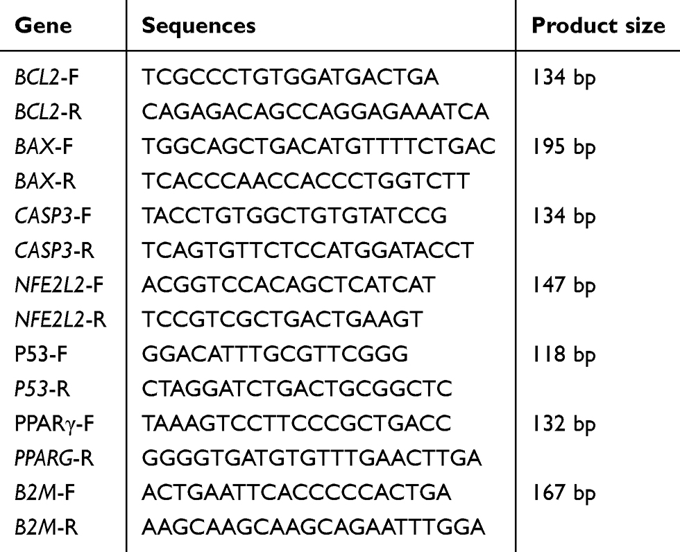 |
Table 1 Forward- and Reverse-Primer Sequences |
Cell Protein–Content Evaluation
In order to investigate the activity of antioxidant enzymes and oxidative indices, after treatment and collectiion of cells using cell-lysis buffer, protein was extracted and the content of each sample measured using the Bradford assay.
Activity of Antioxidant and Malondialdehyde Enzymes
The calorimetric method was used to evaluate the activity of catalase in the enzyme reaction with methanol in the presence of oxygenated water, and the specific activity of the enzyme was calculated in terms of unit/mg protein.17 The activity of superoxide dismutase was analyzed by a Pyrogallol radical superoxide reaction using a spectrophotometer. The specific activity of the enzyme was calculated in terms of unit/mg protein.18 Measurement of glutathione peroxidase activity was performed based on calorimetry and glutathione oxide formation. Specific activity was also calculated in terms of unit/mg protein.19 Malondialdehyde assays were performed based on reactions with a thiobarbituric acid–reactive substrate.20
Data Analysis
REST software was used to analyze the data obtained on gene expression.21 SPSS software was used to analyze the oxidative-stress index and antioxidant-enzyme activity. To describe the data, means, SD, frequency tables, one-way ANOVA, and Tukey’s post hoc test were used.
Results
Evaluation of Gene Expression
The results of this study showed that hydroxytyrosol at all concentrations significantly increased BAX expression (P<0.05, Figure 1A). Expression of BCL2 increased in the treatment group compared to the control, but this increase was not statistically significant (P>0.05, Figure 1B). Hydroxytyrosol increased the BAX:BCL2 ratio at all concentrations compared to the control group (P<0.05, Figure 1C). Hydroxytyrosol increased CASP3 expression at all concentrations compared to the control group (P<0.05) (Figure 1D). Hydroxytyrosol did not significantly affect expression of P53 compared to the control group (P>0.05, Figure 2A). Furthermore, hydroxytyrosol reduced the expression of NFE2L2 in the treated group compared to the control group, which was statistically significant at a concentration of 50 μM (P<0.05, Figure 2B). The treatment group also had a significant reduction in the expression of PPARG at all concentrations by 1.7 (P<0.05, Figure 2C).
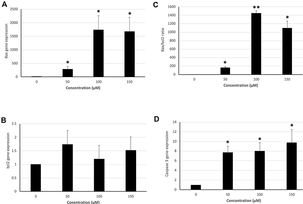 |
Figure 1 Expression of BAX, BCL2, and CASP3 and BAX:BCL2 ratio in groups treated with 50, 100, and 150 μM hydroxytyrosol and controls in the LS180 cell line.Notes: *P<0.05; **P<0.01. BAX:BCL2 ratio was greatest at 100 μM concentration whereas, CASP3 gene activity increased in a dose-dependent manner. (A) BAX expression was significant at all concentrations. (B) BCL2 -expression increase was not significant. (C) The BAX:BCL2 ratio was increased at all concentrations. (D) Hydroxytyrosol increased CASP3 expression at all concentrations compared to control group, which was statistically significant. |
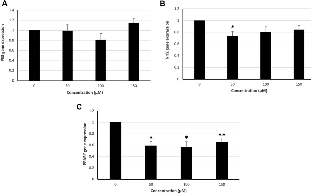 |
Figure 2 Expression of P53, NFE2L2, and PPARG genes in groups treated with 50, 100, and 150 μM hydroxytyrosol and controls in the LS180 cell line.Notes: *P<0.05; **P<0.01. NFE2L2 expression was significant only at 50 μM. (A) The expression of P53 gene compared to control group did not increase significantly. (B) The expression of Nrf2 gene was significant at a concentration of 50 μM. (C) Significant reductions in the of PPARγ expression at all concentrations were found. |
Effect of Hydroxytyrosol on Activity of Antioxidant Enzymes and Thiobarbituric Acid–Reactive Substances
Catalase-enzyme activity at 50, 100, and 150 μM hydroxytyrosol increased in comparison with the control group(P<0.05, Figure 3A). Similarly, all treatment groups had significantly increased activity of glutathione peroxidase and superoxide dismutase compared with the control group (P<0.05,. Figure 3B and C). At all concentrations, hydroxytyrosol decreased levels of thiobarbituric acid–reactive substances compared to the control group (P<0.05, Figure 3D).
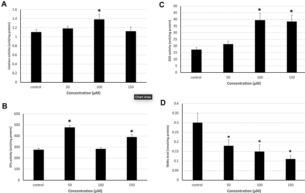 |
Figure 3 Enzymatic activity of catalase, GPx, SOD and TBARS in groups treated with 50, 100, and 150 µM hydroxytyrosol and controls in the LS180 cell line.Notes: *P<0.05. (A) Maximum activity of catalase was seen at 100 µM. (B) GPx activity was greatest at 50 and 150 µM. (C) SOD activity increased in a dose-dependent manner, but at 50 µM did not show a significant increase. (D) TBARS reduced in a dose-dependent manner. |
Discussion
Colorectal cancer is the most common cancer of the digestive tract. In recent years, efforts have been made to find natural products that can fight colorectal cancer. Among these compounds, polyphenols, secondary metabolites of plants that have beneficial effects on human health and reduce the risk of most cancers, have been widely recognized.22,23 Hydroxytyrosol is a natural polyphenol compound and a strong antioxidant. Recent studies have shown that this potent antioxidant has several biological activities, such as oxidative-stress control, cellular efficacy, and induction of apoptosis in several tumor-cell lines.9,24 Our study evaluated the effects of three doses of hydroxytyrosol — 50, 100, and 150 µM — on colorectal cancer cells. Various concentrations of hydroxytyrosol have been reported to have antioxidant,25 antiangiogenic,26 and apoptotic27 effects in different cancer-cell lines.
Several studies have confirmed the antitumor effects of hydroxytyrosol on several cancers, such as breast cancer, leukemia, and melanoma.28 Studies have shown that hydroxytyrosol induces G2/M cell-cycle end points and inhibits cell proliferation. It also induces apoptosis in cancer cells and inhibits Akt activity and the NFKB pathway.29 In a study on HL60 (promyelocytic leukemia) and HT29 (colon adenocarcinoma), it was shown that hydroxytyrosol slowed cell growth and proliferation and stopped the cell cycle and apoptosis in these cells.30 Various proteins are involved in the regulation of apoptosis. The family of BCL2 proteins, which include proapoptotic and antiapoptotic proteins, is one of the major regulators of this process.31 Our study showed that hydroxytyrosol at all concentrations significantly increased expression of BAX compared to the control group. Also, hydroxytyrosol increased BCL2 expression at all concentrations compared to the control. Similarly, the BAX:BCL2 ratio seems to be a more appropriate indicator for inducing apoptosis and determining the cell-death rate toward apoptosis after exposure to cytotoxic agents. Our study showed that hydroxytyrosol at all concentrations significantly increased expression of CASP3 in the LS180 cell line compared to the control group. In a study conducted on uterine (FB2 and TPC1) and follicular thyroid cancer (WRO) cancer-cell lines in 2017, hydroxytyrosol reduced the expression of mRNA and cyclin D1 protein and increased the expression of p21 in uterine cancer cells. Hydroxytyrosol also increased the expression of caspase 3 and PARP1 in these cell lines.27
P53 is the most widely known tumor-suppressor gene, and plays a crucial role in genomic stability and tumor suppression by inducing apoptosis, halting the cell cycle, aging, and angiogenesis.32 Studies have shown that hydroxytyrosol induces apoptosis by effecting the expression of P53. The results of this study showed increased expression of P53 at different concentrations of hydroxytyrosol in the LS180 cell line. In a study on the breast cancer–cell line MCF7, it has been shown that olerupein (from hydrolysis of oleuropein and hydroxytyrosol) increases the expression of P53 in treated groups compared to controls and resulted in apoptosis. It has also been shown in another study on uterine cancer cells (FB2 and TPC1) and thyroid cancer cells (WRO) that hydroxytyrosol increased the expression of p53 and BAD and induced apoptosis.27
Nrf2 is a key factor in mechanisms of cell defense against oxidative stress.33 The results of our study showed that hydroxytyrosol at all concentrations reduced the expression of Nrf2 compared to the control group. A study in 2011 showed that hydroxytyrosol significantly increased the expression of Nrf2 in the normal epithelial cell line MCF10A, but did not increase in the breast cancer–cell lines MDA-MB231 and MCF7 or was not effective.33 Our study results were consistent with this study.
During normal cell activity, reactive oxygen species (ROS) are produced, which also play an important role in apoptosis. Active oxygen species act as a redox messenger in low physiological levels in signaling and intracellular regulation, while at high levels they produce oxidative stress and induce oxidative changes in macromolecules. Each cell is equipped with an antioxidant defense system to prevent the excessive production of active radicals.34 In vitro studies have shown that the beneficial effects of phenolic compounds, such as hydroxytyrosol, on human health are due to their antioxidant properties. In a study on the CACO2 cell line, it was shown that hydroxytyrosol inhibited ROS-induced cytotoxicity in these cells and reduced malondialdehyde levels in dose-dependent groups.35
The results of this study showed that hydroxytyrosol significantly increased the activity of cellular antioxidant enzymes, including catalase, superoxide dismutase, and glutathione peroxidase, in all treatment groups. Also, malondialdehyde concentrations in the treated groups were significantly lower than controls. Studies have shown that anticancer compounds that have antioxidant activity may have beneficial effects by balancing ROS levels, which not only prevent the proliferation of cancer cells but also lead to apoptosis. In a similar study, the semisynthetic derivative of an anticarcinogenic alkaloid, through the production of ROS and increased oxidative stress, led to toxicity to the MCF7 breast cancer–cell line, and upregulated the activity of antioxidant enzymes, such as superoxide dismutase and glutathione peroxidase. The study concluded that this increase in activity of antioxidant enzymes could be a response to increased oxidative stress in treated cells.36 On the other hand, studies have shown that phenolic compounds are not only antioxidants but can also be prooxidants and result in the production of ROS.37 Based on the results of this study, hydroxytyrosol seems to act as a prooxidant in the LS180 cell line of human colorectal cancer and mediates the production of ROS, and thereby the increased activity of the antioxidant enzymes observed in this study could be a response to increased oxidative stress in treated cells.
Conclusion
Hydroxytyrosol can induce apoptosis in the LS180 colorectal cancer–cell line by increasing the expression of proapoptotic genes, such as BAX, CASP3, and P53, increasing the BAX:BCL2 ratio, and downregulating NFE2L2 expression. Also, the treatment of cells with hydroxytyrosol upregulates antioxidative activity in the colorectal cancer–cell line, marked by increased antioxidant enzymes. In vivo studies on the therapeutic effects of hydroxytyrosol on colorectal cancer in animal models can give better conclusions.
Author Contributions
All authors contributed to data analysis, drafting or revising the article, gave final approval to the version to be published, and agree to be accountable for all aspects of the work.
Disclosure
The authors report no conflicts of interest with this work.
References
1. Vargas AJ, Thompson PA. Diet and nutrient factors in colorectal cancer risk. Nutr Clin Pract. 2012;27(5):613–623. doi:10.1177/0884533612454885
2. Haggar FA, Boushey RP. Colorectal cancer epidemiology: incidence, mortality, survival, and risk factors. Clin Colon Rectal Surg. 2009;22(4):191–197. doi:10.1055/s-0029-1242458
3. Fernandez E, La Vecchia C, Talamini R, Negri E. Joint effects of family history and adult life dietary risk factors on colorectal cancer risk. Epidemiology. 2002;13(3):360–363. doi:10.1097/00001648-200205000-00019
4. Santarelli RL, Pierre F, Corpet DE. Processed meat and colorectal cancer: a review of epidemiologic and experimental evidence. Nutr Cancer. 2008;60(2):131–144. doi:10.1080/01635580701684872
5. Hormozi M, Gholami M, Babaniazi A, Gharravi AM. Calendula officinalis stimulate proliferation of mouse embryonic fibroblasts via expression of growth factors TGFβ1 and bFGF. Inflamm Regen. 2019;39(1):7. doi:10.1186/s41232-019-0097-x
6. Visioli F, Bellomo G, Galli C. Free radical-scavenging properties of olive oil polyphenols. Biochem Biophys Res Commun. 1998;247(1):60–64. doi:10.1006/bbrc.1998.8735
7. Rigacci S, Stefani M. Nutraceutical properties of olive oil polyphenols. An itinerary from cultured cells through animal models to humans. Int J Mol Sci. 2016;17(6):843. doi:10.3390/ijms17060843
8. Bouallagui Z, Han J, Isoda H, Sayadi S. Hydroxytyrosol rich extract from olive leaves modulates cell cycle progression in MCF-7 human breast cancer cells. Food Chem Toxicol. 2011;49(1):179–184. doi:10.1016/j.fct.2010.10.014
9. López de Las Hazas M-C, Piñol C, Macià A, Motilva M-J. Hydroxytyrosol and the colonic metabolites derived from virgin olive oil intake induce cell cycle arrest and apoptosis in colon cancer cells. J Agric Food Chem. 2017;65(31):6467–6476. doi:10.1021/acs.jafc.6b04933
10. Terzuoli E, Giachetti A, Ziche M, Donnini S. Hydroxytyrosol, a product from olive oil, reduces colon cancer growth by enhancing epidermal growth factor receptor degradation. Mol Nutr Food Res. 2016;60(3):519–529. doi:10.1002/mnfr.201500498
11. Han J, Talorete TP, Yamada P, Isoda H. Anti-proliferative and apoptotic effects of oleuropein and hydroxytyrosol on human breast cancer MCF-7 cells. Cytotechnology. 2009;59(1):45–53. doi:10.1007/s10616-009-9191-2
12. Hormozi M, Ghoreishi S, Baharvand P. Astaxanthin induces apoptosis and increases activity of antioxidant enzymes in LS-180 cells. Artif Cells Nanomed Biotechnol. 2019;47(1):891–895. doi:10.1080/21691401.2019.1580286
13. Rodríguez-Morató J, Xicota L, Fitó M, Farré M, Dierssen M, de la Torre R. Potential role of olive oil phenolic compounds in the prevention of neurodegenerative diseases. Molecules. 2015;20(3):4655–4680. doi:10.3390/molecules20034655
14. Hashimoto T, Ibi M, Matsuno K, et al. An endogenous metabolite of dopamine, 3, 4-dihydroxyphenylethanol, acts as a unique cytoprotective agent against oxidative stress-induced injury. Free Radic Biol Med. 2004;36(5):555–564. doi:10.1016/j.freeradbiomed.2003.12.003
15. Fabiani R, De Bartolomeo A, Rosignoli P, Servili M, Montedoro GF, Morozzi G. Cancer chemoprevention by hydroxytyrosol isolated from virgin olive oil through G1 cell cycle arrest and apoptosis. Eur J Cancer Care Prev. 2002;11(4):351–358. doi:10.1097/00008469-200208000-00006
16. Zubair H, Bhardwaj A, Ahmad A, et al. Hydroxytyrosol induces apoptosis and cell cycle arrest and suppresses multiple oncogenic signaling pathways in prostate cancer cells. Nutr Cancer. 2017;69(6):932–942. doi:10.1080/01635581.2017.1339818
17. Aebi H. [13] Catalase in vitro. In: Methods in Enzymology. Vol. 105. Elsevier; 1984:121–126.
18. Kuthan H, Haussmann H-J, Werringloer J. A spectrophotometric assay for superoxide dismutase activities in crude tissue fractions. Biochem J. 1986;237(1):175–180. doi:10.1042/bj2370175
19. Flohé L, Günzler WA. [12] Assays of Glutathione Peroxidase. In: Methods in Enzymology. Vol. 105. Elsevier; 1984:114–120.
20. Uchiyama M, Mihara M. Determination of malonaldehyde precursor in tissues by thiobarbituric acid test. Anal Biochem. 1978;86(1):271–278. doi:10.1016/0003-2697(78)90342-1
21. Pfaffl MW, Horgan GW, Dempfle L. Relative expression software tool (REST©) for group-wise comparison and statistical analysis of relative expression results in real-time PCR. Nucleic Acids Res. 2002;30(9):e36–e. doi:10.1093/nar/30.9.e36
22. Zeriouh W, Nani A, Belarbi M, et al. Phenolic extract from oleaster (Olea europaea var. Sylvestris) leaves reduces colon cancer growth and induces caspase-dependent apoptosis in colon cancer cells via the mitochondrial apoptotic pathway. PLoS One. 2017;12(2):e0170823. doi:10.1371/journal.pone.0170823
23. Tresserra-Rimbau A, Lamuela-Raventos RM, Moreno JJ. Polyphenols, food and pharma. Current knowledge and directions for future research. Biochem Pharmacol. 2018;156:186–195. doi:10.1016/j.bcp.2018.07.050
24. Borzì AM, Biondi A, Basile F, Luca S, Vicari ESD, Vacante M. Olive oil effects on colorectal cancer. Nutrients. 2019;11(1):32. doi:10.3390/nu11010032
25. Manna C, Galletti P, Cucciolla V, Montedoro G, Zappia V. Olive oil hydroxytyrosol protects human erythrocytes against oxidative damages. J Nutr Biochem. 1999;10(3):159–165. doi:10.1016/S0955-2863(98)00085-0
26. Fortes C, García-Vilas JA, Quesada AR, Medina MÁ. Evaluation of the anti-angiogenic potential of hydroxytyrosol and tyrosol, two bio-active phenolic compounds of extra virgin olive oil, in endothelial cell cultures. Food Chem. 2012;134(1):134–140. doi:10.1016/j.foodchem.2012.02.079
27. Toteda G, Lupinacci S, Vizza D, et al. High doses of hydroxytyrosol induce apoptosis in papillary and follicular thyroid cancer cells. J Endocrinol Invest. 2017;40(2):153–162. doi:10.1007/s40618-016-0537-2
28. Hormozi M, Baharvand P. Achillea biebersteinni Afan may inhibit scar formation: in vitro study. Mol Genet Genomic Med. 2019;7(5):e640. doi:10.1002/mgg3.640
29. Zhao B, Ma Y, Xu Z, et al. Hydroxytyrosol, a natural molecule from olive oil, suppresses the growth of human hepatocellular carcinoma cells via inactivating AKT and nuclear factor-kappa B pathways. Cancer Lett. 2014;347(1):79–87. doi:10.1016/j.canlet.2014.01.028
30. Granados-Principal S, Quiles JL, Ramirez-Tortosa CL, Sanchez-Rovira P, Ramirez-Tortosa MC. Hydroxytyrosol: from laboratory investigations to future clinical trials. Nutr Rev. 2010;68(4):191–206. doi:10.1111/j.1753-4887.2010.00278.x
31. Goldar S, Khaniani MS, Derakhshan SM, Baradaran B. Molecular mechanisms of apoptosis and roles in cancer development and treatment. Asian Pac J Cancer Prev. 2015;16(6):2129–2144. doi:10.7314/APJCP.2015.16.6.2129
32. Hormozi M, Assaei R, Boroujeni MB. The effect of aloe vera on the expression of wound healing factors (TGFβ1 and bFGF) in mouse embryonic fibroblast cell: in vitro study. Biomed Pharmacother. 2017;88:610–616. doi:10.1016/j.biopha.2017.01.095
33. Warleta F, Quesada CS, Campos M, Allouche Y, Beltrán G, Gaforio JJ. Hydroxytyrosol protects against oxidative DNA damage in human breast cells. Nutrients. 2011;3(10):839–857. doi:10.3390/nu3100839
34. Khoshtabiat L, Mahdavi M. The role of oxidative stress in proliferation and cell death. J Mazandaran Univ Med Sci. 2015;25(127):130–145.
35. Manna C, Della Ragione F, Cucciolla V, et al. Biological effects of hydroxytyrosol, a polyphenol from olive oil endowed with antioxidant activity. In: Advances in Nutrition and Cancer 2. Springer; 1999:115–130.
36. Timur M, Akbas SH, Ozben T. The effect of Topotecan on oxidative stress in MCF-7 human breast cancer cell line. Acta Biochim Pol. 2005;52(4):897–902. doi:10.18388/abp.2005_3404
37. Acquaviva R, Di Giacomo C, Sorrenti V, et al. Antiproliferative effect of oleuropein in prostate cell lines. Int J Oncol. 2012;41(1):31–38.
 © 2020 The Author(s). This work is published and licensed by Dove Medical Press Limited. The full terms of this license are available at https://www.dovepress.com/terms.php and incorporate the Creative Commons Attribution - Non Commercial (unported, v3.0) License.
By accessing the work you hereby accept the Terms. Non-commercial uses of the work are permitted without any further permission from Dove Medical Press Limited, provided the work is properly attributed. For permission for commercial use of this work, please see paragraphs 4.2 and 5 of our Terms.
© 2020 The Author(s). This work is published and licensed by Dove Medical Press Limited. The full terms of this license are available at https://www.dovepress.com/terms.php and incorporate the Creative Commons Attribution - Non Commercial (unported, v3.0) License.
By accessing the work you hereby accept the Terms. Non-commercial uses of the work are permitted without any further permission from Dove Medical Press Limited, provided the work is properly attributed. For permission for commercial use of this work, please see paragraphs 4.2 and 5 of our Terms.