")
Back to Journals » Veterinary Medicine: Research and Reports » Volume 13
Dry Season Eimeria Infection in Dairy Cattle and Sheep in and Around Adama and Bishoftu Towns, Oromia, Ethiopia
Authors Ayana D, Temesgen K, Kumsa B , Alkadir G
Received 13 June 2022
Accepted for publication 10 August 2022
Published 9 September 2022 Volume 2022:13 Pages 235—245
DOI https://doi.org/10.2147/VMRR.S377017
Checked for plagiarism Yes
Review by Single anonymous peer review
Peer reviewer comments 3
Editor who approved publication: Professor Young Lyoo
Dinka Ayana, Kebene Temesgen, Bersissa Kumsa, Gebayehu Alkadir
Department of Pathology and Parasitology, College of Veterinary Medicine and Agriculture, Addis Ababa University, Bishoftu, Ethiopia
Correspondence: Dinka Ayana, Email [email protected]; [email protected]
Introduction: Eimeria infection is one of the protozoal diseases of animals caused by various species of Eimeria (intracellular parasite) and causes reduced productivity and mortality in ruminants, especially in young ones. Despite the fact that the disease is one of the leading causes of economic losses, there is little information in Ethiopia on the occurrence of the infection in cattle and sheep.
Methods: A cross-sectional study was conducted from December 2021 to April 2022 in and around Adama and Bishoftu towns with the objectives to estimate the prevalence of Eimeria infection; identify circulating Eimeria oocysts, the intensity/burden of infection and associated risk factors of Eimeria infection in cattle and sheep. A total of 384 randomly selected (265 cattle and 119 sheep) fecal samples were collected from the rectum and examined by flotation technique using sheather’s sugar solution to detect the oocysts of Eimeria. A 2.5% potassium dichromate solution was added to the positive fecal samples for sporulation of the oocysts.
Results and discussion: The overall prevalence of 48.95% Eimeria infection was recorded during the study. 45.0% and 58% prevalence of the infection was registered in cattle and sheep, respectively. There was a statistically significant difference (P ˂ 0.05) in Eimeria infection between the study animal species, age of the animals, breed, farm hygiene and management system. However, there was no significant difference in Eimeria infection (P > 0.05) in sex, body condition of the animals and fecal consistency. The maximum oocysts per gram of feces was found to be 10,000. Eimeria infection is of great importance to livestock producers and requires serious control and prevention initiatives.
Keywords: Adama, Bishoftu, cattle, sheep, Eimeria, prevalence, risk factors
Introduction
Ethiopia is home to the greatest livestock population in Africa, with an estimated about 65.35 million cattle, 39.89 million sheep and 50.50 million goats. Local breeds account for 97.76% of the overall cattle population, with hybrid and exotic types accounting for the remainder. Almost all of the sheep and goats (99.56% and 99.88%, respectively) are indigenous.19 The livestock sector makes a significant contribution to Ethiopia’s national economy and the livelihoods of many Ethiopians and it continues to promise to rally behind the country’s economic progress.65 Livestock contributes to asset, social, cultural and environmental values, as well as generating revenue for farmers, creating jobs, maintaining food security and providing services.36,65
Despite possessing the world’s largest livestock population, livestock diseases continue to be a serious constraint on productivity and production, resulting in losses for Ethiopian dairy farmers.57 Pneumonia and diarrhea are the most common illness concerns among newborn calves in the area. Rotavirus, Coronavirus, E. coli, Salmonella species and protozoan parasites: Eimeria and Cryptosporidium, are the most common causes of infections related to calf diarrhea.64
Eimeria species oocysts are found in small amounts in the feces of healthy sheep of all ages. When susceptible animals are exposed to pathogenic species, disease outbreaks, also known as coccidiosis, occur. The intensity of the symptoms is determined by the magnitude of the infectious dosage and the host’s sensitivity.55 In calves, Coccidiosis has been identified as a major cause of diarrhea.16,61,62,66
Eimeria infection is one of the protozoal diseases of animals caused by various species of Eimeria (intracellular parasite) and causes reduced productivity and mortality in ruminants, especially in young ones. The disease affects cattle, sheep, goats and other domestic ruminants and is an economically significant disease.22 Coccidiosis is a pathogenic intestinal disease caused by various Eimeria species belonging Protozoa of the phylum Apicomplexa, family Eimeriidae and genus Eimeria.15,20,39
Coccidiosis affects cattle of all ages, but it is more prevalent and serious in young animals. They cause significant financial losses to the cattle industry because of death and morbidity in young animals.26 Adult animals are usually asymptomatic, though they can serve as a reservoir for younger animals.42 Cattle of all ages can become infected, but symptomatic Eimeriosis is most common in young animals.51 The disease is spread through the consumption of contaminated food and water and symptoms include diarrhea with mucus and blood, lack of appetite, weight loss, anemia, fatigue, wool breaking and eventually death of the animal.40
The infections occur in acute, subacute and chronic forms.14 Clinical Coccidiosis in cattle is mostly determined by parameters, such as Eimeria species, age of the diseased animal, number of oocysts consumed, presence of concurrent infections and management approaches. Infection risks are increased by overcrowding and a lack of sanitation20. Since oocysts build up in the environment of confined animals with poor hygiene in the calf pens, Eimeria infections are more common in confined animals than those on pastures. Due to the immunosuppression that inadequate diet and overcrowding create, the disease’s prevalence rises.47 Until recently, more than twelve distinct species of Eimeria have been identified in cattle, among which E. bovis and E. zuernii are highly pathogenic, causing mortality and morbidity.58
The genus Eimeria causes Coccidiosis in sheep; the majority of the animals have coccidia, but no clinical indications. Subclinical Coccidiosis is a type of Coccidiosis that causes significant economic losses due to weight loss, increased susceptibility to certain infections and inefficient feeding. Coccidiosis results in significant financial losses due to treatment expenditures, growth reductions and high mortality.7 Only E. ovinoidalis and E. crandallis, of the several species capable of infecting sheep, cause clinical symptomatology of Eimeriosis; there is minimal evidence that other species are harmful.56
Despite the fact that the disease is one of the leading causes of economic losses, there is little information in Ethiopia on the occurrence of the infection in cattle and sheep. There are no comprehensive data on the prevalence and risk factors of Eimeria infection in dairy cattle and sheep in the study areas; additionally, there is no record of Eimeria infection and prevalence in different breeds, age, sex and management system or agro ecology in both cattle and sheep in the study areas. Therefore, this study was initiated to estimate the prevalence of Eimeria infection, identify Eimeria species and associated risk factors and to estimate the intensity of Eimeria infection in Cattle and Sheep.
Materials and Methods
Description of Study Areas
The study was conducted in Adama and Bishoftu towns and their surroundings, both of which are found in Oromia region, central Ethiopia.
The geographical location of Bishoftu town (Ada’a district) is 9°N latitude and 40°E longitudes, 47 km South-East of Addis Ababa, at an elevation of 1850 meter above sea level. A bimodal rainfall pattern exists in the area, with a short rainy season from March to May and a longer wet season from June to September. It has an annual rainfall of 866 mm of which 84% is in the long rainy season and the remaining in the short rainy season. The dry season extends from October to February. The area’s average annual maximum and lowest temperatures are 26°C and 14°C, respectively, with a 61.3% relative humidity.4 The population of livestock on the basis of species are 160,697 cattle; 22,181 sheep; 37,510 goats; 1660 equines and 191,380 poultry17 (Figure 1).
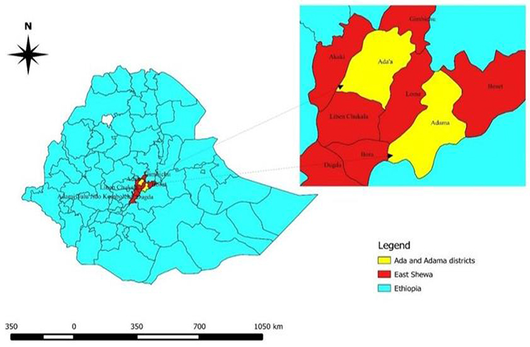 |
Figure 1 Map of Ethiopia showing location of the study areas. Note: Reproduced from Woldemariam T, Mohammed T, Mamo G, Ameni G. An abattoir based study of Bovine Tuberculosis in Adama and Bishoftu Abattoirs, Central Ethiopia. J Sci Dev. 2018;6:1.65 |
The second study area was Adama, and it is located 95 km South Eastern from Addis Ababa at 39.17°N and 8.33°E with an altitude of 1770 meter above sea level, latitude 8.31°N and 39.16°E longitude. Adama is situated in the well-known East African rift valley. It has annual rainfall and temperatures ranging from 400 mm to 800 mm and 13.9°C to 27.7°C, respectively.44 The livestock population on the basis of species are 70, 662 cattle; 36, 142 sheep; 42, 968 goats; 31, 905 equines; 42 camels and 195, 155 poultry3(Figure 1).
Study Animals
The study animals were cattle and sheep, including both sexes and breeds (exotic, cross and local breeds). The animals that were examined were divided into two age groups: group I (young) and group II (adult);27 which was determined by asking the animal owners and records in the farms. The body conditions of the examined animals were also categorized into three: good, medium and poor. It is based on different body structure, visible bone parts and fat deposit.12
Ethics Approval
The approval on animal handling ethics was received before the commencement of the study. All the animal handling and sample collection methods were performed in accordance with the Addis Ababa University College of Veterinary Medicine Research Ethics (AAU-CVMA-REC) and animal welfare guide for the care and use of animals (Ref. No. 01225/2021).
Study Design
A cross-sectional study design was used to determine the prevalence and associated risk factors of Eimeria infection in cattle and sheep from December 2021 to April 2022 on the selected dairy farms and veterinary clinics in the study areas.
Sampling Technique and Sample Size Determination
The sampling method employed for the study involving dairy farms was a purposive sampling technique based on the willingness of farm owners. Systematic random sampling technique was used for animals to be sampled from veterinary clinics. The sample size required for this study was calculated based on sample size determination method for systematic random sampling of infinite population after59 as follows:
n = 1.962Pexp (1-Pexp)
d2
Where
n = required sample size
P exp = expected prevalence
d = desired absolute precision
1.962 = z-value for 95% confidence interval
Since there was no previous study conducted in the study areas, the expected prevalence was 50%. Accordingly, with 5% absolute precision at 95% confidence level, as a result, the required sample size for the study was found to be 384. Since the number of sheep coming to veterinary clinics was few compared to cattle, we collected fecal samples from 265 cattle and 119 sheep.
Sample Collection and Transportation
Fresh fecal samples were collected from the rectum of the animals using a rectal glove. Then, the collected samples were placed in labeled clean plastic containers (universal bottles) and preserved in 2% potassium dichromate or 10% formalin then transported in icebox to CVMA parasitology laboratory, for further processing on the same day of collection. The sample left unprocessed on the first day was stored in the refrigerator at 4°C to be processed in the following days. At the time of sampling, the species, breed, sex, age, fecal consistency, body condition, study site/farm, agro ecology, management system and farm hygiene were recorded on a data recording format. The fecal consistency was determined by physical observation of the feces during sample collection. Hygiene of the dairy farms was assessed based on the frequency of disposal of feces, hygienic status of the dairy cattle and calves, and the frequency at which the farms were cleaned.
Sample Processing
Fecal flotation technique was conducted by using Sheather’s sugar solution to detect the oocysts of Eimeria as depicted by.28 The coprological procedure is described as follows: taking three grams of faeces from each animal and mix with 42 mL of floatation fluid by using pistil and mortar thoroughly, then the suspension was poured through tea strainer. Then, the suspension was poured into the test tube, and the test tube was placed in a rack and the test tube with the suspension was left a convex meniscus at the top of the test tube and cover slip was carefully placed at the top of the tube and let it stand for 15 minutes. Finally, the cover slip was removed with the drop of fluid adhered to it, and immediately the cover slip was put on microscopic slide and examined by 40x magnification to identify Eimeria oocysts.
Estimation of Intensity of Eimeria Infection (Oocyst per Gram of Feces/OPG)
Quantitative fecal examination was performed by floatation technique to determine the number of oocysts per gram of feces (OPG) using Mac master technique. Samples with more than 400 OPG were mixed thoroughly with 2.5% potassium dichromate solution and allowed to sporulate for 10–14 days at room temperature to allow sporulation of Eimeria oocyst and used for oocyst identification. After sporulation, the fecal mixture was processed using the simple test tube flotation procedure using Sheather’s sugar solution with specific gravity of (1.18) to recover the oocysts. The Eimeria oocyst were identified based on the morphology of oocysts and sporocysts (shape, color, micropyle and its cap, presence or absence of residual, polar granule). The size of the oocysts was measured using a calibrated microscope under a 40x objective.25
Data Analysis
Data obtained from all the study were coded and stored in a Microsoft excel spread sheet program v2010 (Microsoft, Redmond WA, USA) and analyzed using IBM SPSS version 20 computer Statistical software for windows. For every data, the prevalence was estimated by dividing the number of infected individual animal by the number of animals sampled and multiplying by 100. Pearson’s chi-square (χ2) was used to assess the relationship between Eimeria infection and the hypothesized risk factors. P-value less than 0.05 (at 5% level of significance) was considered as significant and final results were displayed in the form of tables and figures.
Results
Prevalence and Risk Factors of Eimeria Infection in Cattle and Sheep
Of the total 384 animals examined (48.9%) were found to be infected with Eimeria. The prevalence of Eimeria infection in cattle and sheep was 45% and 58%, respectively. There was a statistically significant (P = 0.003) difference in Eimeria infection between study animal species (cattle and sheep). There was also a significant difference in Eimeria infection between the study sites/farms (P = 0.000) and between agro ecologies (P = 0.000). Similarly, there was a significant difference (P = 0.000) in Eimeria infection among the breeds of cattle with higher prevalence in local cattle (57.4%) than Exotic cattle (49.9%) and cross-bred cattle (18.2%) (Table 1).
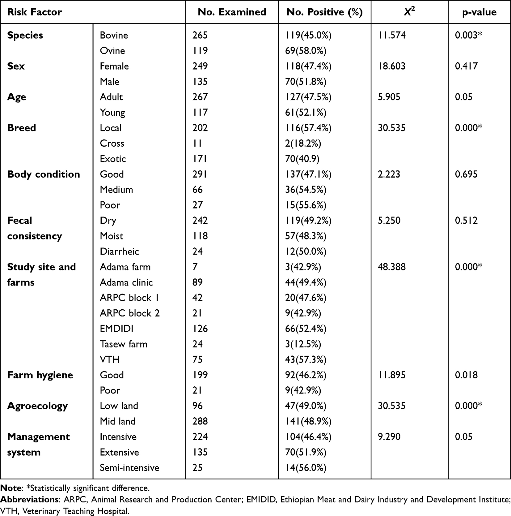 |
Table 1 Overall Prevalence of Eimeria Infection with Respect to Risk Factors |
Among the 384 animals examined, 118 (47.4%) and 70 (51.8%) of female and male animals, were positive with Eimeria infections. Accordingly, the prevalence of Eimeria infection was higher in males than in females; however, the difference was not statistically significant (P = 0.417). The age of the animal was strongly associated with the infection (P = 0.05). Similarly, there was a statistically significant difference in Eimeria infection between breeds of the animals, farm hygiene, between management systems, and between agro ecologies of the study areas.
The intensity of infection, as measured by the number of OPGs in feces, is an important factor that describes the severity of the infection in a particular animal. The mean oocyst per gram of feces ranges from zero to 10,000 in both study animals (Figures 2 and 3).
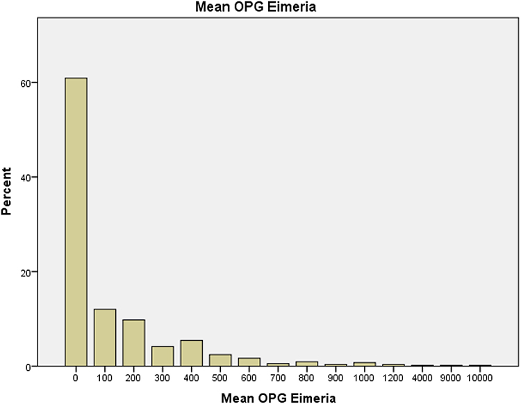 |
Figure 2 Mean oocysts per gram of feces (OPG) of Eimeria infection in cattle. |
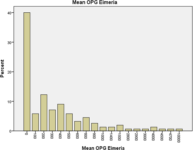 |
Figure 3 Mean oocysts per gram of feces (OPG) of Eimeria infection in sheep. |
Species Identification of Eimeria in Cattle and Sheep
Eimeria species was identified based on the morphological features of the oocysts (Annex 1) as per the guidelines provided by.37 Accordingly, eight Eimeria species were identified in sheep. The three dominant species were Eimeria ovinoidalis (21.7%), Eimeria crandallis (19.6%) and Eimeria parva (14%). While seven Eimeria species were identified in cattle, Eimeria bovis (17.5%), Eimeria subspherica (15.6%) and Eimeria wyomingensis (14.2%) were the dominant species (Table 2).
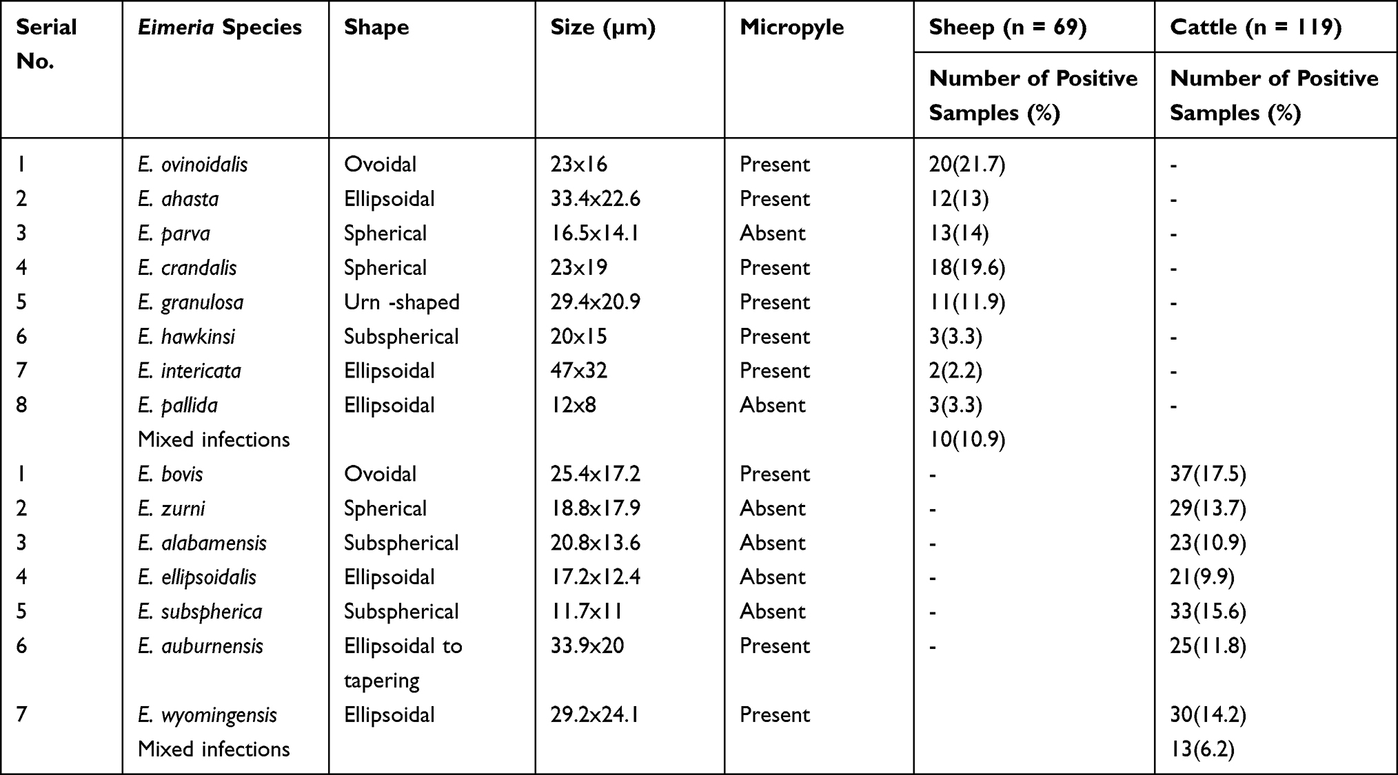 |
Table 2 Eimeria Species Identified from Sheep and Cattle Based on Oocyst Morphology |
Discussion
Eimeria spp. infections are most commonly found in the subclinical stage, without causing clinical diarrhea. Coccidiosis causes economic losses as a result of damage to the intestinal lining resulting in reduced feed consumption, malnutrition, poor growth and weight loss, even though the animals appear healthy.28
Various prevalences of Eimeria infection in cattle and sheep have been reported in different parts of the world. The prevalence of cattle Eimeria infection recorded in Ethiopia was within the range of 5.99% and 72.8%.24,25 In Poland, the prevalence of Eimeria species in cattle was estimated to range from 17.9% to 93%,34,48,49 between 50% and 52% in South Africa;41 29% of infection was reported in Iraq,6 75.5% in Colombia,37 11.97% in India (Assam),20 29–52% in South Africa, 25.9% in Korea,45 46.7% in Egypt.38
The current prevalence of cattle Eimeria infection (45.0%) was lower than the previous reports of 55.0% in Akaki Kality Subcity of Addis Ababa, Ethiopia;10 68.1% in Addis Ababa and Bishoftu, Ethiopia1 and 72.8% in Mekelle, Ethiopia.25 Our current finding was also lower than the reports from different countries including 75.5% prevalence from Colombia37 93% reported in Poland48 and 50% and 52% in South Africa.41 However, the present prevalence of cattle Eimeria infection is higher than 5.99%, 22.7% and 31.9% reported, respectively, by.8,21,24 Previous prevalence reports in Ethiopia were also higher than 29% prevalence which was reported in Iraq;6,33 11.97% in India;20 25.9% in Korea38,45 and 46.7% from Egypt.38
Various reports on the prevalence of Eimeria in sheep were also reported in different parts of Ethiopia. For instance, 66.8% in Bishoftu and central Ethiopia;10 22.4%35 in Addis Zemen, Gondar, Ethiopia and 62.7% in and around Haramaya, Eastern Hararghe, Ethiopia.43 In cattle and sheep; 57.3% Eimeria infection was also reported from Akaki Kality Subcity of Addis Ababa by.10
The present prevalence of sheep Eimeria infection (58.0%) was lower than 66.8% prevalence in Bishoftu and central Ethiopia10,11 and 62.7% in and around Haramaya, Eastern Hararghe, Ethiopia,43,46 68.3% prevalence reported from Brazil,56 97.5% in Tanzania,31,32 91.5% in Western Iran,30 92.9% in Northeastern China.2 However, the present prevalence of sheep Eimeria infection was higher than the one reported by35,60 (22.4%) in Addis Zemen, Gondar, Ethiopia and also higher than the report from India (12.7%).54
The present prevalence of both cattle and sheep Eimeria infection (48.95%) was lower than 57.3% prevalence recorded in Akaki Kality Subcity of Addis Ababa.10 The number of ingested oocysts, the existence of a concurrent microbial infection, weather conditions and seasons, management, the level of immunity and age, techniques of diagnostics and agro-ecology could all have a role in the differences in prevalence estimates.54
The prevalence of Eimeria infection in the present study in the males (51.8%) was slightly higher than in females (47.4%). However, the sex of the animals was not significantly associated (P > 0.05) with the infection by coccidian. This is in agreement with the finding of8,10. There was no evidence of a significant association between sex and the outcome. The lack of a significant association between infection and animal sex could indicate that Eimeria infection affects both male and female animals relatively equally.
There was a statistically significant difference in Eimeria infection between the age categories with higher prevalence in the young (52.1%) than the adult (47.5%). This is in agreement with the findings of other researchers who reported a strong significant association between the age and the infection1,9,10,63. This justifies that young animals are more susceptible to infection than older animals as a result of their lower immunity. If the habitat is heavily contaminated, repeated oocyst uptake could explain the findings, resulting in low frequency from mature animals who would otherwise have developed immunity. Age is a major risk factor in the spread of coccidiosis; morbidity and the risk of infection are greater in young animals. Similarly, there was a significant difference in Eimeria infection among the breeds of cattle with higher prevalence in local cattle (57.4%) than Exotic cattle (49.9%) and cross-bred cattle (18.2%). This could be associated with the low attention given to local cattle in terms of feeding and other management activities compared to the exotic and cross-bred cattle because of the fact that local cattle have lower production potential than the improved breeds.
The prevalence of the present study in young (52.1%) is lower than that of previous prevalence (68.1%) which was overall prevalence of the research conducted in both Addis Ababa (highland) and Bishoftu (midland) by1 and higher than 22.7% and 31.9% previous prevalence reported in Ethiopia by21 in Dire Dawa, Eastern Ethiopia and8 in Kombolcha town, which is located in the North East of Ethiopia in Amhara regional state, respectively. This variation could be due to the differences in climatic conditions of the various study areas.
There was no statistically significant difference in the prevalence of Eimeria between fecal consistency. This was not in agreement with the finding by13. The reason behind could be due to the fact that Coccidiosis mostly occurs in a subclinical form and the thickness of the feces may be related to diet or other infections.
There was a significant difference in Eimeria infection between the dairy farms and agro ecologies where samples were collected. This is most likely due to Bishoftu’s increased rainfall and relative humidity, which provides a far better climatic environment for survival, sporulation and development of the oocysts in Bishoftu than Adama. This observation is in agreement with the work of;1,53 who reported higher infection rates of Eimeria in cattle in high rainfall zones in Addis Ababa and Mexico, respectively.
Eimeria infection was strongly associated with farm hygiene. High prevalence was recorded in good farm than poor farms. Normally, there is more Eimeria infection in poor farms than good farms. The current findings disagree with that of23,50,52 who reported Coccidiosis in cattle is more common in confined herds than animals kept on pastures. This could be related to sampling errors where the samples for the study were not proportional. In the present study, a strong association was also found between Eimeria infection and animal feeding systems. Coccidiosis was also more common in stall fed animals than in free pasture grazing animals.
The Eimeria infection and management system also have a significant association. Previous studies reported 92.7% prevalence of Eimeria species oocysts in an intensive system,1 78.3% in a semi-intensive system,29 and in extensive system 58.9%5) and 90%.56 In extensive systems, limiting factors, such as temperature and management, as well as genetic and immunologic states of the animal, can promote coccidia dissemination and enhance prevalence.18
Conclusion and Recommendations
The current study found a high prevalence of Eimeria infection in cattle and sheep, with an overall prevalence of 48.95%, indicating that Eimeria infection is still prevalent enough to affect domestic ruminant production in the study areas, causing mortality, morbidity, and body condition losses in these animals. This result also indicates that young animals are the most impacted, and that the parasite causes clinical diarrhea in young animals more frequently. There was a significant difference in Eimeria infection in the different agro ecologies. However, no difference was found in Eimeria infection between sex, body condition and fecal consistency.
Therefore, further research is needed to fully understand the economic impact of subclinical infection by Eimeria species in cattle and sheep. More epidemiological investigation involving both the dry and rainy seasons is also required to see the seasonal oocyst shedding of the parasite. Similarly, awareness creation of farmers, dairy personnel and veterinary clinic workers about the occurrence of the parasites is very important.
Disclosure
The authors report no conflicts of interest in this work.
References
1. Abebe R, Wossene A, Kumsa B. Epidemiology of Eimeria infections in calves in Addis-Ababa and Debre-Zeit dairy farms, Ethiopia. Int J Appl Res Vet Med. 2008;6:24–30.
2. Abo-Shehada MN, Abo-Farieha HA. Prevalence of Eimeria species among goats in northern Jordan. Small Rumin Res. 2003;49(2):109–113. doi:10.1016/S0921-4488(03)00078-6
3. ADAO. Adama district Agriculture office, 2003; 2003.
4. ADARDO (Ada’ a District Agricultural and Rural Development Office); 2007.11–12.
5. Ahid SMM, Medeiros VMC, Bezerra ACD, Maia MB, Xavier VM, Vieira LS. Especies de genero Eimeria Schneider,1875. (Apicomplexa: Eimeriidae) empequenosruminantesna mesorregiaooeste do estado do Rio Grande do Norte, Brasil. CiêncAnim Bras. 2009;10(3):984–989.
6. Al-Bakry HS. Diagnosis and classification of Eimeria species in cattle in Mosul. Iraqi Journal of Veterinary Sciences. 2009;23(1):17–21. doi:10.33899/ijvs.2009.5686
7. Albayati HH, Jarad NI, Al-Difaie RS, Albayati HH. Microscopic and molecular diagnosis of Eimeria spp. in sheep as a model of health investigation. Microscope. 2020;12:13.
8. Alemayehu A, Mohammed N, Timketa B. Prevalence of bovine coccidia in Kombolcha district of South Wollo, Ethiopia. J Vet Med Anim Health. 2013;5(2):41–45.
9. Almeida VDA, Magalhaes VCS, Muniz-Neta ES, Munhoz AD. Frequency of species of the genus Eimeria in naturally infected cattle in Southern Bahia, North-east Brazil. Rev Bras Parasitol Vet. 2011;20:78–81. doi:10.1590/s1984-29612011000100017
10. Ayana D, Ayele A, Ashenafi H, Waktole H, Abunna F. Eimeria infection in sheep and dairy cattle in Akaki Kality Subcity of Addis Ababa, Ethiopia. Adv Biol Res. 2017;11(5):233–241.
11. Ayana D, Tilahun G, Wossene A. Study on Eimeria and Cryptosporidium infections in sheep and goats at Elfora export abattoir, Debre-Zeit, Ethiopia. Turkish J Vet Anim. 2009;33(5):367–371.
12. Bajer A. Cryptosporidium and Giardia spp. infections in humans, animals and the environment in Poland. Parasitol Res. 2008;104:1–17. doi:10.1007/s00436-008-1179-x
13. Bangoura B, Mundt HC, Schmäschke R, Westphal B, Daugschies A. Prevalence of Eimeria bovis and Eimeria zuernii in German cattle herds and factors influencing oocyst excretion. Parasitol Res. 2012;110(2):875–881. doi:10.1007/s00436-011-2569-z
14. Bastianetto E, Filho EJF, Lana AMQ, et al. Epidemiology of Eimeria sp. infection in buffaloes (Bubalus bubalis) bred in Minas Gerais, Brazil. Ital J Anim Sci. 2007;6(sup2):911–914. doi:10.4081/ijas.2007.s2.911
15. Bruhn FRP, Lopes MA, Demeu FA, Perazza CA, Pedrosa MF, Guimarães AM. Frequency of species of Eimeria in females of the Holstein-Friesian breed at the post-weaning stage during autumn and winter. Rev Bras Parasitol Vet. 2011;20(4):303–307. doi:10.1590/S1984-29612011000400008
16. Conway P, McKenzie E. Poultry Coccidiosis and Effect of Coccidiosis Diagnostic and Testing Procedures.
17. CACC (Central Agricultural Census Commission of Ethiopia); 2003.14–16
18. Chartier C, Paraud C. Coccidiosis due to Eimeria in sheep and goats, a review. Small Rumin Res. 2012;103(1):84–92. doi:10.1016/j.smallrumres.2011.10.022
19. CSA. Central statistical authority of Ethiopia: report on livestock and livestock characteristics (private peasant holdings); 2020. 1–228.
20. Das M, Deka DK, Sarmah PC, Islam S, Sarma, S. Diversity of Eimeria spp. in dairy cattle of Guwahati, Assam, India. Vet World. 2015;8(8):941. doi:10.14202/vetworld.2015.941-945
21. Dawid F, Amede Y, Bekele M. Calf Coccidiosis in selected dairy farms of Dire Dawa, Eastern Ethiopia. Glob Vet. 2012;9:460–464.
22. Debela N, Habtamu Y, Getachew S. Coccidiosis in domestic ruminants: review, federal Democratic Republic of Ethiopia, Ministry of Agriculture, Addis Ababa, Ethiopia. Acta Parasitol Globalis. 2020;11(3):133–141.
23. Ernst JV, Stewart TB, Witlock DR. Quantitative determination of coccidian oocysts in beef calves from the coastal plain area of Georgia (USA). VetParasitol. 1987;23:1–10.
24. Girma M. Prevalence of Eimeria infection in calves in and around Sekota town, North Wollo. Ethiopia. 2017;4:169–175.
25. Gebeyehu B, Kebede E, Kifleyohannes T, Abebe N, Kumar N. Prevalence of calf Coccidiosis in Mekelle, northern Ethiopia. Ethiop Vet J. 2018;22(2):1–13. doi:10.4314/evj.v22i2.1
26. Hazarika A, Das M. Therapeutic management of winter Coccidiosis in cattle calves of Morigaon, Assam. North-East Vet. 2018;18(1):25–28.
27. Heidari H, Gharekhani J. Detection of Eimeria species in Iranian native cattle. Inter J Adv Res. 2014;2:731–734.
28. Hendrix CM. Diagnostic Veterinary Parasitology.
29. Hamid PH, Kristianingrum YP, Prastowo S. Bovine Coccidiosis cases of beef and dairy cattle in Indonesia. Vet Parasitol. 2019;17:100298.
30. Hassum IC, Menezes RCA. Infeccao natural porespecies do genero Eimeria empequenos ruminantescriadosemdoismunicipios do estado do Rio de Janeiro. Rev Bras Parasitol Vet. 2005;14(3):95–100.
31. Hashemnia M, Rezaei F, Chalechale A, Kakaei S, Gheichivand S. Prevalence and intensity of Eimeria infection in sheep in Western Iran. Int J Livest Res. 2014;4(1):107–112. doi:10.5455/ijlr.20140109084416
32. Kambarage DM, Kimera SI, Kusiluka LJM, Matambo MMA. Prevalence of Eimeria and Cryptosporidium oocysts in cattle, sheep and goats in Morogoro region, Tanzania. J Appl Anim Res. 1996;9:73–78. doi:10.1080/09712119.1996.9706106
33. Kareem SI, Yücel ŞY. Prevalence of Eimeria species in sheep in Sulaimaniya province, Iraq. J Entomol Zool Stud. 2015;3(4):317–322.
34. Kim HC, Choe C, Kim S, et al. Epidemiological survey on Eimeria spp. associated with diarrhea in pre-weaned native Korean calves. Korean J Parasitol. 2018;56(6):619. doi:10.3347/kjp.2018.56.6.619
35. Klockiewicz M, Kaba J, Tomczuk K, et al. The epidemiology of calf Coccidiosis (Eimeria spp.) in Poland. Parasitol Res. 2007;101:121–128. doi:10.1007/s00436-007-0619-3
36. Lakew A, Seyoum Z. Ovine Coccidiosis: prevalence and associated risk factors in and around Addis-Zemen, Northwest Ethiopia. Turk J Vet Anim Sci. 2016;40:645–650. doi:10.3906/vet-1509-18
37. Leta S, Mesele F. Spatial analysis of cattle and shoat population in Ethiopia: growth trend, distribution and market access. SpringerPlus. 2014;3(1):1–10. doi:10.1186/2193-1801-3-310
38. Levine D. Veterinary Protozoology. Ames: Protozoan parasites of domestic animal and man; Iowa state university press; 1985:150–162.
39. Lopez-Osorio S, Villar D, Failing K, Taubert A, Hermosilla C, Chaparro-Gutierrez JJ. Epidemiological survey and risk factor analysis on Eimeria infections in calves and young cattle up to 1 year old in Colombia. Parasitol Res. 2020;119(1):255–266. doi:10.1007/s00436-019-06481-w
40. Malek SS, Kuraa HM. Detection and identification of Eimeria species in naturally infected calves at Assiut Governorate. Zagazig Vet J. 2018;46(1):60–69. doi:10.21608/zvjz.2018.7624
41. Makau DN, Gitau GK, Muchemi GK, et al. Environmental predictors of bovine Eimeria infection in western Kenya. Trop Anim Health Prod. 2017;49(2):409–416. doi:10.1007/s11250-016-1209-0
42. Majeed NM, Aaiz NN, Neama AJ. Molecular study to detect the Eimeria species in sheep in Al-Diwaniyah province, Iraq. Iraqi J Vet Sci. 2020;34(2):377–381. doi:10.33899/ijvs.2019.126064.1225
43. Matjila PT, Penzhorn BL. Occurrence and diversity of bovine coccidia at three localities in South Africa. Vet Parasitol. 2002;104:93–102. doi:10.1016/S0304-4017(01)00605-7
44. Morgoglione ME, Bosco A, Maurelli MP, et al. A 10-year surveillance of Eimeria spp. in cattle and buffaloes in a Mediterranean area. Front Vet Sci. 2020;7:410. doi:10.3389/fvets.2020.00410
45. Muktar Y, Kiltu G, Keffale M. Study on prevalence of small ruminant coccidiosis in and around Haramaya, Eastern Hararghe, Ethiopia. ActaParasitol Glob. 2016;7:07–11.
46. Munyua WK, Ngotho JW. Prevalence of Eimeria species in cattle in Kenya. Vet Parasitol. 1990;35(1–2):163–168. doi:10.1016/0304-4017(90)90126-V
47. NMSA. National Metrology Service Agency Adama. Ethiopia; 2006.
48. Oda K, Nishida Y. Prevalence and distribution of bovine coccidia in Japan, Nippon juigaku. Zasshi. 1990;52(1):71–77.
49. Oddsdóttir C, Pálsdóttir GR. Patterns of Eimeria excretion in young Icelandic calves. Icel Agric Sci. 2021;34:29–39. doi:10.16886/IAS.2021.03
50. Peter SG, Gitau GK, Richards S, et al. Risk factors associated with Cryptosporidia, Eimeria, and diarrhea in smallholder dairy farms in Mukurwe-ini Sub-County, Nyeri County, Kenya. Vet World. 2016;9(8):811–819. doi:10.14202/vetworld.2016.811-819
51. Pilarczyk B, Balicka-Ramisz A, Ramisz A. Studies on Coccidiosis in cattle in Northwest Poland. Electro J Polish Agr Univ. 2000;3:101–103.
52. Pilarczyk B, Balicka-Ramisz A, Kozak W, Ramisz A. Occurrence of endoparasites in heifers imported to Poland from the Netherlands. ArchivTierzucht. 2009;52:265–271.
53. Radostits OM, Gay CC, Hinchcliff KW, Constable PD. Veterinary Medicine. In: A Text Book on Disease of Cattle, Sheep, Pigs and Horse.
54. Rehman TU, Khan MN, Sajid MS, et al. Epidemiology of Eimeria and associated risk factors in cattle of district Toba Tek Singh, Pakistan. Parasitol Res. 2011;108(5):1171–1177. doi:10.1007/s00436-010-2159-5
55. Rodriguez-Vivas RI, Dominguez-Alpizar JL, Torres-Acosta JF. Epidemiological factors associated to bovine Coccidiosis in calves (Bos indicus) in a sub humid tropical climate. Rev Biomed. 1996;7:211–218.
56. Saratsis A, Joachim A, Alexandros S, Sotiraki S. Lamb Coccidiosis dynamics in different dairy production systems. Vet Parasitol. 2011;181(2–4):131–138. doi:10.1016/j.vetpar.2011.04.027
57. Sisodia SL, Pathak KML, Kapoor M, Chauhan PPS. Prevalence and seasonal variation in Eimeria infection in sheep in Western Rajasthan, India. J Vet Parasitol. 1997;11:95–98.
58. Skirnisson KARL. Eimeria spp. (Coccidia, Protozoa) infections in a flock of sheep in Iceland: species composition and seasonal abundance. Icel Agric Sci. 2007;20:73–80.
59. Souza LEBD, Cruz JFD, Teixeira MR, Albuquerque GR, Melo ADB, Tapia DMT. Epidemiology of Eimeria infections in sheep raised extensively in a semiarid region of Brazil. RevistaBrasileira de ParasitologiaVeterinária. 2015;24:410–415.
60. Tamrat H, Mekonnen N, Ferede Y, Cassini R, Belayneh N. Epidemiological study on calf diarrhea and Coccidiosis in dairy farms in Bahir Dar, North West Ethiopia. Ir Vet J. 2020;73(1):1–8. doi:10.1186/s13620-020-00168-w
61. Tigist T, Merzuk H, Gobu B, Mukarim A. Study on prevalence of calves Coccidiosisi in and around Jimma town, Ethiopia. J Vet Med Res. 2017;4(3):1077.
62. Thrusfield M. Veterinary Epidemiology.
63. Urquhart NS, Paulsen SG, Larsen DP. Monitoring for policy‐relevant regional trends over time. Ecol Appl. 1998;8(2):246–257.
64. Verma R, Das G, Saiyam R, Bendigeri S. Clinical Coccidiosis in calves and its treatment. J Entomol Zool Stud. 2018;6(2):2964–2967.
65. Woldemariam T, Mohammed T, Mamo G, Ameni G. An abattoir based study of Bovine Tuberculosis in Adama and Bishoftu Abattoirs, Central Ethiopia. J Sci Dev. 2018;6:1.
66. Yu SK, Gao M, Huang N, Jia YQ, Lin Q. Prevalence of coccidial infection in cattle in Shaanxi province, Northwestern China. J Anim Vet Adv. 2011;1:2716–2719.
 © 2022 The Author(s). This work is published and licensed by Dove Medical Press Limited. The full terms of this license are available at https://www.dovepress.com/terms.php and incorporate the Creative Commons Attribution - Non Commercial (unported, v3.0) License.
By accessing the work you hereby accept the Terms. Non-commercial uses of the work are permitted without any further permission from Dove Medical Press Limited, provided the work is properly attributed. For permission for commercial use of this work, please see paragraphs 4.2 and 5 of our Terms.
© 2022 The Author(s). This work is published and licensed by Dove Medical Press Limited. The full terms of this license are available at https://www.dovepress.com/terms.php and incorporate the Creative Commons Attribution - Non Commercial (unported, v3.0) License.
By accessing the work you hereby accept the Terms. Non-commercial uses of the work are permitted without any further permission from Dove Medical Press Limited, provided the work is properly attributed. For permission for commercial use of this work, please see paragraphs 4.2 and 5 of our Terms.