")
Back to Journals » Drug Design, Development and Therapy » Volume 10
Discovery of a novel anticancer agent with both anti-topoisomerase I and II activities in hepatocellular carcinoma SK-Hep-1 cells in vitro and in vivo: Cinnamomum verum component 2-methoxycinnamaldehyde
Authors Perng D, Tsai Y, Cherng J, Wang J, Chou K, Shih C, Cherng J
Received 2 August 2015
Accepted for publication 15 September 2015
Published 5 January 2016 Volume 2016:10 Pages 141—153
DOI https://doi.org/10.2147/DDDT.S93599
Checked for plagiarism Yes
Review by Single anonymous peer review
Peer reviewer comments 3
Editor who approved publication: Prof. Dr. Wei Duan
Daw-Shyong Perng,1 Yu-Hsin Tsai,2 Jonathan Cherng,3 Jeng-Shing Wang,4 Kuo-Shen Chou,5 Chia-Wen Shih,6 Jaw-Ming Cherng7
1Department of Gastroenterology, E-Da Hospital, I-Shou University, Kaohsiung, Taiwan, Republic of China; 2Sierra College, Rocklin, CA, USA; 3Faculty of Medicine, Medical University of Lublin, Lublin, Poland; 4Department of Internal Medicine, Antai Tian-Sheng Memorial Hospital, Pingtung, 5Department of Family Medicine, Saint Mary’s Hospital Luodong, 6Department of Pathology, Lotung Poh-Ai Hospital, 7Department of Internal Medicine, Saint Mary’s Hospital Luodong, Yilan, Taiwan, Republic of China
Abstract: Cinnamomum verum is used to make the spice cinnamon and has been used as a traditional Chinese herbal medicine for various applications. We evaluated the anticancer effect of 2-methoxycinnamaldehyde (2-MCA), a constituent of the bark of the plant, and its underlying molecular biomarkers associated with carcinogenesis in human hepatocellular carcinoma SK-Hep-1 cell line. The results show that 2-MCA suppressed proliferation and induced apoptosis as indicated by mitochondrial membrane potential loss, activation of caspase-3 and caspase-9, increase in the DNA content in sub-G1, and morphological characteristics of apoptosis, including blebbing of plasma membrane, nuclear condensation, fragmentation, apoptotic body formation, and long comet tail. In addition, 2-MCA also induced lysosomal vacuolation with increased volume of acidic compartments, suppressions of nuclear transcription factors NF-κB, cyclooxygenase-2, prostaglandin E2 (PGE2), and both topoisomerase I and II activities in a dose-dependent manner. Further study reveals the growth-inhibitory effect of 2-MCA was also evident in a nude mice model. Taken together, the data suggest that the growth-inhibitory effect of 2-MCA against SK-Hep-1 cells is accompanied by downregulations of NF-κB-binding activity, inflammatory responses involving cyclooxygenase-2 and PGE2, and proliferative control involving apoptosis, both topoisomerase I and II activities, together with an upregulation of lysosomal vacuolation and volume of acidic compartments. Similar effects (including all of the above-mentioned effects) were found in other tested cell lines, including human hepatocellular carcinoma Hep 3B, lung adenocarcinoma A549, squamous cell carcinoma NCI-H520, colorectal adenocarcinoma COLO 205, and T-lymphoblastic MOLT-3 (results not shown). Our data suggest that 2-MCA could be a potential agent for anticancer therapy.
Keywords: 2-methoxycinnamaldehyde, anticancer, SK-Hep-1 cells, topoisomerase I, topoisomerase II
Introduction
Hepatocellular carcinoma (HCC) is one of the most common malignancies.1 However, HCC is not sensitive to traditional chemotherapeutic agents, and there is a need for better treatment of the disease.
The genus Cinnamomum belongs to the Lauraceae family and comprises over 250 aromatic evergreen trees distributed mostly in Asia. Cinnamomum verum is a small evergreen tree in the genus and native to Sri Lanka. The bark of this plant is used to make the spice cinnamon and has long been used as a traditional Chinese herbal medicine for various conditions, such as improvement of the complexion, making it more youthful, alleviation of fever, inflammation, cough, induction of perspiration, and circulatory disorders.2,3 In our ongoing study to identify anticancer agents from natural resources, 2-methoxycinnamaldehyde (2-MCA), a constituent of the bark of the plant, was discovered to have growth-inhibitory effect in human HCC SK-Hep-1 cells, both in vitro and in vivo.
Cancer is a hyperproliferative disorder. Numerous genetic and epigenetic changes are needed to drive normal cells toward neoplastic transformation. These alterations control various signaling pathways that cooperate to endow cancer cells with a wide range of biological capabilities necessary for growing, disseminating, and finally killing their host.4 Although anticancer drugs may act differently, apoptosis is the most common and preferred mechanism through which many anticancer agents kill and eradicate cancer cells.5
Topoisomerases are enzymes that regulate the topological states of DNA and play an important role in maintaining genomic integrity.6 These enzymes relax supercoiled DNA by transient protein-linked cleavages of either one (topoisomerase I) or both (topoisomerase II) of the sugar–phosphate backbones of double-stranded DNA strands.7 In addition to apoptosis, topoisomerase is another major target of anticancer agents.8–11 The transcription factor nuclear factor κB (NF-κB) plays an important role in the regulation of cell survival and is activated in many malignant tumors. In addition, the inhibition of NF-κB shifts the balance of death/survival toward apoptosis.12
NF-κB pathway is involved in the expression of cyclooxygenase-2 (COX-2).13 Dysregulated expression of COX-2 and prostaglandin cascade plays an important role in carcinogenesis. Expression of constitutive COX-2-catalyzed prostaglandin is induced by most cancer-causing agents, and COX-2 expression is a characteristic feature of all premalignant neoplasms. In addition, COX-2 expression intensifies with stage at detection, cancer progression, and metastasis. Furthermore, various essential features of carcinogenesis are linked to COX-2-driven prostaglandin E2 (PGE2) biosynthesis, and COX-2 inhibitors reduce the risk of human cancer and precancerous lesions.14
This diversity of mechanisms of carcinogenesis suggests that there are probably multiple processes that could be effective targets for the prevention of cancer. In an attempt to understand the effects and underlying mechanisms of 2-MCA in SK-Hep-1 cells, we performed a series of experiments to answer the following questions: 1) what is the effect of 2-MCA on the growth in SK-Hep-1 cells? 2) What are the effects of 2-MCA on topoisomerase I and II activities? 3) How these activities are affected? Our results indicate that 2-MCA inhibited the growth in SK-Hep-1 cells, with decreased NF-κB DNA-binding activity and decreased COX-2 and PGE2 expressions. In addition, 2-MCA inhibited both topoisomerase I and II activities and induced lysosomal vacuolation and increased the volume of acidic compartments (VACs). Finally, 2-MCA induced apoptosis, resulting in the suppression of cell growth, both in in vitro and in vivo.
Materials and methods
Materials
Minimum essential medium and fetal calf serum were purchased from Thermo Fisher Scientific (Waltham, MA, USA). 2-MCA, dimethyl sulfoxide (DMSO), propidium iodide (PI), and RNase were purchased from Sigma-Aldrich (St Louis, MO, USA).
Cell culture
Human HCC cell line, SK-Hep-1 cells (ATCC HTB-52; American Type Culture Collection, Manassas, VA, USA) were cultured in minimum essential medium, supplemented with 1.0 mM sodium pyruvate, 10% (v/v) fetal bovine serum, 10 U/mL penicillin, 10 μg/mL streptomycin, and 0.25 μg/mL amphotericin B at 37°C with 5% CO2. The I-Shou University Institutional Review Board has reviewed and approved the exemption of the protocol regarding human cell lines as this research does not meet the definition of human subject research.
XTT assay for cell viability
Cells were seeded in 96-well culture plates (1×10⁴ cells/well). After incubated for 24 hours, the cells were treated with different concentrations of 2-MCA for 24 hours, 48 hours, and 72 hours. The cell viability was determined by Cell Proliferation Kit II (XTT) (Hoffman-La Roche Ltd., Basel, Switzerland) following the manufacturer’s protocol. The absorbance was measured using Tecan infinite M200 spectrophotometer (Tecan, Männedorf, Switzerland) at 492 nm with a reference wavelength of 650 nm.
Nuclear fragmentation assay
Acridine orange (AO) is a nucleic acid-selective metachromatic dye useful for cell cycle determination. When AO intercalates into dsDNA, the dye fluoresces green. On the contrary, it fluoresces red when interacts with ssDNA or RNA. Apoptotic cells (with a larger fraction of DNA in the denaturated form) display a red fluorescence and a reduced green emission when compared to nonapoptotic interphase cells. In addition, when AO enter acidic compartments, such as lysosomes, the dye become protonated and sequestered. In these low pH conditions, the dye will emit orange light when excited by blue light.15 Nuclear fragmentation assay is based on the characteristics of AO and observed under a fluorescent microscope. Briefly, the cells were treated with different concentrations of 2-MCA for 24 hours and stained with 5 μg/mL AO at room temperature. Then the cells were observed under fluorescent microscope.16
Comet assay
DNA strand breaks were evaluated using single cell gel electrophoresis (comet) assay following the procedure of Olive and Banath.17
Assay for volume of acidic compartment
VAC assay for cell lysosomal vacuolation was carried out as described previously.16
Flow cytometric analysis
To determine the effect of 2-MCA on cell cycle distribution, 5×10⁵ cells were plated in 60 mm dishes and treated with different concentrations of 2-MCA for 24 hours. Then, the cells were harvested by trypsinization, washed with phosphate-buffered saline, and then fixed in chilled 70% ethanol for 2 hours on ice. The cells were then centrifuged to remove the fixative, washed and suspended in phosphate-buffered saline containing 1 mg/mL RNase and 50 μg/mL PI, incubated in the dark at room temperature for 30 minutes, and analyzed by CyFlow SL Flow Cytometer (Cytecs GmbH, Gorlitz, Germany). A total of 10,000 cells were counted for each determination. The data were analyzed using MultiCycle AV DNA Analysis Software (Phoenix Flow System, San Diego, CA, USA).
Assay for caspase activity
The assay is based on the detection of the chromophore AFC after cleavage from the labeled substrate DEVD- and LEHD-AFC by caspase-3 and caspase-9, respectively. Free AFC emits a yellow-green fluorescence (λmax =505 nm). SK-Hep-1 cells were treated with different concentrations of 2-MCA for 24 hours, and caspase-3 and caspase-9 activities were detected using Fluorometric Assay Kit from BioVision (Milpitas, CA, USA) following the manufacturer’s protocol. The AFC light emission was quantified using Tecan infinite M200 spectrophotometer. Results are represented as the percentage of change in activity compared with the untreated control.
Mitochondrial membrane potential assay
Mitochondrial membrane potential was determined using the mitochondrial-specific fluorescent probe JC-1 (Thermo Fisher Scientific) based on the method of Reers et al.18 JC-1 exists as monomer when membrane potentials (ΔΨm) is lower than 120 mV and fluoresces green (540 nm) following excitation by blue light (490 nm) and as dimer (J-aggregate) at membrane potentials >180 mV and fluoresces red (590 nm) following excitation by green light (540 nm). SK-Hep-1 cells were plated in a 96-well plate and treated with different concentrations of 2-MCA for 12 hours, the cells were stained with 25 μM JC-1 at 37°C for 30 minutes. Fluorescence was monitored with the Tecan infinite M200 spectrophotometer. Changes in the ratio of red (590-nm emission) to green (540-nm emission) fluorescence are indicative of the mitochondrial membrane potential changes.19
Assay for topoisomerase I and II activities
These assays were performed by the method of Har-Vardi et al.20
Assay for NF-κB DNA-binding activity
For analyzing transcription factor NF-κB-binding activity to DNA, nuclear proteins were prepared as described previously,21 and binding activity was quantified using TF ELISA kit (Panomics, Fremont, CA, USA) following the manufacturer’s protocol. This method is faster, easier, and significantly more sensitive than the electrophoretic mobility shift assays and does not require the use of radioactivity.22
Assay for COX-2 activity
After incubation, SK-Hep-1 cells were harvested and spun down at 1,500× g at 4°C for 10 minutes and washed once with saline. Then the cells were suspended in cell lysis buffer (Sigma-Aldrich), supplemented with protease and phosphatase inhibitors (Hoffman-La Roche Ltd.), and sonicated before centrifugation at 12,500× g at 4°C for 20 minutes. The supernatants were collected and used for quantitative analysis of COX-2 activity using ELISA kit (USCN LIFE, Wuhan, People’s Republic of China) following the manufacturer’s protocol.
Assay for PGE2 expression
After incubation, the culture medium was collected for measurement of PGE2 expression by using ELISA kit (R&D System, Minneapolis, MN, USA) following the manufacturer’s protocol.
In vivo tumor xenograft study
Male nude mice (BALB/c Nude; 6 weeks old) were purchased from the National Science Council Animal Center (Taipei, Taiwan) and maintained in pathogen-free conditions in accordance with relevant guidelines and regulations for the care and use of laboratory animals of I-Shou University. SK-Hep-1 cells (5×10⁶ cells in 200 μL) were injected subcutaneously into the flanks of nude mice. Tumors were allowed to develop for approximately 20 days until they reached approximately 75 mm3, and then treatment was started. Thirty-two mice were randomly separated into four groups. The mice in the 2-MCA-treated group were injected intratumorally with different concentrations of 2-MCA in a 200 μL volume daily. The control group was treated with an equal volume of vehicle. After transplantation, tumor size was monitored at weekly intervals using calipers, and tumor volume was estimated by the hemiellipsoid model formula: tumor volume = 1/2(4π/3) × (l/2) × (w/2) × h, where l is the length, w is the width, and h is the height.
Specimens were analyzed by fluorescent TUNEL assay using Quick Apoptotic DNA Ladder Detection Kit (Chemicon, Temecuba, CA, USA) following the manufacturer’s protocol.
Statistical analysis
Data were presented as means ± standard error. The evaluation of statistical significance was determined by one-way analysis of variance followed by Bonferroni t-test for multiple comparisons. A P-value <0.05 was considered statistically significant.
Results
Effects of 2-MCA on cell morphological changes
When SK-Hep-1 cells were exposed to 10 μM of 2-MCA, vacuolation of cells was observed, >40 μM of 2-MCA. In addition, plasma membrane blebbing, cell shrinkage, and cell detachment occurred (Figure 1).
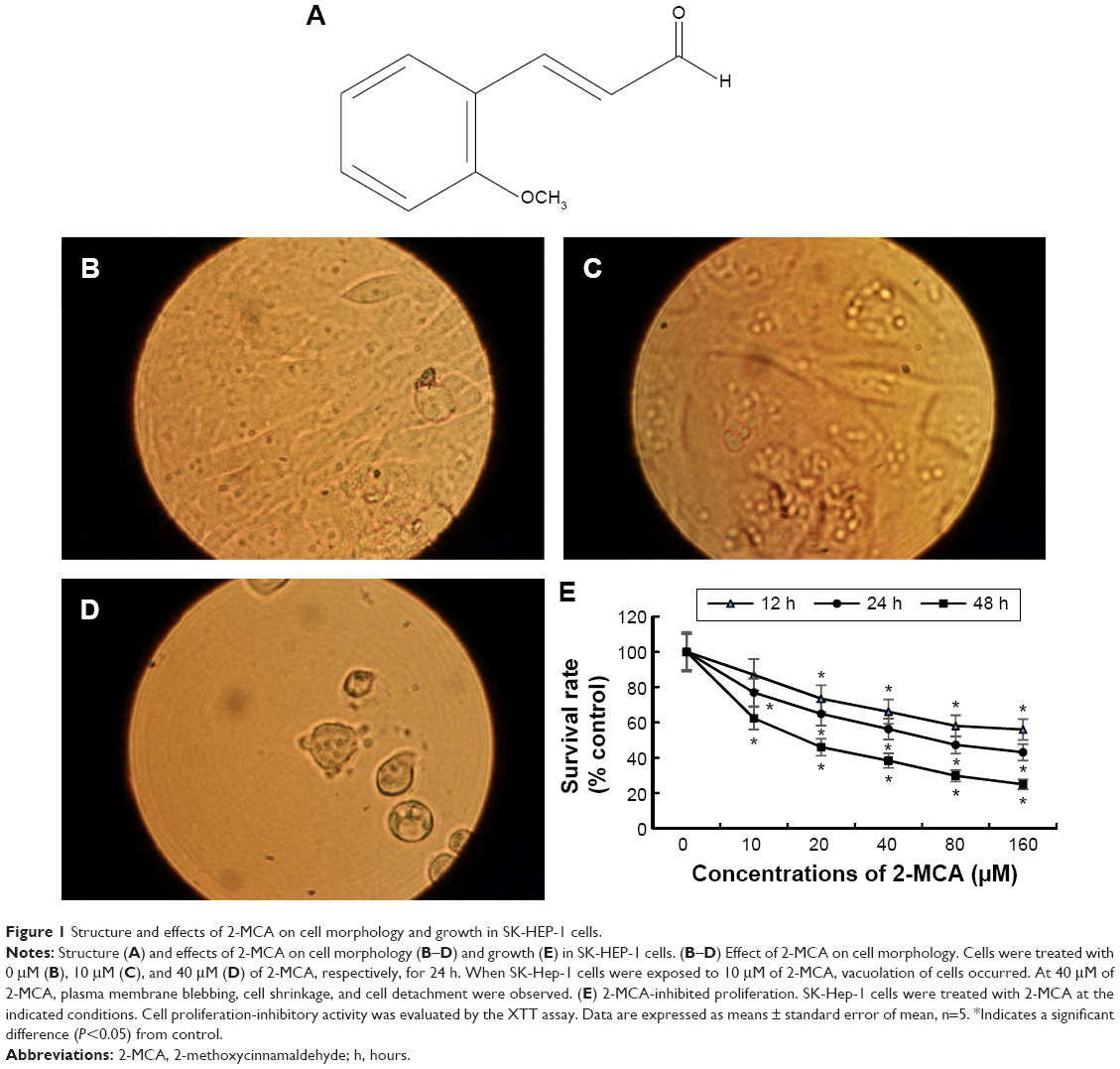 | Figure 1 Structure and effects of 2-MCA on cell morphology and growth in SK-HEP-1 cells. |
2-MCA inhibits SK-Hep-1 cell proliferation
We investigated the potential cell proliferation-inhibitory activity of 2-MCA in SK-Hep-1 cells by the XTT. As shown in Figure 1E, 2-MCA inhibited cell proliferation in SK-Hep-1 cells in a dose- and time-dependent manner. The inhibitory concentration (IC50) value following 48 hours of incubation was 25.72 μM.
Nuclear fragmentation assay
AO is a nucleic acid-selective metachromatic stain useful for cell cycle determination, measuring apoptosis, detecting intracellular pH gradients, and the measurement of proton pump activity.23 The dye differentially stains single-stranded nucleic acids orange and double-stranded nucleic acids green. In addition, in living cells, it serves as a pH indicator, trapped in acidic compartments, such as lysosomes, which then fluoresces to brilliant orange–red.24 When SK-Hep-1 cells were treated with 10 μM of 2-MCA for 24 hours, the result of AO staining demonstrated that a part of SK-Hep-1 cells died by apoptosis with nuclear condensation, fragmentation, and apoptotic bodies. In addition, orange-staining lysosomal vacuoles appeared. No significant nuclear fragmentation in control group was observed.
In addition, DNA strand breakage was investigated by the single cell gel electrophoresis assay (also known as comet assay) at 48 hours following treatment with different concentrations of 2-MCA. Fluorescent comets with tails were evident when SK-Hep-1 cells were treated with 10 μM of 2-MCA for 48 hours. Figure 2 shows representative examples of DNA strand breaks in SK-Hep-1 cells treated with 10 μM of 2-MCA for 48 hours. Treatment with 5 μM of MCA did not show an obvious difference from controls, which mostly appeared spherical (result not shown).
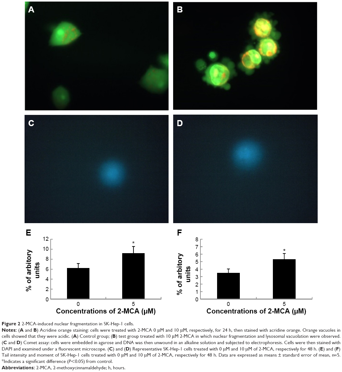 | Figure 2 2-MCA-induced nuclear fragmentation in SK-Hep-1 cells. |
Blebbing of plasma membrane, nuclear condensation, fragmentation, and apoptotic body formation are characteristic morphologic features of apoptosis.25 The morphological changes observed in our study suggest that 2-MCA did induce apoptosis in SK-Hep-1 cells (Figures 1D and 2B and D).
2-MCA increases volume of acidic compartments in SK-Hep-1 cells
Neutral Red has been used to stain lysosomes and quantify the VAC in cells.16,26,27 Figure 3 demonstrates that 2-MCA treatment resulted in acidic vacuoles in SK-Hep-1 cells with positive neutral red staining. As shown in Figure 3D, the VAC of 2-MCA-treated SK-Hep-1 cells increased in a dose-dependent manner.
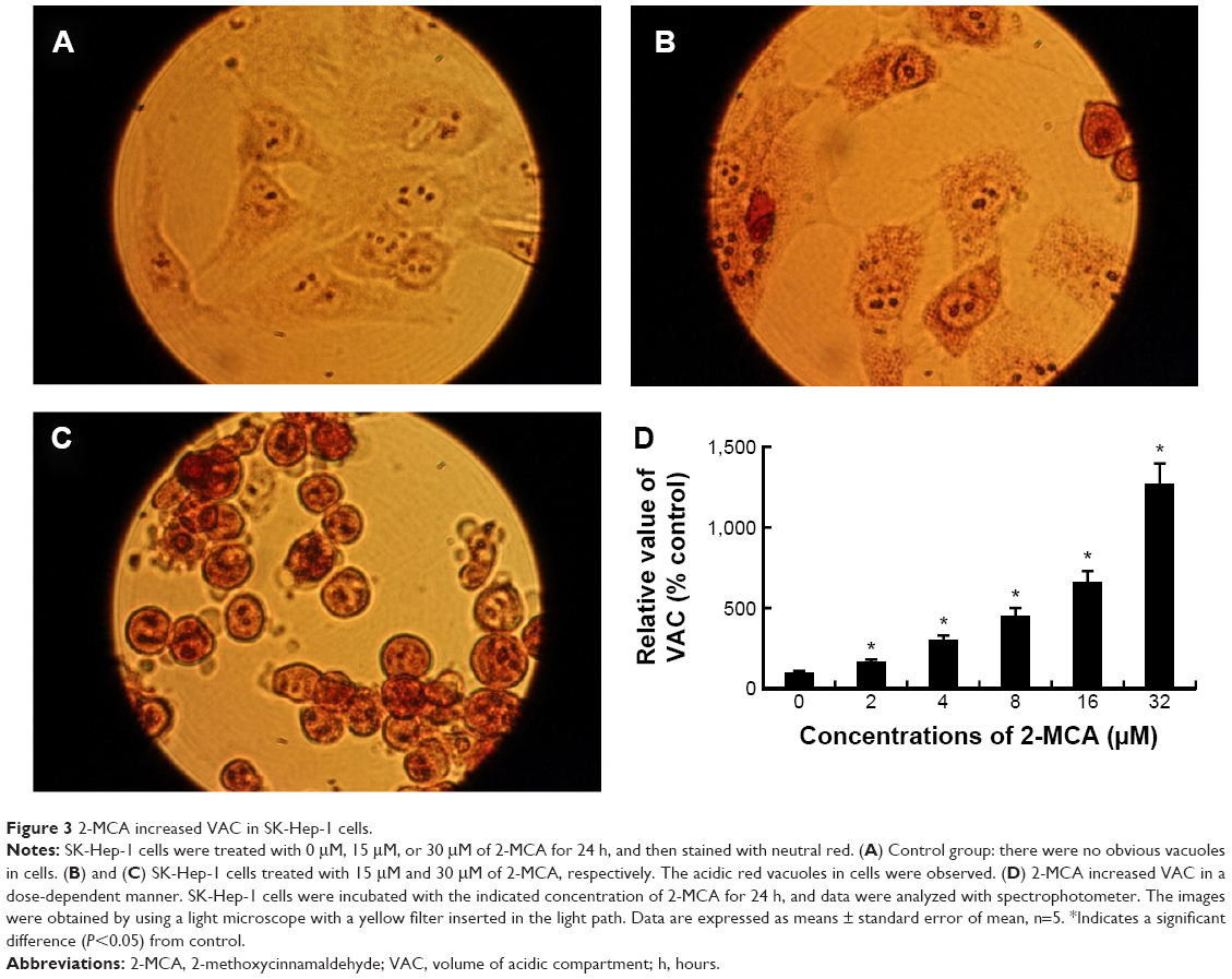 | Figure 3 2-MCA increased VAC in SK-Hep-1 cells. |
2-MCA induces apoptosis in SK-Hep-1 cells
Flow cytometer was used to examine the mechanism responsible for the inhibition of cell proliferation by 2-MCA. DNA content histogram analysis obtained from PI-stained SK-Hep-1 cells demonstrated that treatment with 2-MCA led to elevated sub-G1. The results shown in Figure 4 reveal that the percentage of cell population with reduced (hypodiploid) DNA content increased from untreated cells to cells exposed to 20 μM 2-MCA for 24 hours in a dose-dependent manner. The percentage DNA content in sub-G1 region increased from 1.602% in untreated control to 21.923% in cells treated with 20 μM 2-MCA for 24 hours as mentioned earlier (Figure 4B).
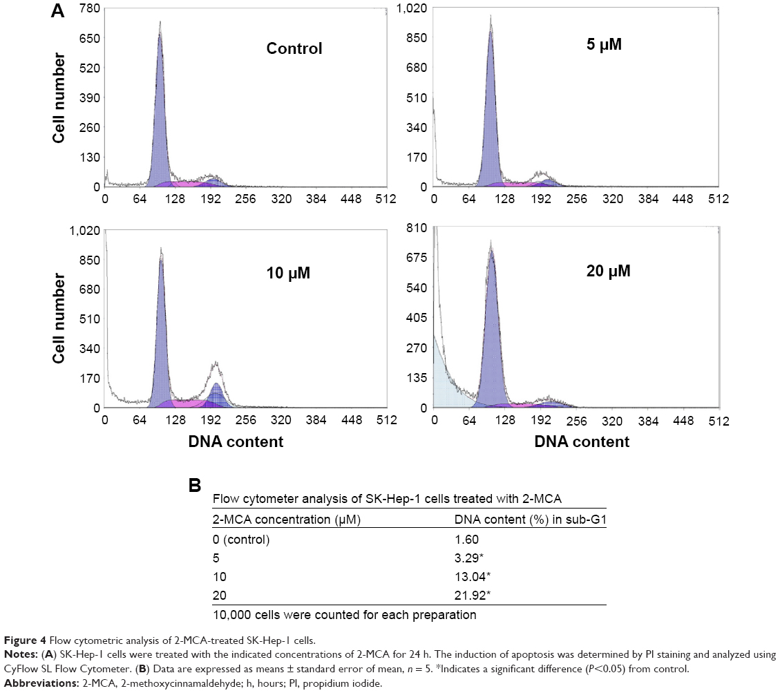 | Figure 4 Flow cytometric analysis of 2-MCA-treated SK-Hep-1 cells. |
Then, we further investigated the role of mitochondria in the 2-MCA-induced apoptosis in SK-Hep-1 cells. Since early apoptotic cell death often involves mitochondrial depolarization and release of cytochrome c from mitochondria into cytosol, we initially investigated mitochondrial dysfunction, by measuring mitochondrial membrane potential ΔΨm in 2-MCA-treated SK-Hep-1 cells using the mitochondria-specific dye JC-1, both microscopically and spectrophotometrically. As shown in Figure 5, 2-MCA induced loss of mitochondrial membrane potential as indicated by decreased ΔΨm in a dose-dependent manner.
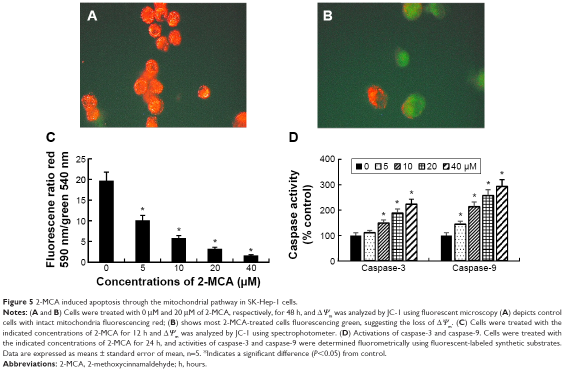 | Figure 5 2-MCA induced apoptosis through the mitochondrial pathway in SK-Hep-1 cells. |
Caspases, or cysteine-aspartic proteases, are a family of cysteine proteases that play important roles in apoptosis. As shown in Figure 5D, the activities of both caspase-3 and caspase-9 increased in a dose-dependent manner in 2-MCA-treated SK-Hep-1 cells. This is consistent with the mitochondrial depolarization and release of cytochrome c from mitochondria into the cytosol.
2-MCA inhibits topoisomerase I activity in SK-Hep-1 cells
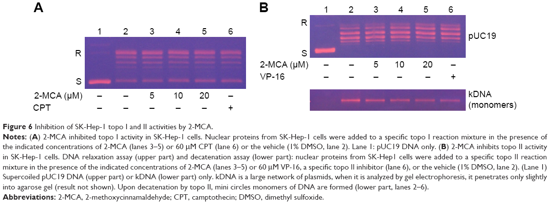
Inhibition of topoisomerase I activity in SK-Hep-1 cells by 2-MCA was performed in the presence of increasing concentration of 2-MCA (Figure 6) or camptothecin (CPT) (lane 6), a known specific inhibitor of topoisomerase I.28–30 Figure 6A shows that the conversion of the supercoiled plasmid pUC 19 to the relaxed form decreased in a dose-dependent manner in the presence of 2-MCA or CPT (compare lanes 3–6 with lane 2). These results show that the DNA relaxation activity of SK-Hep-1 cell nuclear proteins is inhibited by 2-MCA.
 | Figure 6 Inhibition of SK-Hep-1 topo I and II activities by 2-MCA. |
2-MCA inhibits topoisomerase II activity in SK-Hep-1 cells
Inhibition of topoisomerase II activity in SK-Hep-1 cells by 2-MCA was examined in the presence of increasing concentration of 2-MCA (Figure 6B, lanes 3–5) or 60 μM VP-16 (lane 6), a known inhibitor of topoisomerase II.29 Figure 6B, upper part, shows that the conversion of the supercoiled plasmid pUC 19 to the relaxed form decreased in a dose-dependent manner in the presence of 2-MCA or VP-16 (compare lanes 3–6 with lane 2). These results show that the DNA relaxation activity of SK-Hep-1 cell nuclear proteins is inhibited by 2-MCA. In addition, the effect of 2-MCA on topoisomerase II in SK-Hep-1 cells was further evaluated by decatenation assay. Decatenation activity is the releasing of monomers (minicircle DNA) from the kDNA (a large network of plasmid). Nuclear proteins extract from SK-Hep-1 cells contained topoisomerase II, which converted a kinetoplast DNA to monomer DNA molecules (Figure 6B, lower part, compare lane 2 with lane 1). The conversion of kinetoplast DNA to monomers decreased in a dose-dependent manner in the presence of 2-MCA (compare lanes 3–5 with lane 2) or VP-16 (compare lane 6 with lane 2). These results show that the decatenation activity of SK-Hep-1 cell nuclear proteins is inhibited by 2-MCA.
2-MCA inhibits NF-κB, COX-2, and PGE2 levels
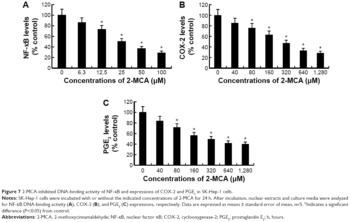
We then examined the effect of 2-MCA treatment on cellular factors associated with tumorigenesis in SK-Hep-1 cells. SK-Hep-1 cells were incubated without or with various concentrations of 2-MCA for 24 hours. After incubation, the NF-κB DNA-binding activity was quantified by ELISA. As shown in Figure 7, 2-MCA decreased NF-κB DNA-binding activity in a dose-dependent manner. The IC50 value of 2-MCA in inhibiting the binding activity was 42.16 μM.
 | Figure 7 2-MCA-inhibited DNA-binding activity of NF-κB and expressions of COX-2 and PGE2 in SK-Hep-1 cells. |
To investigate the effects of 2-MCA on COX-2 activity in SK-Hep-1 cells, SK-Hep-1 cells were incubated with different concentrations of 2-MCA for 24 hours. Then, the COX-2 activity was determined by ELISA. As shown in Figure 7B, 2-MCA decreased COX-2 activity in a dose-dependent manner. The IC50 value of 2-MCA in inhibiting COX-2 activity was 407.92 μM.
In addition, as shown in Figure 7C, 2-MCA decreased PGE2 level in a dose-dependent manner. The IC50 value of 2-MCA in inhibiting PGE2 expression was 415.31 μM.
2-MCA inhibits growth of SK-Hep-1 xenograft in nude mice
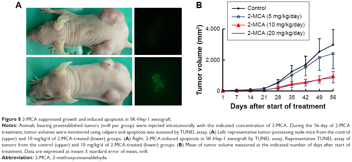
To determine whether 2-MCA suppresses growth of SK-Hep-1 xenograft, equal numbers of SK-Hep-1 cells were injected subcutaneously into both flanks of the nude mice. Tumor growth suppression was noticed in all groups of 2-MCA-treated (5 mg/kg/d, 10 mg/kg/d, or 20 mg/kg/d of 2-MCA, respectively) mice. However, significant growth suppression was observed only in mice treated with 10 mg/kg/d or 20 mg/kg/d of 2-MCA, where ~70% reductions in tumor size were found. No significant difference between these two groups was found (Figure 8). None of the 2-MCA treatments caused any significant decrease in diet consumption or body weight change (data not shown) compared with control mice. To gain insight into the mechanism of antitumor effect of 2-MCA in vivo, we harvested the SK-Hep-1 xenograft from vehicle- and 2-MCA-treated mice and assessed cell death by TUNEL analysis. As shown in Figure 8A, right parts, compared with tumors of vehicle-treated mice (upper part), elevated TUNEL-positive cells, suggesting apoptosis, were found in tumors of the 2-MCA-treated mice (lower part).
 | Figure 8 2-MCA suppressed growth and induced apoptosis in SK-Hep-1 xenograft. |
Discussion
Epidemiological and experimental studies have consistently shown that there is a correlation between regular consumption of fruits and vegetables and prevention of developing lifestyle disorders, such as cardiovascular disorders and cancer.31,32 Phytochemicals, such as polyphenols and flavonoids that are abundant in fruits and vegetables, seem to possess many of the desirable qualities for anticancer and could have great potential as chemopreventive and antiproliferative agents.33–38 C. verum has been traditionally used for treating dyspepsia, blood circulation, and inflammatory disorders, including gastritis.39,40 2-MCA, a constituent of the bark of the plant, could to be such a natural agent. Very few studies about 2-MCA have been reported. Moreover, to the best of our knowledge, there has been no report to date with regard to its effects on topoisomerase I and II activities. The current study was aimed at investigating the antiproliferative activity of 2-MCA and elucidating the underlying mechanisms of action.
In this study, we first elaborately examined the effects of 2-MCA on the growth of human HCC SK-Hep-1 cells. We found that 2-MCA suppressed the proliferation of SK-Hep-1 cells in a dose- and time-dependent manner. Although cells can die by nonapoptotic mechanisms, apoptosis is the most common and preferred mechanism through which many chemotherapeutic agents kill and eradicate tumor cells.5 In addition, apoptosis has been reported as the major mechanism of cancer cell death induced by selected polyphenols.41–44 Our results suggested that 2-MCA did induce apoptosis as indicated by loss of ΔΨm, activations of caspase-3 and caspase-9 (Figure 5), increased DNA content in sub-G1 region as shown in flow cytometric analysis (Figure 4), and morphological characteristics of apoptosis, including blebbing of plasma membrane, nuclear condensation, fragmentation, and apoptotic body formation as shown in various stainings and comet assay (Figures 1–3).
Mitochondria are crucial to multicellular life and apoptosis-inducing agents that target mitochondria may affect them in various ways. They may induce the formation of membrane pores, leading to mitochondrial swelling, or increase the permeability of mitochondrial membrane, resulting in releasing of apoptotic effectors from mitochondrial into cytosol. The released cytochrome c binds to apoptotic protease activating factor-1 and dATP, which then bind to pro-caspase-9 to create a protein complex called apoptosome. The apoptosome cleaves the pro-caspase-9 to its active form of caspase-9. The activated caspase-9 in turn activates effector caspase-3, thereby initiating a cascade of proteolytic events.45–47 The present study with regard to the key events in induction of apoptosis demonstrate that 2-MCA induced the collapse of ΔΨm, upregulated activities of most upstream protease of intrinsic apoptotic pathway, caspase-9, and the effector caspase-3 suggested the involvement of these proteins in 2-MCA-induced apoptotic cell death.
In addition, our results suggested that 2-MCA induced vacuolation with elevated VAC. Increase of VAC has been reported to be a common phenomenon of cells that undergo either apoptotic or necrotic cell death and could be a hallmark of dying cells.48 Since apoptosis is an ordered process, the increase of VAC could be responsible for the self-digestion during the course of cell death.48
Type I topoisomerases act by creating a transient single-stranded break in the DNA double helix molecule, followed by either a single-stranded DNA passage or controlled rotation about the break. Type I topoisomerases are involved in all DNA processes that involve tracking systems and play important roles in maintaining genomic integrity.6 Furthermore, elevated levels of topoisomerase I mRNA, protein, and catalytic activity are seen across human tumors.49
Type II topoisomerases act by generating a transient double-stranded DNA break, followed by a double-stranded DNA passage event. Type II topoisomerases function in numerous DNA processes and are required for recombination, the separation of daughter chromosomes, and proper chromosome structure, condensation, and decondensation.6 The enzyme is increased drastically during cell proliferation and peak in G2/M. The resulting transient double-stranded break could lead to fragmentation of the genome with chromosomal translocations and other DNA aberrations.7,50
In addition to cell cycle regulation, topoisomerase is another major target of anticancer agents.8–11 Chemotherapeutic agent etoposide kills cells by stabilizing the transient intermediate cleavage complex. The accumulation of cleavage complexes leads to the generation of permanent DNA strand breaks that fragments the genome, resulting in the activation of death pathways.51 Apoptosis has been demonstrated to be the most efficient death pathway in tumor cells after topoisomerase II had been inhibited.52 Our results documented that 2-MCA inhibited topoisomerase I and II activities in a concentration-dependent manner (Figure 6), which in parts, could be a mechanism driving the cells to apoptosis. While the majority of topoisomerase inhibitors are selectivity against either topoisomerase I or II,53 our study obviously shows that 2-MCA inhibited both topoisomerase I and II activities in SK-Hep-1 cells. However, further works are needed to elucidate the specific underlying mechanism of the inhibition, possible mutagenic effect, and others for clinical usage as a chemopreventive or anticancer agent against HCC and/or other malignances.
It is generally accepted that carcinogenesis is a multistep process. Studying the effects of 2-MCA on both topoisomerase I and II in SK-Hep-1 cells in this process may provide new information on the pathological process of cancer.
Many diseases are due to the aberrant activation and expression of genes involved in the initiation and progression of pathogenesis. In general, these genes are quiescent or have low activity in normal physiological status, but under certain conditions are turned on by preexisting genetic switches.40 These genetic switches are partially controlled by transcription factor NF-κB. NF-κB, a heterometric complex consisting of p50, p65, and IκBα, is present in its inactive state in the cytoplasm. When NF-κB is activated, IκBα is degraded and p50-p65 heterodimer is translocated to the nucleus, binds the κB-regulatory elements at the promoter region, and activates genes. NF-κB is involved in the regulation of cell proliferation, differentiation, immunity, inflammation, and apoptosis. The aberrant activation of NF-κB signaling results in the transcription of genes, generating biologically active proteins such as mitogen-activated protein kinase and COX-2. Cohesive scientific evidence from molecular, animal, and human studies suggests the hypothesis that aberrant induction of COX-2 and prostaglandin cascade play an important role in inflammation, aging, and carcinogenesis. Therefore, inhibition of the process has strong potential for cancer prevention and treatment.14 Our results in this study clearly demonstrate that 2-MCA suppressed NF-κB DNA-binding activity and COX-2 and PGE2 levels in SK-Hep-1 cells in a dose-dependent manner. It has been shown that COX-2 was highly expressed in the HCC tissue.54 Treatment that inhibits COX-2 may be a promising targeted approach in HCC.
Therapy-induced cytotoxicity and other associated side effects of anticancer drugs are major concerns of chemotherapy. Therefore, ideal drugs should selectively kill cancer cells and not damage the healthy. None of the 2-MCA treatments caused any significant decrease in diet consumption or body weight change compared with control mice. These results convincingly indicate the protective effect of 2-MCA treatment against SK-Hep-1 xenograft growth in nude mice without any observable toxicity. Indeed, similar effects (including all of the above-mentioned effects) were found in other tested cell lines, including human HCC Hep 3B, lung adenocarcinoma A549, squamous cell carcinoma NCI-H520, colorectal adenocarcinoma COLO 205, and T-lymphoblastic MOLT-3 (results not shown), suggesting an antiproliferative action of 2-MCA in SK-Hep-1 cells and the agent as a potential source of antiproliferative agent for cancer.
Conclusion
Collectively, our data clearly indicate that 2-MCA induced apoptosis, suppressed tumor cells growth and the associated biomarkers. The molecular events associated with the tumor suppression effect of 2-MCA including downregulation of cell proliferative controls, involving apoptosis, transcription factor NF-κB, both topoisomerase I and II, and inflammatory responses involving COX-2 and PGE2. The 2-MCA efficacy observed in the present study in terms of a shrinkage of tumor size would have potential clinical significance.
In conclusion, the present study provides fundamental information on the tumor suppression effect of 2-MCA in SK-Hep-1 cells, both in vitro and in vivo, suggesting a short-term model for evaluation of potential chemopreventive pharmacological modulators against hepatoma. Our results provide a focus for the rational development of 2-MCA as an anticancer agent against HCC.
Acknowledgments
This work was supported by grants from E-Da Hospital (no EDAH-2014-0001-001-02).
Disclosure
The authors report no conflicts of interest in this work.
References
Bruix J, Sherman M, Llovet JM, et al; EASL Panel of Experts on HCC. Clinical management of hepatocellular carcinoma. Conclusions of the Barcelona-2000 EASL conference. European association for the study of the liver. J Hepatol. 2001;35:421–430. | ||
Hwa JS, Jin YC, Lee YS, et al. 2-methoxycinnamaldehyde from Cinnamomum cassia reduces rat myocardial ischemia and reperfusion injury in vivo due to HO-1 induction. J Ethnopharmacol. 2012;139:605–615. | ||
Duke JA, Duke P-AK, duCellier JL. Duke’s Handbook of Medicinal Plants of the Bible. Boca Raton, London, New York: CRC Press; 2008. | ||
Artandi SE, DePinho RA. Telomeres and telomerase in cancer. Carcinogenesis. 2010;31:9–18. | ||
Aleo E, Henderson CJ, Fontanini A, Solazzo B, Brancolini C. Identification of new compounds that trigger apoptosome-independent caspase activation and apoptosis. Cancer Res. 2006;66:9235–9244. | ||
McClendon AK, Osheroff N. DNA topoisomerase II, genotoxicity, and cancer. Mutat Res. 2007;623:83–97. | ||
Heck MM, Earnshaw WC. Topoisomerase II: a specific marker for cell proliferation. J Cell Biol. 1986;103:2569–2581. | ||
Naowaratwattana W, De-Eknamkul W, De Mejia EG. Phenolic-containing organic extracts of mulberry (Morus alba L.) leaves inhibit HepG2 hepatoma cells through G2/M phase arrest, induction of apoptosis, and inhibition of topoisomerase IIalpha activity. J Med Food. 2010;13:1045–1056. | ||
Baikar S, Malpathak N. Secondary metabolites as DNA topoisomerase inhibitors: a new era towards designing of anticancer drugs. Pharmacogn Rev. 2010;4:12–26. | ||
Bandele OJ, Clawson SJ, Osheroff N. Dietary polyphenols as topoisomerase II poisons: B ring and C ring substituents determine the mechanism of enzyme-mediated DNA cleavage enhancement. Chem Res Toxicol. 2008;21:1253–1260. | ||
Sudan S, Rupasinghe HP. Flavonoid-enriched apple fraction AF4 induces cell cycle arrest, DNA topoisomerase II inhibition, and apoptosis in human liver cancer HepG2 cells. Nutr Cancer. 2014;66:1237–1246. | ||
Rayet B, Gelinas C. Aberrant rel/nfkb genes and activity in human cancer. Oncogene. 1999;18:6938–6947. | ||
Vandoros GP, Konstantinopoulos PA, Sotiropoulou-Bonikou G, et al. PPAR-gamma is expressed and NF-kB pathway is activated and correlates positively with COX-2 expression in stromal myofibroblasts surrounding colon adenocarcinomas. J Cancer Res Clin Oncol. 2006;132:76–84. | ||
Harris RE. Cyclooxygenase-2 (cox-2) and the inflammogenesis of cancer. Subcell Biochem. 2007;42:93–126. | ||
Darzynkiewicz Z. Differential staining of DNA and RNA in intact cells and isolated cell nuclei with acridine orange. Methods Cell Biol. 1990;33:285–298. | ||
Fan C, Wang W, Zhao B, Zhang S, Miao J. Chloroquine inhibits cell growth and induces cell death in A549 lung cancer cells. Bioorg Med Chem. 2006;14:3218–3222. | ||
Olive PL, Banath JP. The comet assay: a method to measure DNA damage in individual cells. Nat Protoc. 2006;1:23–29. | ||
Reers M, Smiley ST, Mottola-Hartshorn C, Chen A, Lin M, Chen LB. Mitochondrial membrane potential monitored by JC-1 dye. Methods Enzymol. 1995;260:406–417. | ||
Martin EJ, Forkert PG. Evidence that 1,1-dichloroethylene induces apoptotic cell death in murine liver. J Pharmacol Exp Ther. 2004;310:33–42. | ||
Har-Vardi I, Mali R, Breietman M, et al. DNA topoisomerases I and II in human mature sperm cells: characterization and unique properties. Hum Reprod. 2007;22:2183–2189. | ||
Cherng JM, Lin HJ, Hung MS, Lin YR, Chan MH, Lin JC. Inhibition of nuclear factor kappaB is associated with neuroprotective effects of glycyrrhizic acid on glutamate-induced excitotoxicity in primary neurons. Eur J Pharmacol. 2006;547:10–21. | ||
Benotmane AM, Hoylaerts MF, Collen D, Belayew A. Nonisotopic quantitative analysis of protein-DNA interactions at equilibrium. Anal Biochem. 1997;250:181–185. | ||
White K, Grether ME, Abrams JM, Young L, Farrell K, Steller H. Genetic control of programmed cell death in Drosophila. Science. 1994;264:677–683. | ||
Mpoke SS, Wolfe J. Differential staining of apoptotic nuclei in living cells: application to macronuclear elimination in tetrahymena. J Histochem Cytochem. 1997;45:675–683. | ||
Wyllie AH, Kerr JF, Currie AR. Cell death: the significance of apoptosis. Int Rev Cytol. 1980;68:251–306. | ||
Cover TL, Puryear W, Perez-Perez GI, Blaser MJ. Effect of urease on HeLa cell vacuolation induced by Helicobacter pylori cytotoxin. Infect Immun. 1991;59:1264–1270. | ||
Patel HK, Willhite DC, Patel RM, et al. Plasma membrane cholesterol modulates cellular vacuolation induced by the Helicobacter pylori vacuolating cytotoxin. Infect Immun. 2002;70:4112–4123. | ||
Pommier Y. Diversity of DNA topoisomerases I and inhibitors. Biochimie. 1998;80:255–270. | ||
Li TK, Liu LF. Tumor cell death induced by topoisomerase-targeting drugs. Annu Rev Pharmacol Toxicol. 2001;41:53–77. | ||
Pommier Y, Pourquier P, Urasaki Y, Wu J, Laco GS. Topoisomerase I inhibitors: selectivity and cellular resistance. Drug Resist Updat. 1999;2:307–318. | ||
Willett WC. Balancing life-style and genomics research for disease prevention. Science. 2002;296:695–698. | ||
Block G, Patterson B, Subar A. Fruit, vegetables, and cancer prevention: a review of the epidemiological evidence. Nutr Cancer. 1992;18:1–29. | ||
Shukla S, Meeran SM, Katiyar SK. Epigenetic regulation by selected dietary phytochemicals in cancer chemoprevention. Cancer Lett. 2014;355:9–17. | ||
Priyadarsini RV, Nagini S. Cancer chemoprevention by dietary phytochemicals: promises and pitfalls. Curr Pharm Biotechnol. 2012;13:125–136. | ||
Surh YJ. Cancer chemoprevention with dietary phytochemicals. Nat Rev Cancer. 2003;3:768–780. | ||
Yang CS, Landau JM, Huang MT, Newmark HL. Inhibition of carcinogenesis by dietary polyphenolic compounds. Annu Rev Nutr. 2001;21:381–406. | ||
Watson WH, Cai J, Jones DP. Diet and apoptosis. Annu Rev Nutr. 2000;20:485–505. | ||
Middleton E Jr, Kandaswami C, Theoharides TC. The effects of plant flavonoids on mammalian cells: implications for inflammation, heart disease, and cancer. Pharmacol Rev. 2000;52:673–751. | ||
Tanaka S, Yoon YH, Fukui H, et al. Antiulcerogenic compounds isolated from Chinese cinnamon. Planta Med. 1989;55:245–248. | ||
Reddy AM, Seo JH, Ryu SY, et al. Cinnamaldehyde and 2-methoxycinnamaldehyde as NF-kappaB inhibitors from Cinnamomum cassia. Planta Med. 2004;70:823–827. | ||
Miura T, Chiba M, Kasai K, et al. Apple procyanidins induce tumor cell apoptosis through mitochondrial pathway activation of caspase-3. Carcinogenesis. 2008;29:585–593. | ||
Liu JR, Dong HW, Chen BQ, Zhao P, Liu RH. Fresh apples suppress mammary carcinogenesis and proliferative activity and induce apoptosis in mammary tumors of the Sprague-Dawley rat. J Agric Food Chem. 2009;57:297–304. | ||
Yoon H, Liu RH. Effect of selected phytochemicals and apple extracts on NF-kappaB activation in human breast cancer MCF-7 cells. J Agric Food Chem. 2007;55:3167–3173. | ||
Zheng CQ, Qiao B, Wang M, Tao Q. Mechanisms of apple polyphenols-induced proliferation inhibiting and apoptosis in a metastatic oral adenoid cystic carcinoma cell line. Kaohsiung J Med Sci. 2013;29:239–245. | ||
Pop C, Timmer J, Sperandio S, Salvesen GS. The apoptosome activates caspase-9 by dimerization. Mol Cell. 2006;22:269–275. | ||
Zou H, Henzel WJ, Liu X, Lutschg A, Wang X. Apaf-1, a human protein homologous to C. elegans CED-4, participates in cytochrome c-dependent activation of caspase-3. Cell. 1997;90:405–413. | ||
Li P, Nijhawan D, Budihardjo I, et al. Cytochrome c and dATP-dependent formation of Apaf-1/caspase-9 complex initiates an apoptotic protease cascade. Cell. 1997;91:479–489. | ||
Ono K, Wang X, Han J. Resistance to tumor necrosis factor-induced cell death mediated by PMCA4 deficiency. Mol Cell Biol. 2001;21:8276–8288. | ||
Husain I, Mohler JL, Seigler HF, Besterman JM. Elevation of topoisomerase I messenger RNA, protein, and catalytic activity in human tumors: demonstration of tumor-type specificity and implications for cancer chemotherapy. Cancer Res. 1994;54:539–546. | ||
Hsiang YH, Wu HY, Liu LF. Proliferation-dependent regulation of DNA topoisomerase II in cultured human cells. Cancer Res. 1988;48:3230–3235. | ||
Baldwin EL, Osheroff N. Etoposide, topoisomerase II and cancer. Curr Med Chem Anticancer Agents. 2005;5:363–372. | ||
El-Awady RA, Ali MM, Saleh EM, Ghaleb FM. Apoptosis is the most efficient death-pathway in tumor cells after topoisomerase II inhibition. Saudi Med J. 2008;29:558–564. | ||
Denny WA, Baguley BC. Dual topoisomerase I/II inhibitors in cancer therapy. Curr Top Med Chem. 2003;3:339–353. | ||
Yang Y, Zhu J, Gou H, Cao D, Jiang M, Hou M. Clinical significance of Cox-2, survivin and Bcl-2 expression in hepatocellular carcinoma (HCC). Med Oncol. 2011;28:796–803. |
 © 2016 The Author(s). This work is published and licensed by Dove Medical Press Limited. The full terms of this license are available at https://www.dovepress.com/terms.php and incorporate the Creative Commons Attribution - Non Commercial (unported, v3.0) License.
By accessing the work you hereby accept the Terms. Non-commercial uses of the work are permitted without any further permission from Dove Medical Press Limited, provided the work is properly attributed. For permission for commercial use of this work, please see paragraphs 4.2 and 5 of our Terms.
© 2016 The Author(s). This work is published and licensed by Dove Medical Press Limited. The full terms of this license are available at https://www.dovepress.com/terms.php and incorporate the Creative Commons Attribution - Non Commercial (unported, v3.0) License.
By accessing the work you hereby accept the Terms. Non-commercial uses of the work are permitted without any further permission from Dove Medical Press Limited, provided the work is properly attributed. For permission for commercial use of this work, please see paragraphs 4.2 and 5 of our Terms.