")
Back to Journals » Journal of Asthma and Allergy » Volume 12
Detection of pepsin and IL-8 in saliva of adult asthmatic patients
Authors Marshall S, McCann AJ , Samuels TL, Blair A, Bonne V , Johnston N , Koufman J
Received 15 February 2019
Accepted for publication 29 April 2019
Published 28 May 2019 Volume 2019:12 Pages 155—161
DOI https://doi.org/10.2147/JAA.S205482
Checked for plagiarism Yes
Review by Single anonymous peer review
Peer reviewer comments 5
Editor who approved publication: Dr Luis Garcia-Marcos
Samuel Marshall,1 Alec J McCann,1 Tina L Samuels,1 Amy Blair,2 Valerie Bonne,2 Nikki Johnston,1,3 Jamie Koufman4
1Department of Otolaryngology and Communication Sciences, Medical College of Wisconsin, Milwaukee, WI, USA; 2Department of Medicine, Medical College of Wisconsin, Milwaukee, WI, USA; 3Department of Microbiology and Immunology, Medical College of Wisconsin, Milwaukee, WI, USA; 4Voice Institute of New York, New York, NY, USA
Objective: Asthma and gastric reflux disease are widespread and often coexisting diseases with complex interactions, leading some to suspect that asthma symptoms of patients with reflux may improve with anti-reflux therapy. The objective of this study was to determine whether pepsin in saliva, indicative of airway reflux, could be detected in patients with asthma of varying severity and test the requirement of citric acid as a pepsin preservative.
Methods: Saliva samples were collected in the clinic (with/without citric acid) and upon waking the following morning from 25 asthmatic patients. Enzyme-linked immunosorbent assay was performed for pepsin and interleukin-8 (IL-8), an inflammatory cytokine induced by pepsin in other airway epithelia. Pepsin induction of IL-8 was tested in a lung epithelial cell culture model.
Results: Pepsin was detected in saliva from 14/25 patients (56%; mean concentration of pepsin in specimens where observed ±SD =80.3±87.5 ng/mL); significant agreement was found between samples collected in the presence/absence of citric acid. No significant associations were found with pepsin and clinical measures of asthma severity. IL-8 was detected in saliva from 22/25 patients (88%; mean IL-8 in all specimens where observed =3.27±3.91 ng/mL). IL-8 was significantly upregulated in human lung epithelial cells exposed to pepsin at pH7 in vitro (P=0.041).
Conclusion: In summary, more than half of the asthma patients in this study were found to have pepsin in their saliva, indicative of airway reflux. These data support the use of salivary pepsin as a noninvasive tool for future investigation of airway reflux in a larger cohort. The data further suggest that collection in citric acid as a sample preservative is not warranted and that pooling of multiple saliva samples collected at various timepoints may improve sensitivity of pepsin detection and reduce costs incurred by multiple sample analysis in future studies.
Keywords: extraesophageal reflux, laryngopharyngeal reflux, asthma, salivary diagnostics, IL-8
Introduction
Asthma is a common chronic disease of the airways described as a history of discrete attacks.1 During an asthma attack, the airway becomes inflamed causing, airflow obstruction and symptoms including wheezing, coughing, shortness of breath, and chest tightness/pain. The causes of asthma are unknown, but thought to be due to host and environmental factors such as airway infections, airborne allergens, occupational exposures, and air pollution. The Center for Disease Control (CDC) reports the incidence of asthma in both adults and children at 8.3% in the US, costing over $56 billion per year.2,3
Asthma symptoms are not well controlled in approximately of 10% patients, despite advances in current medical therapy.4 This may be due to poor inhaler skills and adherence or confounding comorbidities such as obesity and gastroesophageal reflux disease (GERD). Several studies have demonstrated an increased prevalence of GERD in asthma patients and highlighted its impact on asthma control.5,6 Studies are needed to identify subgroups of asthmatic patients that may benefit from anti-reflux therapy.
Reflux of gastric contents into the esophagus, termed gastroesophageal reflux (GER), is a normal physiological phenomenon. Brief and infrequent exposure of the esophagus to gastric contents does not result in injury and disease. In fact, up to 50 reflux episodes a day (below pH 4) are considered normal. Esophageal symptoms and complications are thought to arise when reflux is prolonged and/or there is a breakdown in the defense mechanisms, making an individual more susceptible to reflux-mediated esophageal injury, known as GERD. The most common symptom of GERD is heartburn. Diagnosis of GERD is made by endoscopy, and pH-monitoring of GERD is thought to affect approximately 30% of the Western populations. When gastric reflux travels more proximally into the laryngopharynx, it is termed extraesophageal or airway reflux. The airways are considerably more sensitive to damage by reflux. A single episode of reflux into the airways is considered pathological. Airway reflux contributes to several otolaryngologic symptoms and inflammatory disorders, and perhaps to neoplastic diseases of the laryngopharynx. The most common symptoms of airway reflux are hoarseness, sore throat, globus, chronic cough, and dysphagia. It is estimated that 10% of patients visiting otolaryngology clinics have reflux-attributed disease, and most patients with airway reflux do not have GERD. The airways are sensitive to weak or non-acid reflux, therefore pH-monitoring alone is insufficient for detection of symptom-associated airway reflux. Combined multichannel intraluminal impedance and pH-monitoring (MII-pH) which detects both acid and non-acid reflux is currently considered the gold standard for reflux detection; however, this test is expensive and invasive. Pepsin A is only produced in the stomach and, thus, is a specific biomarker for reflux.7–9 Further, pepsin is present in all refluxate in contrast to the other gastric components such as acid or bile, which may or may not be present.
In a study of the prevalence of airway reflux in children with reflux-associated pulmonary disease, pepsin was detected in the bronchoalveolar lavage (BAL) of more than 70% (47/65) patients with pulmonary disease who underwent bronchoscopy and tracheostomy compared to 0/11 controls who had elective surgery with no history of pulmonary disease.10 The authors found detection of pepsin in BAL samples to be more sensitive and specific than the current method of measuring lipid laden macrophages for the detection of aspiration. Hayat et al11 assayed salivary pepsin, indicative of airway reflux, in 111 patients with heartburn and 100 asymptomatic controls who underwent MII-pH demonstrating that findings of >210 ng/mL pepsin in one sample was 98.2% specific for diagnosis of GERD and hypersensitive esophagus. The authors concluded that a positive salivary pepsin test may obviate the need for more invasive and expensive diagnostic testing.
Hunt et al12 detected pepsin in BAL samples from 46/78 (58.9%) asthmatic patients, but found no association between pepsin levels and asthma severity or control. Despite the high prevalence of GER in asthma, a causal link is lacking. However, the literature does support GER as a risk factor for chronic obstructive pulmonary disease (COPD) exacerbations,13–15 and pepsin concentration in BAL fluid has been shown to correlate with local neutrophil counts, which predicts more severe pulmonary symptoms.16 We recently found that gastric juice from patients taking proton pump inhibitors increased neutrophil migration across lung epithelial monolayers independently of pH17 and that pepsin triggered neutrophil migration across acid damaged lung epithelium.18 We suspect that pepsin-induced neutrophil migration may be a result of pepsin-induced interleukin 8 (IL-8) expression. Pepsin elicits expression of the neutrophil chemoattractant and proinflammatory cytokine IL-8 in airway epithelia cells, and concentrations of pepsin parallel those of IL-8 in BAL of cystic fibrosis patients suggesting a similar relationship exists in the lung.19–21 Elevation of IL-8 in pepsin-treated lung epithelial cells would provide support for a mechanism whereby pepsin could contribute to inflammation, and thereby the pathophysiology of asthma. The response of IL-8 to pepsin has not been examined in lung cells to date.
Evidence of pepsin exposure leading to airway epithelial damage, barrier leakiness, and/or neutrophil breach of the epithelium suggests a potential for the capacity of pepsin to exert a pathological response. Given that salivary pepsin appears to be a sensitive and specific biomarker for airway reflux,11,22 the objective of this study was to measure salivary pepsin in 25 consecutive established asthma patients and compare levels with severity of disease. IL-8 was measured in patient saliva and the effect of pepsin on IL-8 levels in a human lung epithelial cell line was investigated.
Materials and methods
Human specimens
This study was approved by the MCW Institutional Review Board (protocol PRO00031392). All participants provided written informed consent, and this study was conducted in accordance with the Declaration of Helsinki. Study participants included 25 consecutive patients with clinically diagnosed asthma seen by the recruiting physician in the asthma clinic at Froedtert Hospital. Patients demographics and clinical data related to their asthma, including day of visit spirometry, smoking history, medications including prednisone for physician-determined exacerbations, body mass index, complete blood count, and IgE were collected for descriptive statistics. Patients provided a throat-clearing saliva sample during their routine clinic visit; half of this specimen was immediately transferred to a tube containing 0.5 mL 0.01 M citric acid and both were placed at 4°C. Patients provided a second sample on waking, prior to brushing their teeth, drinking or eating in the morning the day after their clinic visit which they collected in a provided tube and shipped back to the research laboratory via USPS in provided packaging. Once received in the laboratory, an aliquot of each specimen was transferred to -80°C for subsequent IL-8 analysis. The remainder of all specimens were stored for pepsin analysis for up to 14 days at 4°C.
Cell culture
Lung epithelial cells were treated with pepsin to determine whether pepsin induced expression of neutrophil chemoattractant and marker of inflammation, IL-8. Treatment conditions were based on the median pepsin concentration observed in BAL of asthmatics (4 ng/mL median concentration of pepsin in 180-fold diluted bronchial specimen)12 and persistence of pepsin in the lung of a rat model of aspiration (up to 50 hours following aspiration).23 A human epithelial cell line derived from a lymph node metastasis of a pulmonary mucoepidermoid carcinoma (H292; American Type Culture Collection, Manassas, VA) was cultured in RPMI with 10% fetal bovine serum and Antibiotic-Antimycotic (Thermo Fisher Scientific, Waltham, MA) at 37°C and 5% CO2 were grown to 75% confluence and treated with normal growth media at pH 7 with 1 mg/mL porcine pepsin (Sigma Aldrich, St Louis, MO) for 24 hours at 37°C and 5% CO2 in triplicate reactions. At the end of 24 hours, culture media was harvested, aliquoted, and stored at -80°C.
Enzyme-linked immunosorbent assay (ELISA)
The concentration of pepsin in human saliva specimens was determined by non-competitive indirect sandwich ELISA, as previously described.24,25 Human IL-8/CXCL8 DuoSet ELISA (R&D Systems, Minneapolis, MN) was performed on human saliva specimens and H292 culture media per manufacturer instructions.
Statistical analyses
To evaluate the effect of time of saliva collection on pepsin detection, Cohen’s Kappa was used to measure the agreement between pepsin presence or absence outcomes, with a value of 0 indicating agreement consistent with chance and a value of 1 indicating perfect agreement. McNemar’s test was used to evaluate the bias between the two outcomes. To evaluate the effect of citric acid as a preservative of pepsin in human saliva, the measured level of pepsin in saliva specimens was compared via a paired t-test among the saliva samples collected in clinic with citric acid (CA) and without citric acid (C) in patients for which pepsin was detected in at least one of the samples. For descriptive statistics of the study population and outcomes by the presence of pepsin in any sample, Wilcoxon rank-sum test was used for continuous data and Fisher's exact test was used for categorical data. Spearman’s correlation was used to assess the relationship between pepsin and IL-8 protein expression in human saliva specimens due to the skewness of their distributions.
ANOVA with Tukey’s adjustment for multiple testing was used to compare H292 IL-8 concentrations between treatment conditions. Concentrations were log transformed for the analysis to stabilize variability and improve normality; the estimates were back-transformed for reporting.
Results
Patient demographics are reported in Table 1. Twenty-five subjects were recruited and provided at least one specimen. Of these, 24 patients had a saliva specimen collected in the clinic, a portion of each were diluted in citric acid; 22 patients had a specimen collected on waking. Pepsin was present in at least one of the three specimens in 14/25 (56%) patients (mean±SD=66.7±76.5 ng/mL), see Table 2. No significant difference was observed in age, sex, race, body-mass index, asthma severity, smoking status, number of exacerbations in the last 12 months, reflux medication status, ACT, or FEV1 (post %) between patients with at least one pepsin positive specimen and those with none (Table 3; see
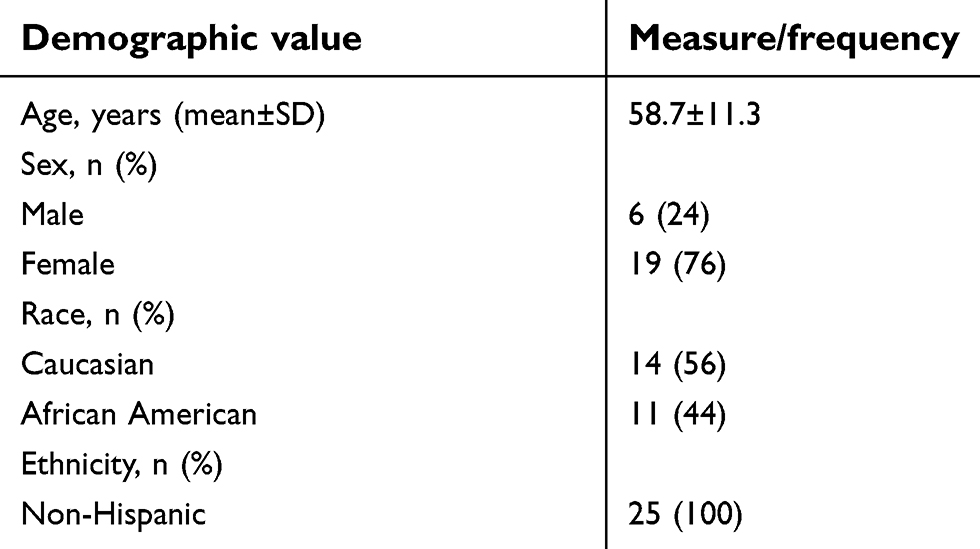 | Table 1 Subject demographics (n=25) |
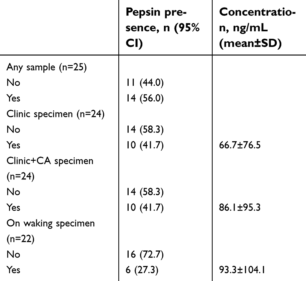 | Table 2 Pepsin in saliva of asthma patients |
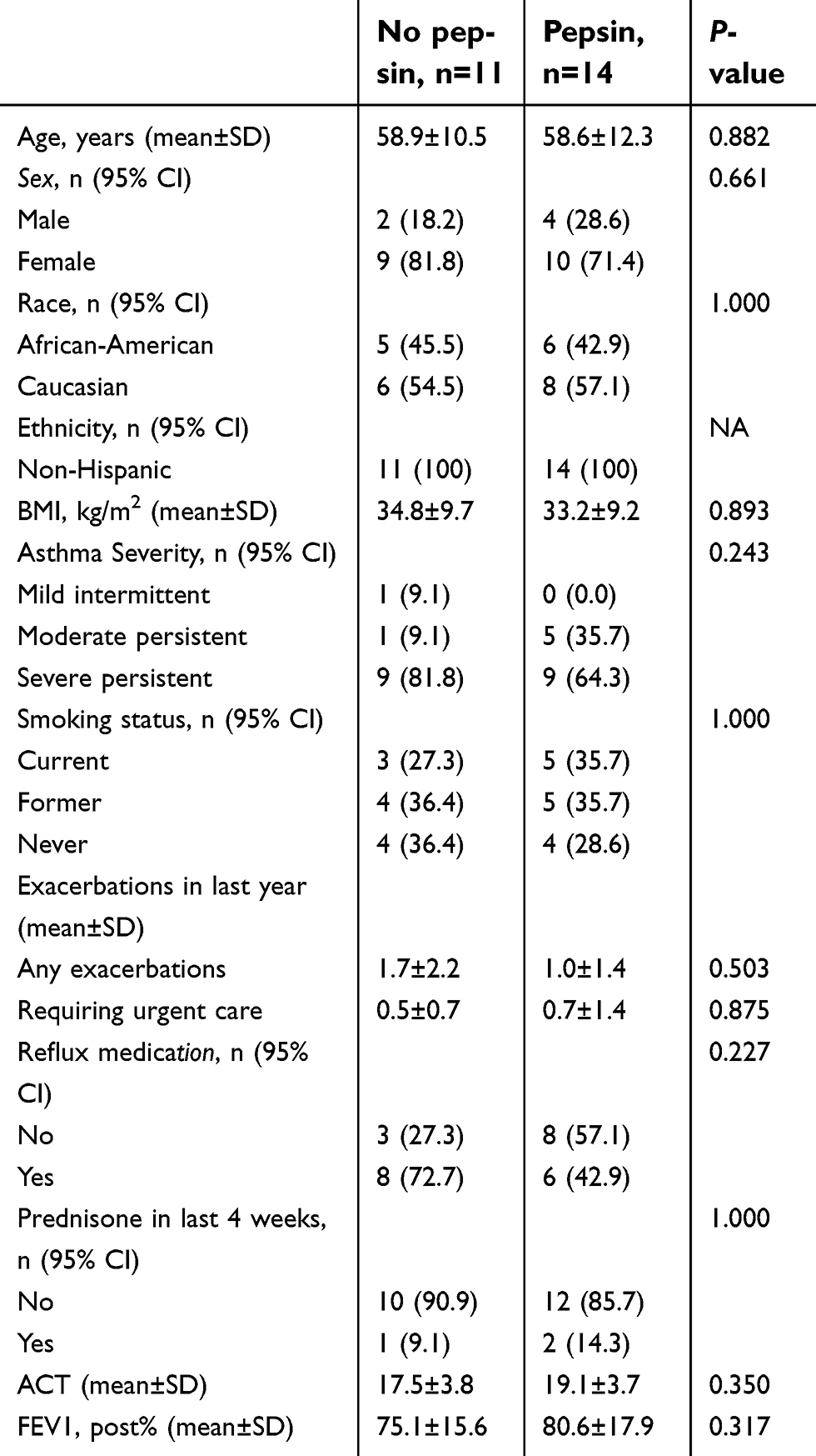 | Table 3 Descriptive statistics of the study population and outcomes by the presence of pepsin in any sample |
Pepsin was observed in 10/25 clinic-collected specimens without citric acid (mean±SD=66.7±76.5 ng/mL), in 10/25 clinic-collected specimens with citric acid (mean±SD=86.1±95.3 ng/mL), and in 6/25 specimens collected on waking (mean±SD=93.3±104.1 ng/mL), see Table 2. Cohen’s Kappa test demonstrated high agreement between clinic specimens diluted in citric acid and undiluted (Kappa=0.83; Table 4). Pepsin results from specimens collected from in clinic and on waking demonstrated poor agreement (Kappa=0; Table 4). No evidence of bias was observed between outcomes in clinic vs on-waking specimens (McNemar’s test P-value=1.000; Table 4). IL-8 was observed in 22/25 patients (mean concentration in all samples where observed±SD=3.27±3.91 ng/mL). No correlation was observed between pepsin and IL-8 protein levels in saliva specimens (P=0.1182).
 | Table 4 Agreement and bias of pepsin presence between samples |
IL-8 was significantly elevated in human lung epithelial H292 cells exposed to 1 mg/mL pepsin at pH7 for 24 hours (185±41.62 pg/mL) relative to cells maintained in media pH7 alone (75.66±7.57 pg/mL; P=0.003).
Discussion
Using salivary pepsin as a biomarker for reflux and aspiration, we report a similar prevalence of reflux in asthmatic patients (56%) as that reported by others in the literature.12,26 Collection of saliva is non-invasive and, thus, would be preferred over more invasive sampling methods such as BAL as a tool to diagnose airway reflux and assess its role in disease. Evidence of reflux has not been correlated with asthma severity to date.12 Accordingly, while more than half of the asthma patients in this study were found to have reflux, consistent with the findings reported by Hunt et al,12 the presence and levels of salivary pepsin did not correlate with clinical measures of disease severity.
It is important to remember that pepsin may only be transiently present in saliva because of the intermittent nature of reflux and influence of swallowing, both of which vary with food intake. Therefore, optimization of timing and method of salivary sampling to yield the greatest balance of sensitivity and specificity is essential if pepsin is to be used clinically as a marker of airway reflux. Although certain recommendations for collection time, such as waiting at least 1hour after meals to avoid detection of postprandial reflux events, have been suggested in previous studies,11 large-scale studies have not been performed to determine the optimal timing or sampling method. Larger studies with more frequent sampling may allow determination of the ideal time for sample collection, which might in turn facilitate the use of salivary pepsin as a more sensitive and specific tool for diagnosing airway reflux. Salivary pepsin should, therefore, be measured in broader studies to determine the role of airway reflux in the etiology and evaluation of asthma, potentially identifying a specific subset of asthma patients in whom reflux and aspiration contributes to disease and who might, therefore, benefit from lifestyle modifications or surgery for reflux. In follow-up to the study herein, we are currently collecting saliva samples from asthma patients, adding collection when the patient experiences a symptom (coughing, wheezing, shortness of breath, etc.) up to six times during a 24 hour period, to the sampling times utilized herein (ie, during their routine appointment to the pulmonology outpatient clinic in which collection technique is demonstrated, and when the patient wakes the following morning before eating or brushing their teeth). The samples are being pooled in a single vial without citric acid. Although some studies report collection of saliva in citric acid for pepsin stability,11,27 this pilot study revealed a significant agreement between samples collected in the presence/absence of citric acid. We, therefore, conclude that use of citric acid for sample collection is not warranted and would reduce assay sensitivity by diluting the sample. Further, given the levels of pepsin detected in the study herein (mean concentration of pepsin in specimens in which it was detected±SD=80.3±87.5 ng/mL) and LOD of our pepsin assay (0.1 ng/mL),24,25 dilution caused by pooling samples would not be expected to result in false negatives. We hypothesize that pooled saliva samples from multiple timepoints over a 24 hour period will increase the sensitivity of pepsin detection and decrease the cost of specimen analysis, thereby improving the utility of salivary pepsin ELISA as a tool to measure airway reflux for association with clinical measures of asthma severity.
IL-8 is a suspected mediator of reflux-induced esophagitis,28,29 and exposure of hypopharyngeal cells to pepsin at pH7 has been shown to induce production of the neutrophil chemoattractant IL-8 in vitro,19 suggesting a potential role for IL-8 in reflux-mediated inflammation. IL-8 is also elevated in airway secretions in acute severe asthma and observations of negative correlation between sputum IL-8 and forced expiratory volume (FEV1) have been reported in patients with severe asthma.30,31 Salivary IL-8 was detected in 22/25 patients in our cohort (3267.88±3908.44 pg/mL). Although salivary IL-8 levels did not correlate with pepsin levels, pepsin exposure was found to significantly induce IL-8 in human lung epithelial cells in vitro, supporting a role for refluxed pepsin in lung epithelial damage. In previous studies, non-acidic gastric juice and pepsin were found to increase neutrophil migration across the lung epithelium.17,18 These data suggest that pepsin may not simply be a biomarker of reflux-related disease, but it also may play a proinflammatory and damaging role in the lung. Although pepsin-mediated IL-8 elevation in vitro provides support for a potential mechanism by which pepsin could cause inflammation in the lung and worsen asthma, the importance of this finding has yet to be established. Interestingly, there are reports of occupational asthma thought to be caused by inhaled pepsin.32,33 Given the high percentage of asthma patients which have airway reflux and pepsin in a BAL,12 it is important to elucidate the effect of pepsin at a cellular level.
One limitation of this study is the small sample size. This study was a pilot to determine whether pepsin could be detected via ELISA in the saliva of patients with asthma of varying severity and test the requirement of citric acid as a pepsin preservative. A larger population will be required to provide an accurate measure of prevalence of airway reflux in asthma patients and identify a subgroup that may benefit from anti-reflux therapy. In addition, although the in vitro pepsin dose and treatment times herein were based on concentrations of pepsin in BAL and persistence of gastric contents in the lung of an in vivo model of aspiration, caution should generally be exercised in making inferences regarding pathophysiological processes from in vitro studies. Further experiments in in vivo models and in clinical specimens should be performed to verify the significance of pepsin-mediated IL-8 elevation in lung cells observed herein.
Conclusion
Airway reflux is prevalent in asthmatics across the spectrum of disease severity. Salivary pepsin can be used to diagnose airway reflux, although the optimal timing of sample collection needs further investigation. Although a correlation between salivary pepsin levels and asthma severity was not found in this study, optimization of saliva collection timing may reveal an association. Pepsin increases the neutrophil chemoattractant IL-8 and induces neutrophil migration in lung epithelial cells in vitro. Thus, the role of pepsin in the pathophysiology of asthma warrants further investigation.
Acknowledgments
This work was funded by MCW Department of Otolaryngology and Communication Sciences. The authors would like to thank Aniko Szabo, PhD, and Joy Liu of the Medical College of Wisconsin (MCW) Institute for Health and Equity Division of Biostatistics for assistance with statistical analyses.
Disclosure
The authors report no conflicts of interest in this work. The authors alone are responsible for the content and writing of the paper.
References
1. Cropp GJA, Bernstein IL, Boushey HA, Hyde RW. Guidelines for Bronchial Inhalation Challenges with Pharmacologic and Antigenic Agents. 1980.
2.
3.
4.
5. Ciprandi G, Gallo F, Gelardi M. Impact of gastric reflux on asthma in clinical practice. Respirology (Carlton, Vic). 2018;23(2):230–231. doi:10.1111/resp.13149
6. Porsbjerg C, Menzies-Gow A. Co-morbidities in severe asthma: clinical impact and management. Respirology (Carlton, Vic). 2017;22(4):651–661. doi:10.1111/resp.13026
7. Johnston N, Dettmar PW, Lively MO, et al. Effect of pepsin on laryngeal stress protein (Sep70, Sep53, and Hsp70) response: role in laryngopharyngeal reflux disease. Ann Otol Rhinol Laryngol. 2006;115(1):47–58. doi:10.1177/000348940611500108
8. Johnston N, Knight J, Dettmar PW, Lively MO, Koufman J. Pepsin and carbonic anhydrase isoenzyme III as diagnostic markers for laryngopharyngeal reflux disease. Laryngoscope. 2004;114(12):2129–2134. doi:10.1097/01.mlg.0000149445.07146.03
9. Samuels TL, Johnston N. Pepsin as a marker of extraesophageal reflux. Ann Otol Rhinol Laryngol. 2010;119(3):203–208.
10. Kelly EA, Samuels TL, Johnston N. Chronic pepsin exposure promotes anchorage-independent growth and migration of a hypopharyngeal squamous cell line. Otolaryngology Head Neck Surg. 2014;150(4):618–624. doi:10.1177/0194599813517862
11. Hayat JO, Gabieta-Somnez S, Yazaki E, et al. Pepsin in saliva for the diagnosis of gastro-oesophageal reflux disease. Gut. 2015;64(3):373–380. doi:10.1136/gutjnl-2014-307049
12. Hunt EB, Ward C, Power S, et al. The potential role of aspiration in the asthmatic airway. Chest. 2017;151(6):1272–1278. doi:10.1016/j.chest.2017.03.005
13. Donaldson GC, Seemungal TA, Bhowmik A, Wedzicha JA. Relationship between exacerbation frequency and lung function decline in chronic obstructive pulmonary disease. Thorax. 2002;57(10):847–852.
14. Kim J, Lee JH, Kim Y, et al. Association between chronic obstructive pulmonary disease and gastroesophageal reflux disease: a national cross-sectional cohort study. BMC Pulm Med. 2013;13:51. doi:10.1186/1471-2466-13-51
15. Sakae TM, Pizzichini MM, Teixeira PJ, Silva RM, Trevisol DJ, Pizzichini E. Exacerbations of COPD and symptoms of gastroesophageal reflux: a systematic review and meta-analysis. Jornal Brasileiro De Pneumologia. 2013;39(3):259–271. doi:10.1590/S1806-37132013000300002
16. Rosen R, Johnston N, Hart K, Khatwa U, Nurko S. The presence of pepsin in the lung and its relationship to pathologic gastro-esophageal reflux. Neurogastroenterology Motil. 2012;24(2):
17. Hurley B, Jugo R, Johnston N, Rosen R. Tu1742 gastric fluid from patients receiving proton pump inhibitors triggers increased neutrophil migration across lung epithelial monolayers independently of pH. Gastroenterology. 2016;150(4):S931. doi:10.1016/S0016-5085(16)33153-5
18. Hurley BP, Yonker LM, Mou H, Johnston N. Pepsin triggers neutrophil migration across acid damaged lung epithelium. Sci Rep. 2019.
19. Samuels TL, Johnston N. Pepsin as a causal agent of inflammation during nonacidic reflux. Otolaryngology Head Neck Surg. 2009;141(5):559–563. doi:10.1016/j.otohns.2009.08.022
20. McNally P, Ervine E, Shields MD, et al. High concentrations of pepsin in bronchoalveolar lavage fluid from children with cystic fibrosis are associated with high interleukin-8 concentrations. Thorax. 2011;66(2):140–143. doi:10.1136/thx.2009.130914
21. Tan JJ, Wang L, Mo TT, Wang J, Wang MG, Li XP. Pepsin promotes IL-8 signaling-induced epithelial-mesenchymal transition in laryngeal carcinoma. Cancer Cell Int. 2019;19:64. doi:10.1186/s12935-019-0772-7
22. Saritas Yuksel E, Hong SK, Strugala V, et al. Rapid salivary pepsin test: blinded assessment of test performance in gastroesophageal reflux disease. Laryngoscope. 2012;122(6):1312–1316. doi:10.1002/lary.23252
23. Leung JH, Chang JC, Foltz E, et al. Clearance of bile and trypsin in rat lungs following aspiration of human gastric fluid. Exp Lung Res. 2016;42(1):37–43. doi:10.3109/01902148.2016.1139213
24. Knight J, Lively MO, Johnston N, Dettmar PW, Koufman JA. Sensitive pepsin immunoassay for detection of laryngopharyngeal reflux. Laryngoscope. 2005;115(8):1473–1478. doi:10.1097/01.mlg.0000172043.51871.d9
25. Luebke K, Samuels TL, Chelius TH, et al. Pepsin as a biomarker for laryngopharyngeal reflux in children with laryngomalacia. Laryngoscope. 2017;127(10):2413–2417. doi:10.1002/lary.26537
26. Broers C, Tack J, Pauwels A. Review article: gastro-oesophageal reflux disease in asthma and chronic obstructive pulmonary disease. Aliment Pharmacol Ther. 2018;47(2):176–191. doi:10.1111/apt.14416
27. Lee D, Lee YJ, Eun YG, Lee GJ. Label-free detection of salivary pepsin using gold nanoparticle/polypyrrole nanocoral modified screen-printed electrode. Sensors (Basel). 2018;18:6.
28. Altomare A, Luca Guarino Sara Emerenziani MP, Cicala M, et al. Gastrointestinal sensitivity and gastroesophageal reflux disease. Ann N Y Acad Sci. 2013;1300:80–95. doi:10.1111/nyas.12236
29. Isomoto H, Inoue K, Kohno S. Interleukin-8 levels in esophageal mucosa and long-term clinical outcome of patients with reflux esophagitis. Scand J Gastroenterol. 2007;42(3):410–411. doi:10.1080/00365520600931469
30. Ordonez CL, Shaughnessy TE, Matthay MA, Fahy JV. Increased neutrophil numbers and IL-8 levels in airway secretions in acute severe asthma: clinical and biologic significance. Am J Respir Crit Care Med. 2000;161(4 Pt 1):1185–1190. doi:10.1164/ajrccm.161.4.9812061f suppl
31. Morjaria JB, Babu KS, Vijayanand P, Chauhan AJ, Davies DE, Holgate ST. Sputum IL-6 concentrations in severe asthma and its relationship with FEV1. Thorax. 2011;66(6):537. doi:10.1136/thx.2010.136523
32. Anibarro Bausela B, Fontela JL. Occupational asthma in a cheese worker. Allergy. 1996;51(12):960–961.
33. Cartier A, Malo JL, Forest F, et al. Occupational asthma in snow crab-processing workers. J Allergy Clin Immunol. 1984;74(3 Pt 1):261–269.
 © 2019 The Author(s). This work is published and licensed by Dove Medical Press Limited. The full terms of this license are available at https://www.dovepress.com/terms.php and incorporate the Creative Commons Attribution - Non Commercial (unported, v3.0) License.
By accessing the work you hereby accept the Terms. Non-commercial uses of the work are permitted without any further permission from Dove Medical Press Limited, provided the work is properly attributed. For permission for commercial use of this work, please see paragraphs 4.2 and 5 of our Terms.
© 2019 The Author(s). This work is published and licensed by Dove Medical Press Limited. The full terms of this license are available at https://www.dovepress.com/terms.php and incorporate the Creative Commons Attribution - Non Commercial (unported, v3.0) License.
By accessing the work you hereby accept the Terms. Non-commercial uses of the work are permitted without any further permission from Dove Medical Press Limited, provided the work is properly attributed. For permission for commercial use of this work, please see paragraphs 4.2 and 5 of our Terms.