")
Back to Journals » Infection and Drug Resistance » Volume 13
Detection of Extended-Spectrum β-Lactamase and Carbapenemase Activity in Gram-Negative Bacilli Using Liquid Chromatography – Tandem Mass Spectrometry
Authors Serafim V , Shah AJ , Licker M , Horhat FG , Vulpie S, Musuroi C, Muntean D
Received 19 June 2020
Accepted for publication 7 September 2020
Published 10 November 2020 Volume 2020:13 Pages 4021—4029
DOI https://doi.org/10.2147/IDR.S267160
Checked for plagiarism Yes
Review by Single anonymous peer review
Peer reviewer comments 2
Editor who approved publication: Dr Sahil Khanna
Vlad Serafim,1,2 Ajit J Shah,3 Monica Licker,4,5 Florin George Horhat,4 Silvana Vulpie,5 Corina Musuroi,4,5 Delia Muntean4,5
1Genetics Discipline, “Victor Babes” University of Medicine and Pharmacy, Timisoara 300041, Romania; 2The National Institute of Research and Development for Biological Sciences, Bucharest 060031, Romania; 3Department of Natural Sciences, Middlesex University, London NW4 4BT, UK; 4Department of Microbiology, “Victor Babes” University of Medicine and Pharmacy, Timisoara 300041, Romania; 5“Pius Brînzeu” Emergency Clinical County Hospital, Timișoara 300723, Romania
Correspondence: Monica Licker
Department of Microbiology, “Victor Babes” University of Medicine and Pharmacy, 2 E. Murgu, Timisoara 300041, Romania
Tel +40 0256-707513
Email [email protected]
Purpose: Several mass spectrometry-based methods for antimicrobial sensitivity testing have been described in recent years. They offer an alternative to commercially available testing systems which were considered to have disadvantages in terms of cost- and time-efficiency. The aim of this study was to develop an LC-MS/MS-based antibiotic hydrolysis assay for evaluating antimicrobial resistance (AMR) of Gram-negative bacteria.
Materials and Methods: Four species of Gram-negative bacilli (Klebsiella pneumoniae, Escherichia coli, Providencia stuartii and Acinetobacter baumannii) were tested against six antibiotics from three different classes: ampicillin, meropenem, imipenem, ceftazidime, ceftriaxone and cefepime. Bacterial suspensions from each species were incubated with a mixture of the six antibiotics. Any remaining antibiotic following incubation was measured using LC-MS/MS. The results were interpreted using measurements obtained for an E. coli strain sensitive to all antibiotics and expressed as percentage of hydrolyzed antibiotic. These were subsequently compared to commercially-available system for the bacteria identification and susceptibility testing.
Results: Overall, LC-MS/MS assay and commercial antimicrobial susceptibility platform results showed good agreement in terms of an organism being resistant/sensitive to an antibiotic. The time required to complete the LC-MS/MS-based hydrolysis test was under 5 h, significantly shorter that commercially available susceptibility testing platforms.
Conclusion: By using a sensitive strain for results interpretation and simultaneous use of multiple antibiotics, the proposed protocol offers improved robustness and multiplexing over previously described methods for antibiotic sensitivity testing. Nevertheless, further research is needed before routine assimilation of the method, especially for strains with intermediate resistance.
Keywords: antimicrobial, resistance, β-lactamases, LC-MS/MS, Gram-negative, mass spectrometry
Introduction
Antimicrobial resistance (AMR) is an increasing health concern. It is regarded as a global threat which will have a severe negative impact on health management. The increased β-lactam resistance in Gram-negative pathogens makes the optimizing of antibiotic therapies against carbapenemases or extended-spectrum β-lactamases (ESBL) producing bacteria extremely challenging.1–3 Systemic infections due to these β-lactamase producing strains are associated with higher morbidity and mortality.4
A fast and accurate method is required for detection of carbapenemases or ESBL producing organisms is required in medical laboratories. Commercial automated systems that can be used for both microorganism identification and antimicrobial susceptibility testing (AST) are commonly used nowadays.5,6 Depending on the organism, the results are obtained between 3 and 24 h.6–8
To reduce the turnaround time for AST, and to clarify the resistance mechanism, several mass spectrometry-based methods have been developed. The success of matrix-assisted laser desorption time-of-flight mass spectrometry (MALDI-TOF MS) for microbial identification has drawn interest in using the technique for AST. Different approaches have been reported using this technique, these include: comparing differences in protein expression,9 detection of β-lactamase itself,10 and antibiotic hydrolysis assay.11,12 Of these, the β-lactamase hydrolysis assay has been commercialized.13 The principle of the method is based on analyzing the mass spectra of β-lactam antibiotics before and following incubation with a bacterial culture.
Although it has been shown that MALDI-TOF can be used for detection of β‐lactamase activity, the assay is susceptible to interferences that can arise from matrix ions and it cannot be used effectively for quantitative analysis.14 As precise quantitation of unhydrolyzed antibiotic will reflect the level of enzyme activity more accurately, a better suited method is needed. Liquid chromatography coupled with tandem mass spectrometry (LC-MS/MS) is ideally suited for quantitative analysis of small molecules from complex matrices, thus it can be used to accurately quantitate the level of antibiotic remaining after hydrolysis.15–19
In this study, we aimed to determine AMR activity of β-lactamase producing Gram-negative bacteria using a LC-MS/MS-based antibiotic hydrolysis assay, and to compare the LC-MS/MS results to a widely used commercial antimicrobial susceptibility platform.
Materials and Methods
Standards and Reagents
Meropenem, imipenem, ceftazidime, ceftriaxone and ampicillin (all of analytical standards or of certified reference standards) were purchased from Sigma-Aldrich (St. Louis, MO, USA) and cefepime hydrochloride monohydrate powder was acquired from Alfa Aesar (Ward Hill, MA, USA).
Acetonitrile (HPLC-grade) was purchased from AppliChem GmbH (Darmstadt, Germany) while formic acid (HPLC-grade), di-sodium hydrogen phosphate, sodium dihydrogen phosphate dihydrate and zinc chloride were obtained from Merck (Darmstadt, Germany). HPLC-grade water was purchased from VWR International (Radnor, Pennsylvania, USA) while Columbia 5% sheep blood agar was from Sanimed (Bucharest, Romania).
Bacterial Strains
The bacterial strains used in this study were obtained from the routine clinical activity of the “Pius Brînzeu” Emergency Clinical County Hospital, Timișoara (PBECCHT) Clinical Laboratory. The reference strains were purchased from Thermo Scientific (Lenexa, Kansas, USA). Identification of all clinical isolates was performed using a commercial antimicrobial susceptibility platform (Vitek® 2 System, bio-Mérieux, Marcy l’Etoile, France).
AST was tested according to the Clinical Laboratory and Standards Institute (CLSI) criteria, which is by determining the minimum inhibitory concentration (MIC) using the commercial antimicrobial susceptibility platform.20 Phenotypic confirmation of ESBL production was done using the synergy test between extended-spectrum cephalosporins and clavulanic acid and the double disk synergy test with cefotaxime, ceftazidime and cefepime with and without clavulanic acid.21 Carbapenemase production was demonstrated by the modified Hodge test and by combined disk diffusion method (KPC, MBL and OXA-48 Confirm kit, Rosco Diagnostica, Denmark).12,22 Table 1 gives a description of the strains used.
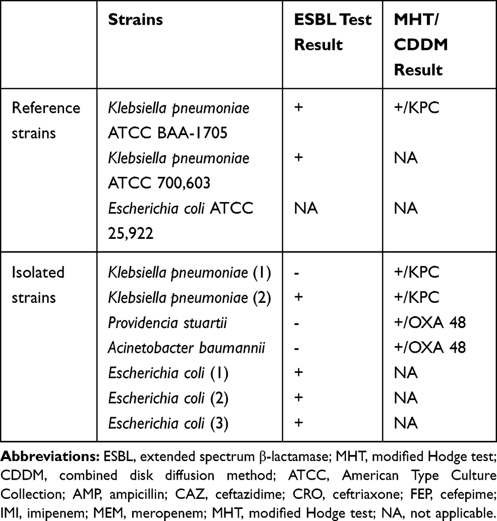 |
Table 1 Strain Description with Phenotypic Confirmation of ESBL and Carbapenemase Production |
Hydrolysis Test
The bacterial strains were grown on Columbia 5% sheep blood agar for 24 h at 37°C. Suspensions of bacteria culture were prepared in 100 mM phosphate buffer pH 7, containing 10 µg/mL ZnCl2. The turbidity was adjusted equivalent to McFarland 4 using a densitometer (Densimat, bioMérieux, Italy). In a 1.5 mL centrifuge tube, 100 µL of bacteria suspension was mixed with 10 µL of antibiotic reaction mix (containing 5 mg/mL ampicillin and 0.5 mg/mL ceftriaxone, ceftazidime, cefepime, meropenem and imipenem) and then incubated for 2 h at 37°C. A negative control was also prepared by incubating, under the same conditions, 100 µL of phosphate buffer with 10 µL of the mixture of antibiotics. An aliquot (200 µL) of ice-cold acetonitrile was subsequently added and the samples were centrifuged at 10,000 g for 5 min. The supernatant (100 µL) was mixed with 300 µL HPLC-grade water and an aliquot was analyzed using LC-MS/MS.
LC-MS/MS Analysis
The LC-MS/MS system used comprised of a Shimadzu Nexera X2 UHPLC hyphenated to a LCMS-8045 triple quadrupole mass spectrometer (Shimadzu Corporation, Kyoto, Japan). The chromatographic separation was achieved using an Onyx Monolithic C18 column (100 x 3 mm) from Phenomenex (Torrance, CA, USA) equipped with an Onyx Monolithic C18 guard cartridge (5 x 3 mm) also from Phenomenex. A binary gradient consisting of “A” - 0.1% formic acid in water and “B” - 0.1% formic acid in acetonitrile was used. The elution profile started at 15% B then increased to 75% over 1 min, it was maintained at this level for 2 min and then returned to 15% B over 30 sec. The column was equilibrated at this level of B for 6.5 min between injections. The flow rate was set to 0.3 mL/min. Column oven and autosampler temperature was set to 40 and 5°C respectively. An injection volume of 5 µL used was. The mass spectrometer was equipped with an electrospray ionization (ESI) source and operated in a positive mode. The MRM transitions (Table 2) were optimized by direct infusion of the standards at a concentration of 10 µg/mL.
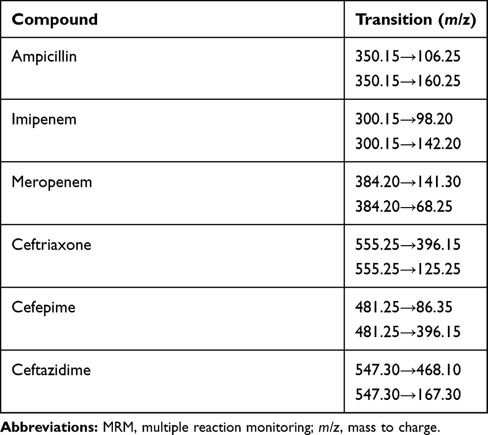 |
Table 2 The Optimized MRM Transitions for Each Antibiotic |
Calibration standards were prepared in the concentration range 0.625 to 10 µg/mL for ampicillin; and 0.0625 to 1 µg/mL for meropenem, imipenem, ceftazidime, ceftriaxone and cefepime.
The MS data were acquired and processed using LabSolution® software (v 5.91/2017, Shimadzu Corporation, Kyoto, Japan). Integration of HPLC-peaks was performed using the i-PeakFinder algorithm for all compounds. Spectra were smoothed (standard method - 5 counts and width - 5 sec) and correction was applied to the baseline (1-degree baseline following degree). Peak identification was performed using absolute retention time (RT). Reference ion ratio allowance was set to 30%. Quantification was performed using an external calibration with linear fit and not forced though zero.
Test Validation
The LC-MS/MS validation procedure included linearity, limit of detection (LOD), limit of quantification (LOQ), accuracy, precision. The LOD and LOQ were automatically calculated using the instrument software. The lowest concentration detectable with a signal-to-noise (S/N) ratio of ≥ 3:1 was used to establish the LOD, whilst the lowest concentration measurable with a signal-to-noise (S/N) ratio of ≥ 10:1 was used to establish the LOQ.
Accuracy was determined by carrying out ten repeated injections of each calibration standards. Precision was assessed by carrying out five repeated injection of one sample. Repeatability of the hydrolysis test was assessed by repeating the incubation five times using E. coli ATCC 25,922 and K. pneumoniae ATCC BAA-1705 strains and determining the antibiotic concentration from each test tube. The relative standard deviation (RSD%) of peak area was calculated for each antibiotic. This was used to estimate the standard uncertainty of the measurement. All values were calculated using LabSolution software (v 5.91/2017, Shimadzu, Corporation, Kyoto, Japan).
Data Interpretation
The sensitive E. coli ATCC 25,922 strain was used as negative control, the antibiotic concentrations obtained after incubation with this strain were considered as reference. The level of antibiotic detected in samples where bacteria were incubated with mixture of antibiotics was calculated relative to the negative control and expressed as percentage of hydrolyzed antibiotic.
The RSD% values from test validation was used to establish the limit where hydrolysis is considered present. For this purpose, the precision test results were used to establish the range of uncertainty out of which the hydrolysis was considered to be present. For example, for an antibiotic in which the accuracy of the test result was 10%, β-lactamase catalyzed hydrolysis was considered present if less than 90% could be detected. If more than 90% of the antibiotic could be detected, we deemed the organism not to have any β-lactamase activity.
The specificity of the LC-MS/MS antibiotic hydrolysis assay were evaluated using IBM-SPSS version 25 (IBM, Armonk, New York, USA) using Crosstabs. The MIC test results were used as reference.
Ethics Statement
This study was approved by the Ethics Committee of the “Pius Branzeu” Timisoara Emergency Clinical County Hospital (ref. no. 175/18 Oct. 2019).
Results
A multiple reaction monitoring (MRM) method was developed for all antibiotics. Two transitions per compound were used: one was used for quantification ion and the other for confirmation. All antibiotics eluted within 3 min and chromatograms showed good peak shapes. The MRM chromatograms for standards solution (5 µg/mL for ampicillin, and 0.5 µg/mL for imipenem, meropenem, ceftriaxone, cefepime and ceftazidime) are shown in Figure 1. LODs (S/N ≥ 3:1) were situated in 0.05–2.38 ng/mL interval while LOQs (S/N ≥ 3:1) were in 0.015–7.2 ng/mL interval. The five-point calibration curves showed a very good concentration/response correlation with R-squared value for all analytes being greater than 0.98. The retention time, LODs, LOQs, and results for accuracy and precision tests are presented in Table 3.
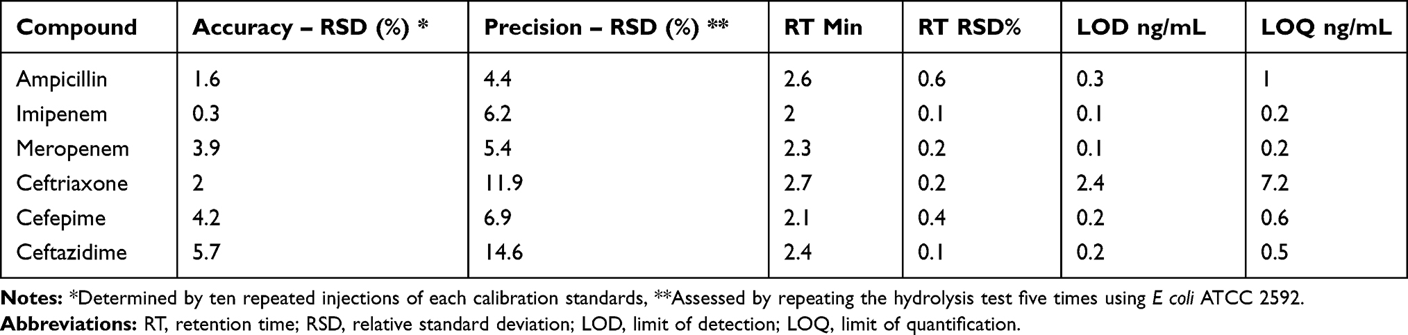 |
Table 3 Accuracy, Precision, Retention Time, LODs and LOQs for Each Antibiotic |
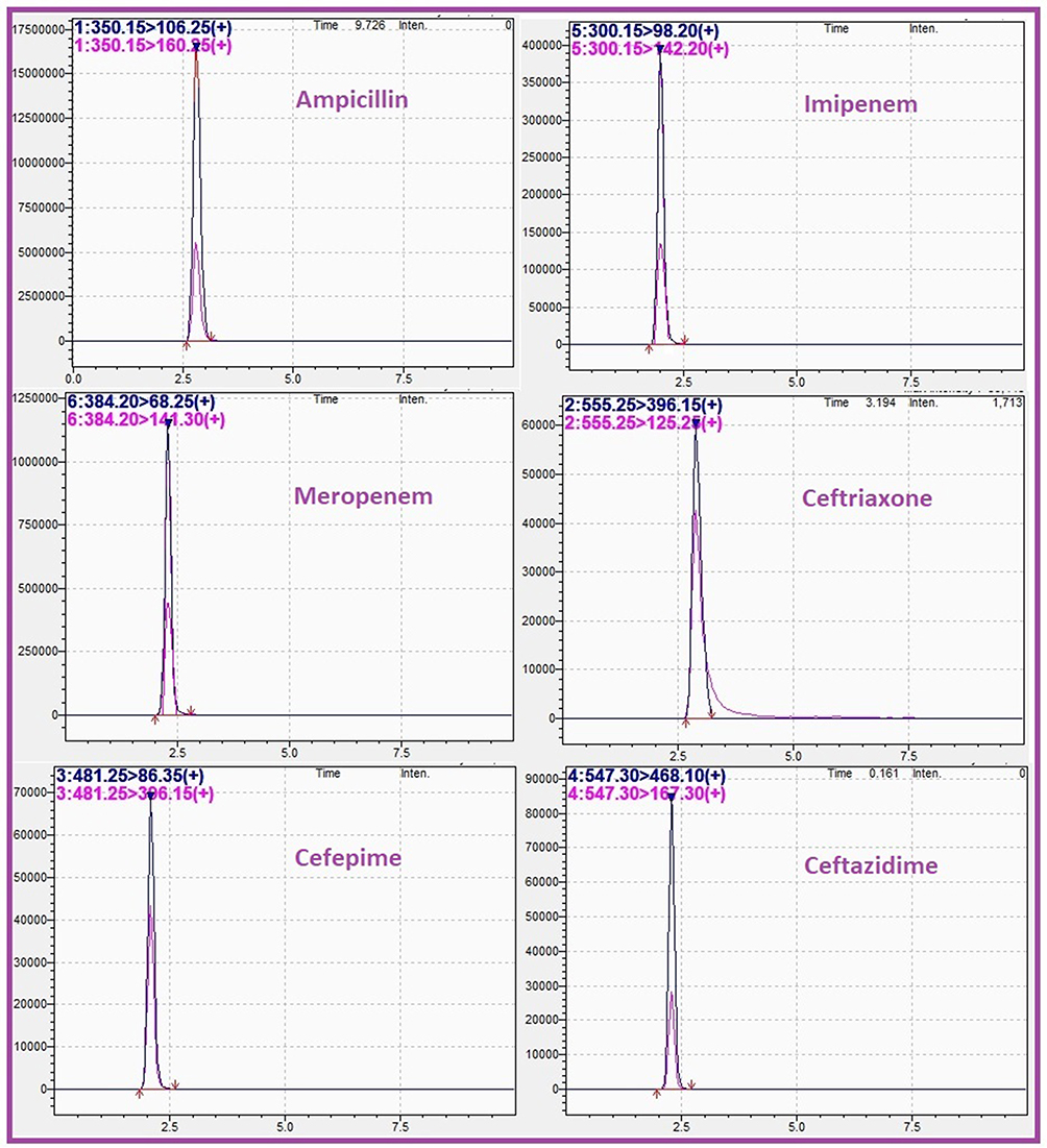 |
Figure 1 MRM chromatograms of mixture of ampicillin (5 µg/mL), imipenem (0.5 µg/mL), meropenem (0.5 µg/mL), ceftriaxone (0.5 µg/mL), cefepime (0.5 µg/mL), and ceftazidime (0.5 µg/mL) dissolved in phosphate buffer. |
Using precision test (RSD%), the presence of β-lactamase activity was deemed present when more than 10% of antibiotic was hydrolyzed for ampicillin, imipenem, meropenem and cefepime; and more than 15% for ceftriaxone and ceftazidime. The results from the LC-MS/MS antibiotic hydrolysis assay compared with the minimum inhibitory concentration test for all organisms tested are shown in Table 4.
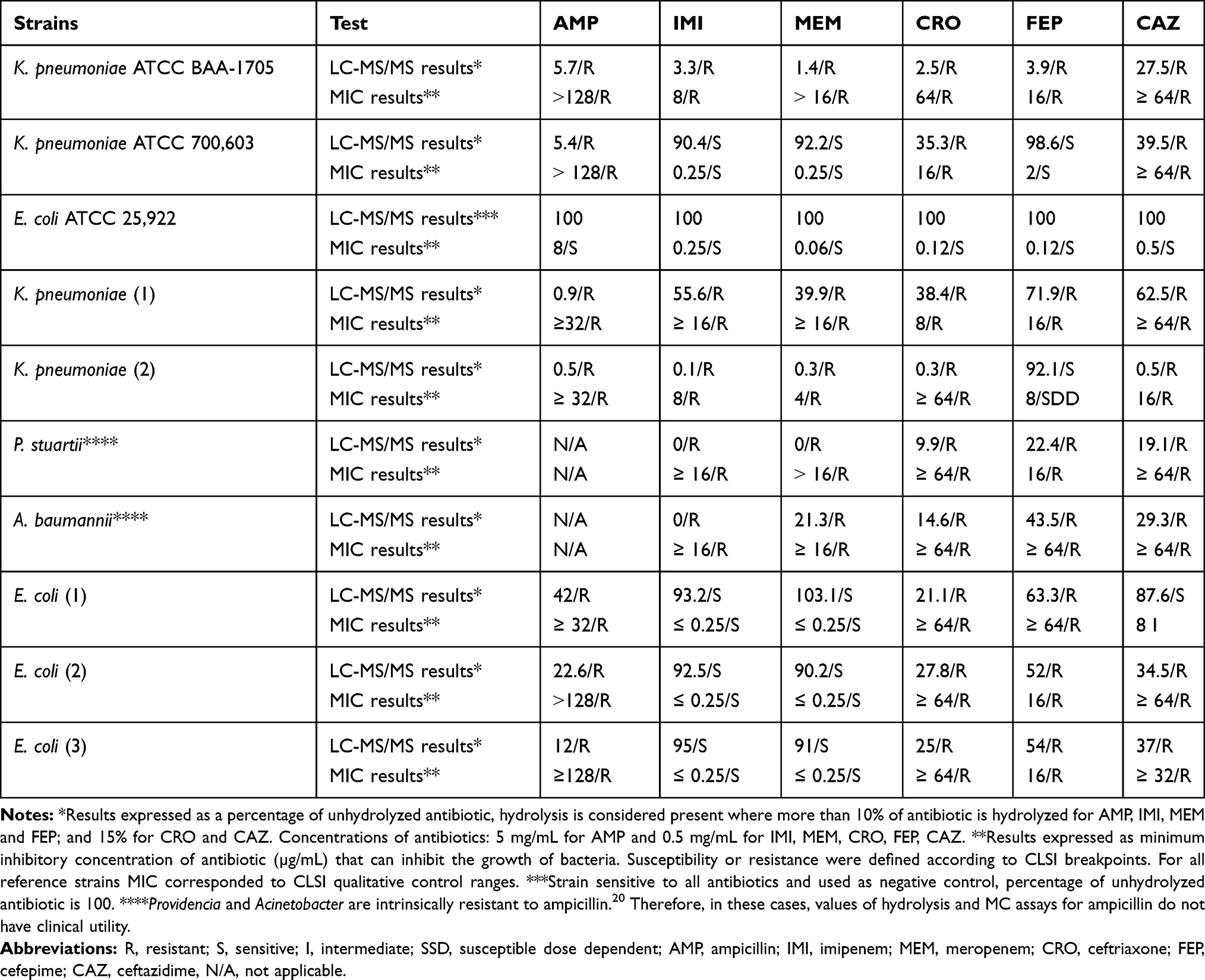 |
Table 4 LC-MS/MS Antibiotic Hydrolysis Assay Compared with the Minimum Inhibitory Concentration Test. For Antibiotic Hydrolysis Assay, The Strains Were Incubated for 2 h at 37°C with a Mixture of Antibiotics |
A comparison between hydrolysis levels of each antibiotic for three representative strains (ESBL- and carbapenemase-producing K. pneumoniae, carbapenemase-producing A. baumannii and ESBL-producing E. coli), are displayed in form of bar chart in Figure 2.
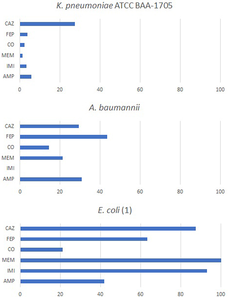 |
Figure 2 Hydrolysis level of each antibiotic for Klebsiella pneumoniae ATCC BAA-1705, Acinetobacter baumannii and Escherichia coli. |
The LC-MS/MS results showed good agreement with MIC test, except for one K. pneumoniae strain for which the LC-MS/MS result was sensitive to cefepime compared to MIC test where the result was sensitive dose dependent (SDD), and one E. coli strain for which the LC-MS/MS result was sensitive to ceftazidime compared to MIC test where the result was intermediate. The assay specificity was 100% for ampicillin, meropenem, imipenem and ceftriaxone; and 87.5% for cefepime and ceftazidime.
Discussion
The evidence of a high risk of increased mortality among patients with systemic infections, in the event of a failure to control the pathogen during the first 24–48h23–25 was an important reason for conducting this study. For this purpose, a fast LC-MS/MS assay was developed and applied to detect β-lactamases activity in Escherichia coli, Klebsiella pneumoniae, Providencia stuartii and Acinetobacter baumannii clinical isolates based on their ability to hydrolyze β-lactams in solution.
Although detection of resistance to carbapenems and cephalosporins in Gram-negative bacilli using LC-MS/MS was previously described,16–19 the present study brings a new approach for results interpretation and a simultaneous detection of hydrolysis of six antibiotics from three different classes of β-lactams. Selection of antibiotics and bacterial strains was based on the findings from previous studies in which numerous strains of Enterobacterales and Acinetobacter baumannii with acquired resistance to broad-spectrum cephalosporins and carbapenems were reported.1,2 We also used two strains with intermediate resistance to highlight the limitation of the assay.
During method validation stage, our key aim was to assess the precision of the assay. This was considered important mainly for avoiding false positive results. For this purpose and to ensure that the results were reproducible, we repeated the hydrolysis experiment and subsequent LC-MS/MS analysis on different days. The range of uncertainty established in the validation study was used for result interpretation to help decide if hydrolysis is present. The current study proposed a protocol which implies the use of a reference strain sensitive to all antibiotics used. Furthermore, the degree of β-lactamase activity for an organism was calculated relative to this negative control, thus accounting for auto hydrolysis and matrix interferences. The interpretation of results can be easily automated by using a formula that uses test sample and control measurement together with range of uncertainty (%), thus eliminating any subjective interpretation.
The concentration of antibiotics used for determining β-lactamase activity that had been established in previous work15 was used with minor changes. A less concentrated bacterial suspension (McFarland 1) and a shorter incubation (1h) were also evaluated in the current study (results not shown). We found that using less concentrated bacterial suspension and shorter incubation as previously reported16–18 did not either produce detectable level of hydrolysis of any of the antibiotics or the result was not conclusive. The reason for this may be attributable to different experiment conditions, such as the number of antibiotics used. In the present study, a greater number of antibiotics were used for the hydrolysis test which required more enzyme and thus a more concentrated bacteria suspension. Although a longer incubation time is required in the current study, it can be used to measure enzyme activity of an organism towards several antibiotics simultaneously.
The protocol developed in the current study could reduce antibiotic resistance testing turnaround time by several hours, time that could be decisive in disease outcome. We estimated that the time required for completing the test, including sample preparation and incubation, is between 3 to 5 h, time that could be influenced by the number of samples and operator experience. In comparison, MIC test using the commercial antimicrobial susceptibility platform can take up to 8 h to complete.26 Estimated advantages and disadvantages of the two methods are shown in Table 5.
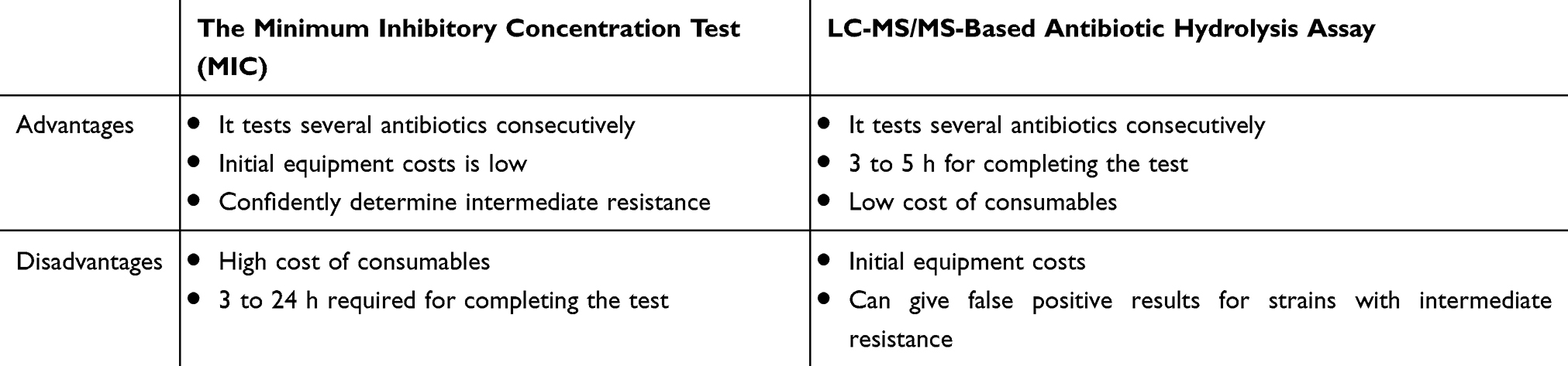 |
Table 5 Advantages and Disadvantages of Minimum Inhibitory Concentration Test and LC-MS/MS-Based Antibiotic Hydrolysis Assay |
The discrepancies with the MIC test for intermediate and SDD strains could be explained by the fact that the MIC is a dynamic test where the bacteria are maintained in a nutrient environment, where the enzyme is constantly being produced. In comparison, the sample of bacteria used in the hydrolysis test contains a fixed level of enzyme. The finding that one K. pneumoniae SDD to cefepime strain was diagnosed as sensitive using the LC-MS/MS test could represent a limitation especially because it is known that a high proportion of K. pneumoniae strains fall into SDD category.27 Therefore, we consider that further research should be done using several intermediate and SDD strains in combination with incubation in nutrient media to confirm if this is a limitation. This could provide more consistent results that meet the CLSI criteria-compliant MIC testing. The calculated specificity in the case of cefepime and ceftazidime may not reflect the performance of the assay very well since the intermediate and SSD strains give a different result. Therefore, future studies should include an appropriate statistical method to assess the assay performance. Also, the fact that we did not have reference strains for every isolated species could be a limitation.
Conclusions
The present study proposes a protocol for detecting β-lactamase activity in Gram-negative bacilli with improved robustness and multiplexing over previously described LC-MS/MS- and MALDI-based protocols. This has been achieved by using sensitive strains to validate the results in every experiment and using six antibiotics from three different classes. However, we estimate that for a broad assimilation of the method for routine testing, further research is needed, especially for strains with intermediate resistance.
Disclosure
The authors report no conflicts of interest in this work.
References
1. Muntean D, Horhat FG, Badiţoiu L, et al. Multidrug-resistant gram-negative bacilli: a retrospective study of trends in a tertiary healthcare unit. Medicina. 2018;54(6):92 doi:10.3390/MEDICINA54060092
2. Muntean D, Licker M, Horhat F, et al. Extensively drug-resistant Acinetobacter baumannii and proteeae association in a Romanian intensive care unit: risk factors for acquisition. Infect Drug Resist. 2018;11:2187–2197. doi:10.2147/IDR.S171288
3. Licker M, Sorescu T, Rus M, et al. Extensively drug-resistant Myroides odoratimimus – a case series of urinary tract infections in immunocompromised patients. Infect Drug Resist. 2018;11:743–749. doi:10.2147/IDR.S161069
4. Marchaim D, Navon-Venezia S, Schwaber MJ, Carmeli Y. Isolation of imipenem-resistant Enterobacter species: emergence of KPC-2 carbapenemase, molecular characterization, epidemiology, and outcomes. Antimicrob Agents Chemother. 2008;52(4):1413–1418. doi:10.1128/AAC.01103-07
5. Zhou M, Wang Y, Liu C, et al. Comparison of five commonly used automated susceptibility testing methods for accuracy in the China antimicrobial resistance surveillance system (CARSS) hospitals. Infect Drug Resist. 2018;11:1347–1358. doi:10.2147/IDR.S166790
6. Bobenchik AM, Deak E, Hindler JA, Charlton CL, Humphries RM, Carroll KC. Performance of Vitek 2 for antimicrobial susceptibility testing of Enterobacteriaceae with Vitek 2 (2009 FDA) and 2014 CLSI breakpoints. J Clin Microbiol. 2015;53(3):816–823. doi:10.1128/JCM.02697-14
7. Ligozzi M, Bernini C, Bonora MG, De Fatima M, Zuliani J, Fontana R. Evaluation of the VITEK 2 system for identification and antimicrobial susceptibility testing of medically relevant gram-positive cocci. J Clin Microbiol. 2002;40(5):1681–1686. doi:10.1128/JCM.40.5.1681-1686.2002
8. Mittman SA, Huard RC, Della-Latta P, Whittier S. Comparison of BD phoenix to Vitek 2, MicroScan MICroSTREP, and Etest for antimicrobial susceptibility testing of Streptococcus pneumoniae. J Clin Microbiol. 2009;47(11):3557–3561. doi:10.1128/JCM.01137-09
9. Idelevich EA, Sparbier K, Kostrzewa M, Becker K. Rapid detection of antibiotic resistance by MALDI-TOF mass spectrometry using a novel direct-on-target microdroplet growth assay. Clin Microbiol Infect. 2018;24(7):738–743. doi:10.1016/j.cmi.2017.10.016
10. Camara JE, Hays FA. Discrimination between wild-type and ampicillin-resistant Escherichia coli by matrix-assisted laser desorption/ionization time-of-flight mass spectrometry. Anal Bioanal Chem. 2007;389(5):1633–1638. doi:10.1007/s00216-007-1558-7
11. Carvalhaes CG, Cayô R, Visconde MF, et al. Detection of carbapenemase activity directly from blood culture vials using MALDI-TOF MS: a quick answer for the right decision. J Antimicrob Chemother. 2014;69(8):2132–2136. doi:10.1093/jac/dku094
12. Hrabák J, Chudáčková E, Papagiannitsis CC. Detection of carbapenemases in Enterobacteriaceae: a challenge for diagnostic microbiological laboratories. Clin Microbiol Infect. 2014;20(9):839–853. doi:10.1111/1469-0691.12678
13. Dortet L, Tandé D, De Briel D, et al. MALDI-TOF for the rapid detection of carbapenemase-producing Enterobacteriaceae: comparison of the commercialized MBT STAR®-Carba IVD Kit with two in-house MALDI-TOF techniques and the RAPIDEC® CARBA NP. J Antimicrob Chemother. 2018;73(9):2352–2359. doi:10.1093/jac/dky209
14. Hrabák J, Walková R, Študentová V, Chudáčková E, Bergerová T. Carbapenemase activity detection by matrix-assisted laser desorption ionization-time of flight mass spectrometry. J Clin Microbiol. 2011;49(9):3222–3227. doi:10.1128/JCM.00984-11
15. Shah AJ, Vlad S, Xu Z, Mkrtchyan HV, Shah HN. Determination of antimicrobial resistance using tandem mass spectrometry. In: Shah HN, Gharbia SE, editors. MALDI-TOF and Tandem MS for Clinical Microbiology.
16. Tadros M, Goneau L, Romaschin A, Jarvis M, Matukas L, Gonzalez-Bello C. Rapid detection of resistance to carbapenems and cephalosporins in Enterobacteriaceae using liquid chromatography tandem mass spectrometry. PLoS One. 2018;13(11):e0206842. doi:10.1371/journal.pone.0206842
17. Kulkarni MV, Zurita AN, Pyka JS, Murray TS, Hodsdon ME, Peaper DR. Use of imipenem to detect KPC, NDM, OXA, IMP, and VIM carbapenemase activity from gram-negative rods in 75 minutes using liquid chromatography-tandem mass spectrometry. J Clin Microbiol. 2014;52(7):2500–2505. doi:10.1128/JCM.00547-14
18. Peaper DR, Kulkarni MV, Tichy AN, Jarvis M, Murray TS, Hodsdon ME. Rapid detection of carbapenemase activity through monitoring ertapenem hydrolysis in Enterobacteriaceae with LC–MS/MS. Bioanalysis. 2013;5(2):147–157. doi:10.4155/bio.12.310
19. Carricajo A, Verhoeven PO, Guezzou S, Fonsale N, Aubert G. Detection of carbapenemase-producing bacteria by using an ultra-performance liquid chromatography-tandem mass spectrometry method. Antimicrob Agents Chemother. 2014;58(2):1231–1234. doi:10.1128/AAC.01540-13
20. Wayne P. Clinical and Laboratory Standards Institute. Performance Standards for Antimicrobial Susceptibility Testing; 29th Ed. CLSI Supplement M100. Clin Lab Stand Institute; 2019.
21. Miriagou V, Tzelepi E, Kotsakis SD, Daikos GL, Bou Casals J, Tzouvelekis LS. Combined disc methods for the detection of KPC- and/or VIM-positive Klebsiella pneumoniae: improving reliability for the double carbapenemase producers. Clin Microbiol Infect. 2013;19(9):E412–E415. doi:10.1111/1469-0691.12238
22. Clinical and Laboratory Standards Institute. 2019. doi:10.1007/978-3-662-48986-4_300416
23. Blot S. Limiting the attributable mortality of nosocomial infection and multidrug resistance in intensive care units. Clin Microbiol Infect. 2008;14(1):5–13. doi:10.1111/j.1469-0691.2007.01835.x
24. Depuydt P, Blot S. Antibiotic therapy for ventilator-associated pneumonia: de-escalation in the real world. Crit Care Med. 2007;35(2):632–633. doi:10.1097/01.CCM.0000254049.23884.D5
25. Colardyn F. Appropriate and timely empirical antimicrobial treatment of ICU infections - a role for carbapenems. Acta Clin Belg. 2005;60(2):51–62. doi:10.1179/acb.2005.011
26. Truant AL, Yi‐Wei Tang KB. Manual of Commercial Methods in Clinical Microbiology: International Edition.
27. Menegucci TC, Bastos MDS, Viana GF, et al. What does the susceptible-dose dependent interpretive category for cefepime in ESBL-producing bacteria? J Chemother. 2017;29(3):189–194. doi:10.1080/1120009X.2015.1120407
 © 2020 The Author(s). This work is published and licensed by Dove Medical Press Limited. The full terms of this license are available at https://www.dovepress.com/terms.php and incorporate the Creative Commons Attribution - Non Commercial (unported, v3.0) License.
By accessing the work you hereby accept the Terms. Non-commercial uses of the work are permitted without any further permission from Dove Medical Press Limited, provided the work is properly attributed. For permission for commercial use of this work, please see paragraphs 4.2 and 5 of our Terms.
© 2020 The Author(s). This work is published and licensed by Dove Medical Press Limited. The full terms of this license are available at https://www.dovepress.com/terms.php and incorporate the Creative Commons Attribution - Non Commercial (unported, v3.0) License.
By accessing the work you hereby accept the Terms. Non-commercial uses of the work are permitted without any further permission from Dove Medical Press Limited, provided the work is properly attributed. For permission for commercial use of this work, please see paragraphs 4.2 and 5 of our Terms.