")
Back to Journals » Clinical, Cosmetic and Investigational Dermatology » Volume 12
Dermoscopic pattern of pityriasis versicolor
Authors Mathur M , Acharya P , Karki A, KC N, Shah J
Received 25 November 2018
Accepted for publication 5 April 2019
Published 30 April 2019 Volume 2019:12 Pages 303—309
DOI https://doi.org/10.2147/CCID.S195166
Checked for plagiarism Yes
Review by Single anonymous peer review
Peer reviewer comments 4
Editor who approved publication: Dr Jeffrey Weinberg
Mahesh Mathur,* Prakash Acharya,* Alina Karki, Nisha KC, Jyoti Shah
Department of Dermatology, College of Medical Sciences, Bharatpur, Nepal
*These authors contributed equally to this work
Background: Pityriasis versicolor (PV) is essentially a clinical diagnosis characterized by hypopigmented or hyperpigmented patches on the skin. Dermoscopy is gaining popularity as a noninvasive procedure for the diagnosis of different pigmentary and inflammatory disorders. However, scarce evidence exists on the dermoscopic pattern of PV.
Objective: To describe the dermoscopic features of hypopigmented and hyperpigmented lesions of PV.
Methods: Dermoscopic images of PV lesions located on different body sites were retrospectively evaluated for the presence of predefined criteria.
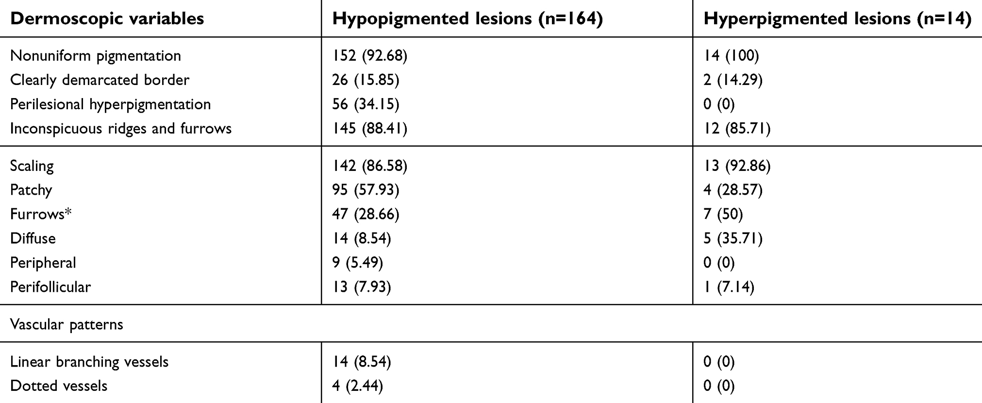
Results: A total of 178 lesions from 125 patients were included in the study among which 164 lesions were hypopigmented and 14 lesions were hyperpigmented. Nonuniform pigmentation was the most common dermoscopic feature seen in both hypopigmented lesions (n=152, 92.68%) and hyperpigmented lesions (n=14, 100%). Scales were seen in 142 hypopigmented lesions (86.56%) and 13 hyperpigmented lesions (92.86%). Patchy scaling was more common in hypopigmented lesions (n=95, 57.92%) while scaling in the furrows was more common in the dermoscopy of hyperpigmented lesions (n=5, 35.71%). Inconspicuous ridges and furrows and perilesional hyperpigmentation were other significant features seen in dermoscopy of the lesions.
Conclusion: To our knowledge, this is the first study describing the dermoscopic features of PV in such a large number of patients. Description of these new features adds valuable information and may help to establish dermoscopy as an important auxiliary tool for the diagnosis of PV.
Keywords: dermoscopy, dermatoscopy, tinea versicolor, diagnosis, hypopigmented patches
Introduction
Pityriasis versicolor (PV) or tinea versicolor is a fungal infection of the superficial layer of skin caused by Malassezia yeasts. It is clinically characterized by hyperpigmented or hypopigmented, round to oval lesions commonly found on the trunk, upper arms and face.1 Although some patients may complain of mild pruritus, the disease is often asymptomatic. The patients usually seek medical consultation for the cosmetic appearance associated with this condition.2
PV is usually a clinical diagnosis and microscopic examination is useful when the diagnosis is uncertain.3 Microscopic examination of the slides prepared with potassium hydroxide reveal characteristic “spaghetti and meatball appearance” which represent hyphae and spores of the fungus.3 The list of differential diagnosis is broad and includes disorders like vitiligo, pityriasis rosea, seborrhoeic dermatitis, pityriasis alba, chloasma, tinea corporis, erythrasma, secondary syphilis, confluent and reticulated papillomatosis, pityriasis rotunda, and pinta.2
Dermoscopy is emerging as a useful tool for the noninvasive diagnosis of various dermatological disorders.4 However, with the exception of a case report and a personal observation,4,5 no studies have been performed to describe the dermoscopic features of PV to our knowledge.
The aim of our study was to describe the dermoscopic pattern of hypopigmented and hyperpigmented lesions of PV.
Material and methods
This was a retrospective hospital-based study carried out at the dermatology department of College of Medical Sciences, Bharatpur, Nepal, from April 2018 to September 2018. This study was conducted in accordance with the declaration of Helsinki and was approved by the institutional ethical committee. The patients were explained about the study and written informed consent was obtained from all the included patients before study procedures. All clinically suspected cases whose diagnosis was confirmed by the potassium hydroxide (KOH) examination were included in the study. Patients who did not consent to the study and those who had a history of a topical application within six months prior to the dermoscopic evaluation were excluded from the study.
Dermoscopic images were taken using a Firefly DE300 handheld dermoscope (Firefly Global, MA, USA) attached to a MacBook Pro 2013 (Apple Inc., CA, USA) in a polarized mode at 10× magnification without a contact medium. Two independent dermoscopists (M.M.,P.A.) analyzed the dermoscopic patterns in the images. Due to the lack of literature describing dermoscopic patterns of PV, the dermoscopic variables were selected on the basis of published literature for other pigmentary disorders and the dermoscopists’ experience. These variables included nonuniform pigmentation, clearly demarcated border, perilesional hyperpigmentation, inconspicuous ridges and furrows, scaling and vascularity. More than one image was taken from a single patient only if a different dermoscopic pattern was seen.
Results
A total of 125 patients having PV were included in the study. A male predominance (n=74, 59.2%) with the male:female ratio of 1.45:1 was observed. The patients were between 3 and 68 years of age with the mean age of 23.21 years. Of the total 178 lesions evaluated from these patients, 164 lesions were hypopigmented and 14 lesions were hyperpigmented. Eighty-six of 178 lesions (48.31%) were located on the trunk, 55 (30.9%) on the extremities and 37 (20.79%) on the face. Dermoscopic features of the lesions seen in the study are shown in Table 1, and some common patterns are illustrated in Figure 1.
 | Table 1 Frequency of dermoscopic variables in the lesions of pityriasis versicolor |
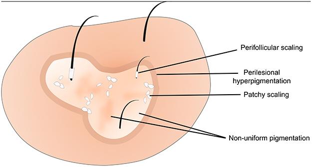 | Figure 1 Illustration depicting dermoscopic features of a typical hypopigmented lesion of pityriasis versicolor. |
Nonuniform pigmentation (Figures 2–5) was the most common dermoscopic feature seen in both hypopigmented lesions (n=152, 92.68%) and hyperpigmented lesions (n=14, 100%). Among the various patterns of scaling (Figure 3), patchy scaling (Figure 3A) was more frequently observed in hypopigmented lesions (n=95, 57.92%) while scaling in the furrows (Figure 3B) was more frequent in the dermoscopy of hyperpigmented lesions (n=5, 35.71%). Inconspicuous ridges and furrows (Figures 2A, 3A, D, and 4), perilesional hyperpigmentation (Figures 2A, 3A, and 4A) and varied vascular patterns (Figure 4) were the other dermoscopic features of the lesions.
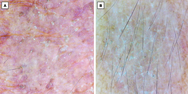
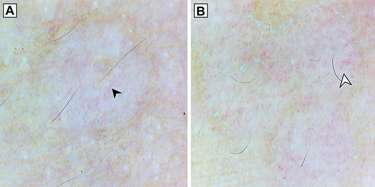
 | Figure 2 (A) Dermoscopy (original magnification 10×) of hypopigmented lesion showing nonuniform pigmentation, inconspicuous ridges and furrows, perilesional hyperpigmentation (white arrowhead) and patchy scaling (black arrowhead). (B) Dermoscopy (original magnification 10×) of hyperpigmented lesion showing non-uniform pigmentation. |
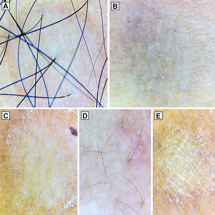 | Figure 3 Dermoscopy (original magnification 10×) showing different patterns of scaling in pityriasis versicolor. (A) Patchy scaling in hypopigmented lesion. (B) Diffuse scaling in hyperpigmented lesion with the simultaneous presence of scaling in the furrows. (C) Peripheral scaling. (D) Perifollicular scaling. (E) Scaling in the furrows. Note the nonuniform pigmentation in all the lesions, perilesional hyperpigmentation in (A) and inconspicuous ridges and furrows in (A), (D). |
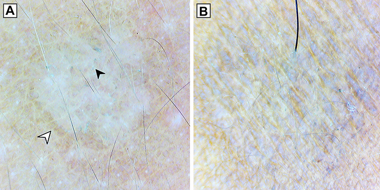
 | Figure 4 Dermoscopy (original magnification 10×) showing different vascular patterns in pityriasis versicolor. (A) Linear branching vessels (black arrowhead). (B) Dotted vessels (white arrowhead). Note perilesional hyperpigmentation in (A), peripheral scaling in (B) and inconspicuous ridges and furrows in both (A) and (B). |
Discussion
Malassezia yeasts are the members of the normal skin flora which may be associated with different skin dermatoses like PV, malassezia folliculitis, seborrheic dermatitis, dandruff and atopic dermatitis under favorable conditions.6 Akaza et al6 in their study of cutaneous Malassezia microbiota showed that the number of cutaneous Malassezia species varied according to sex and body part due to the differences in properties of sebum and sweat. They reported that the increased number of living Malassezia cells during summer may be the reason for the increased incidence of PV during summer months and in tropical regions.
PV is usually a clinical diagnosis because of the characteristic clinical appearance and distribution of the lesions.3 However, atypical cases may pose a diagnostic dilemma in an inexperienced physician.2 The present study describes the various features observed in the dermoscopy of both the hypopigmented and hyperpigmented lesions of PV.
Dermoscopy of PV is not well described in the literature. The presence of fine scales in the dermoscopy of PV lesions has been reported by Zhou et al5 and Errichetti and Stinco4. Zhou et al5 described the ability of a dermatoscope to visualize the fine scales in PV lesions which could help to differentiate PV from other hyperpigmented disorders.
In our study, nonuniform pigmentation within a lesion was the most common dermoscopic feature seen in both the hypopigmented and hyperpigmented lesions. This was characterized by lighter and darker areas within a lesion. A clearly demarcated border separating the lesion from normal skin was present in only 15.85% (n=26) of hypopigmented lesions and 15.28% (n=2) of hyperpigmented lesions. This finding is consistent with the observation made by Errichetti et al4 who described fairly demarcated white area in the hypopigmented lesions of PV compared to the clearly demarcated lesions of guttate vitiligo. In addition, we found that the lesions seemed to radially distribute from the hair follicles and as they progressed, they coalesced with other lesions to form different shapes. Satellite phenomenon was also observed in which a larger lesion was surrounded by smaller lesions (Figure 5).
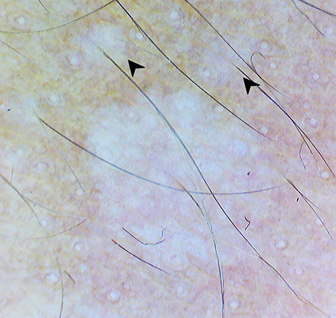 | Figure 5 Dermoscopy (original magnification 10×) showing radial distribution of the lesion from hair follicle and satellite lesions (black arrowhead). |
Perilesional or marginal hyperpigmentation was another feature noted in the hypopigmented lesions observed in our study. Kumar Jha et al7 reported this feature in 23.3% of the lesions of stable and repigmenting vitiligo and described it as a hallmark feature of repigmenting vitiligo. Similar frequency (34.15%) was noted in the hypopigmented lesions of PV in our study. The frequency of this feature in PV has not been reported in literature.
Presence of scales was another prominent dermoscopic finding in both the hypopigmented and hyperpigmented lesions in our study. Mild patchy scaling was the most frequent type of scaling followed by a more severe diffuse kind of scaling. White scales in the furrows were present either as isolated scaling pattern or simultaneously present with patchy or diffuse scaling pattern. The other scaling patterns noted in our study were perilesional scaling and perifollicular scaling. Perilesional scaling pattern may also be seen in the lesions of pityriasis rosea and secondary syphilis which are among the important differential diagnosis of PV.8
Hyperpigmented lesions are characterized by a higher number of organisms and more pronounced inflammatory infiltrate compared to the hypopigmented lesions.9 In our study, we found that the diffuse kind of scaling was more frequently seen in hyperpigmented lesions compared to the hypopigmented lesions (35.71% vs 8.54%). This increased severity of scaling in the hyperpigmented lesions probably suggests that the disease activity can be correlated with the amount of scaling in the lesions. A case of chronic renal failure (male of 68 years of age) and another case of systemic lupus erythematosus (female of 32 years of age) who were taking oral steroids for their disease displayed extensive scaling on the lesions of PV on face and trunk, respectively (Figure 6). Malassezia lies within the stratum corneum where the immune response is likely minimal; however, it has been suggested that the patients receiving oral corticosteroids or immunocompromised patients may have superficial fungal infections that are more prevalent or severe.2 This could explain the extensive scaling in the lesions of immunocompromised individuals in our study.
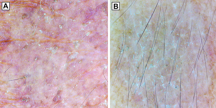 | Figure 6 Dermoscopy (original magnification 10×) showing diffuse scaling pattern in cases of (A) chronic renal failure and (B) systemic lupus erythematosus taking oral corticosteroids. |
Fine scales in the PV lesions are not easily visible to an unaided eye.3 Similarly, perifollicular and perilesional scaling are also difficult to appreciate without aid. As seen in our study, dermoscopy can be a useful tool to demonstrate such scales and provide an important clue to differentiate PV from the other pigmentary disorders like vitiligo, pityriasis alba and progressive macular hypomelanosis which lack scale.3,10 However, fine scales have been reported as a less common finding in dermoscopy of idiopathic guttate hypomelanosis.11
In our study, inconspicuous skin ridges and furrows was another common dermoscopic finding seen in both the hypopigmented and hyperpigmented lesions. Although common, this finding has not been reported or quantified in literature.
A visible vascular pattern within the lesion was less frequently seen in our study compared to the other dermoscopic patterns. Only hypopigmented lesions displayed this feature. In the absence of a history of topical application, this might probably be due to the ongoing inflammatory reaction to the causative agent.
Limitations
The lesions evaluated in our study were mostly from dark-skinned individuals of Fitzpatrick skin type IV and V. The lesions in light-skinned individuals may show different dermoscopic characteristics. We included only those cases who were confirmed by KOH examination; however, KOH examination is not 100% sensitive for PV.12 This could result in the exclusion of features seen in KOH-negative lesions. Another limitation of our study was the absence of a control group.
Conclusion
We describe new dermoscopic patterns in both the hypopigmented and hyperpigmented lesions of PV. Important features like scaling patterns, pigmentation patterns and border patterns are better visualized with a dermatoscope. These patterns could be useful to differentiate PV from other similar appearing disorders. Scaling patterns could also be used as a measure to assess the severity of the disease and might help the clinicians to decide the form of treatment. We conclude by saying that this study adds new information to the dermoscopic features of PV and may set the foundation for further studies on a larger and more diverse population with control groups.
Acknowledgments
Our sincere thanks to Dr. Ayush Jha who reviewed the dermoscopic features during the revision phase of this manuscript and Mr. Sishir Bhusal who revised the English language during the revision process.
Disclosure
The authors report no conflicts of interest in this work.
References
1. Pedrosa AF, Lisboa C, Rodrigues AG. Malassezia infections: a medical conundrum. J Am Acad Dermatol. 2014;71(1):170–176. doi:10.1016/j.jaad.2013.12.022
2. Gupta AK, Bluhm R, Summerbell R. Pityriasis versicolor. J Eur Acad Dermatol Venereol. 2002;16(1):19–33. doi:10.1046/j.1468-3083.2002.00378.x
3. Renati S, Cukras A, Bigby M. Pityriasis versicolor. BMJ. 2015;350:h1394. doi:10.1136/bmj.h1394
4. Errichetti E, Stinco G. Dermoscopy in general dermatology: a practical overview. Dermatol Ther (Heidelb). 2016;6(4):471–507. doi:10.1007/s13555-016-0141-6
5. Zhou H, Tang XH, De Han J, Chen M-K. Dermoscopy as an ancillary tool for the diagnosis of pityriasis versicolor. J Am Acad Dermatol. 2015;73(6):e205–e206. doi:10.1016/j.jaad.2015.08.058
6. Akaza N, Akamatsu H, Sasaki Y, et al. Cutaneous Malassezia microbiota of healthy subjects differ by sex, body part and season. J Dermatol. 2010;37(9):786–792. doi:10.1111/j.1346-8138.2010.00913.x
7. Kumar Jha A, Sonthalia S, Lallas A, Chaudhary RKP. Dermoscopy in vitiligo: diagnosis and beyond. Int J Dermatol. 2018;57(1):50–54. doi:10.1111/ijd.13795
8. Tognetti L, Sbano P, Fimiani M, Rubegni P. Dermoscopy of Biett’s sign and differential diagnosis with annular maculo-papular rashes with scaling. Indian J Dermatol Venereol Leprol. 2017;83(2):270–273. doi:10.4103/0378-6323.196318
9. Galadari I, El-Komy M, Mousa A, et al. Tinea versicolor: histologic and ultrastructural investigation of pigmentary changes. Int J Dermatol. 1992;31(4):253–256. doi:10.1111/j.1365-4362.1992.tb03565.x
10. Schwartz RA. Superficial fungal infections. Lancet. 2004;364(9440):1173–1182. doi:10.1016/S0140-6736(04)17107-9
11. Errichetti E, Stinco G. Dermoscopy of idiopathic guttate hypomelanosis. J Dermatol. 2015;42(11):1118–1119. doi:10.1111/jde.2015.42.issue-11
12. Shah A, Koticha A, Ubale M, Wanjare S, Mehta P, Khopkar U. Identification and speciation of Malassezia in patients clinically suspected of having pityriasis versicolor. Indian J Dermatol. 2013;58(3):239. doi:10.4103/0019-5154.110841
 © 2019 The Author(s). This work is published and licensed by Dove Medical Press Limited. The full terms of this license are available at https://www.dovepress.com/terms.php and incorporate the Creative Commons Attribution - Non Commercial (unported, v3.0) License.
By accessing the work you hereby accept the Terms. Non-commercial uses of the work are permitted without any further permission from Dove Medical Press Limited, provided the work is properly attributed. For permission for commercial use of this work, please see paragraphs 4.2 and 5 of our Terms.
© 2019 The Author(s). This work is published and licensed by Dove Medical Press Limited. The full terms of this license are available at https://www.dovepress.com/terms.php and incorporate the Creative Commons Attribution - Non Commercial (unported, v3.0) License.
By accessing the work you hereby accept the Terms. Non-commercial uses of the work are permitted without any further permission from Dove Medical Press Limited, provided the work is properly attributed. For permission for commercial use of this work, please see paragraphs 4.2 and 5 of our Terms.