")
Back to Journals » Drug Design, Development and Therapy » Volume 14
Curcumin Protects Against Radiotherapy-Induced Oxidative Injury to the Skin
Authors Shabeeb D , Musa AE , Abd Ali HS, Najafi M
Received 29 May 2020
Accepted for publication 24 July 2020
Published 5 August 2020 Volume 2020:14 Pages 3159—3163
DOI https://doi.org/10.2147/DDDT.S265228
Checked for plagiarism Yes
Review by Single anonymous peer review
Peer reviewer comments 2
Editor who approved publication: Professor Anastasios Lymperopoulos
Dheyauldeen Shabeeb,1,2 Ahmed Eleojo Musa,3 Hayder Shabeeb Abd Ali,4 Masoud Najafi5
1Department of Physiology, School of Medicine, University of Misan, Misan, Iraq; 2Misan Radiotherapy Center, Ministry of Health, Misan, Iraq; 3Department of Medical Physics, Tehran University of Medical Sciences, Tehran, Iran; 4Department of Physiology, Ministry of Education, Misan, Misan, Iraq; 5Department of Radiology and Nuclear Medicine, Kermanshah University of Medical Sciences, Kermanshah, Iran
Correspondence: Dheyauldeen Shabeeb
Department of Physiology, School of Medicine, University of Misan, Misan, Iraq
Tel +9647717879332
Email [email protected]
Ahmed Eleojo Musa
Department of Medical Physics, Tehran University of Medical Sciences, Tehran, Iran
Tel +989012843317
Email [email protected]
Objective: Side-effects to normal tissues reduce the therapeutic window of radiotherapy. During radiotherapy, the skin is inevitably exposed to doses of ionizing radiation, leading to varying degrees of skin damage. Natural antioxidants have been explored for their radioprotective potentials. Thus, the present study aimed to investigate the protective effect of curcumin against radiotherapy-induced oxidative damage to the skin.
Methods: Forty rats were divided into four groups as follows: vehicle control (without irradiation or drug treatment), treatment with 150 mg/kg curcumin, 10 Gy single dose irradiation only, and 150 mg/kg curcumin plus 10 Gy radiation (RC). In the treatment groups, each rat was treated orally with 150 mg/kg curcumin 1 day before irradiation to 3 consecutive days after irradiation. Weeks 1, 2, or 4 after irradiation, all rats were sacrificed and their skin tissues collected and frozen at − 80°C for the determination of malondialdehyde (MDA), catalase (CAT), superoxide dismutase (SOD), and glutathione peroxidase (GSH-Px) activity in skin tissues.
Results: Radiotherapy-induced oxidative injury to the skin was evidenced by elevated MDA levels as well as depleted CAT, SOD, and GSH-Px activities. However, the administration of curcumin before and after irradiation prevented radiotherapy-induced oxidative damage by significantly elevating the activities of antioxidant enzymes.
Conclusion: From the findings of the present study, curcumin showed potential for protection against radiotherapy-induced oxidative injury to the skin. However, future studies are required to evaluate its clinical efficacy.
Keywords: curcumin, radiotherapy, ionizing radiation, radiation protection, skin
Introduction
It is estimated that more than half of cancer patients receive radiation during their course of therapy, a process known as radiotherapy.1 In spite of the fact that radiotherapy has become a mainstay in cancer therapy, it could also lead to normal tissue toxicity. Ionizing radiation can generate reactive oxygen species (ROS) to attack genetic contents of cells including the nucleus and DNA. Normal tissue toxicity is a limiting factor for receiving sufficient radiation doses to kill tumors, thereby reducing the therapeutic window of radiotherapy.
During radiotherapy, the skin is usually the first organ of contact with ionizing radiation, hence, it is inevitably exposed to ionizing radiation. It has been estimated that about 90–95% of cancer patients experience acute skin reactions after radiotherapy.2,3 Thus, radiation-induced damage to the skin is a major complication from radiotherapy. The mechanism of ionizing radiation-induced skin toxicities has been attributed to apoptosis, mitotic catastrophe, and necrosis, which gives rise to the appearance of skin reactions such as inflammation, occurring a few weeks after radiotherapy.4 Apart from being a dose-limiting factor, this has a negative effect on patient’s quality-of-life post radiotherapy. Therefore, it is imperative that this important organ is protected from the deleterious effects of ionizing radiation.
In a bid to ensure protection of normal tissues against radiotherapy-induced toxicities, several studies have been conducted to evaluate the protective effects of different agents. However, due to side-effects from some of these chemical agents, there is a growing interest towards natural agents. Amongst the different types of natural agents, polyphenols such as curcumin are the most important and well known. Curcumin has been traditionally used for healing some diseases such as inflammation, thrombosis, and preventing hepatic disorders.5 Curcumin has shown potential as a chemopreventive agent in a Phase 1 clinical trial without side-effects.6 Thus, based on the promising protective effects of curcumin, the present study aimed to investigate its potential for radioprotection of the skin.
Methods
Ethics
The conducts of this experimental study was subjected to approval by the ethical committee of Tehran University of Medical Sciences (approval number: 35,116). Thus, their guidelines for the care and use of laboratory animals were strictly followed in this study.
Animals
Forty male wistar rats (8-week-old and weighing 180–210 g) were procured from the animal laboratory of the School of Medicine, Tehran University of Medical Sciences, Tehran, Iran. Before commencement of experiments, all animals were kept in plexiglas cages for 1 week for acclimatization under the following conditions: feeding with rodent chow diet and water, ambient temperature (21°C), relative humidity (50–70%), air-flow rate (15 exchanges per hour), in addition to a 12-hour light and dark cycle (7 am to 7 pm).
Study Design and Irradiation
Forty rats were randomly allocated into 4 groups (10 rats each) as follows; G1: vehicle control (VC) without drug treatment or irradiation, treatment with curcumin (C), treatment with radiation only (R), treatment with curcumin plus radiation (RC). Curcumin was dissolved in 20% ethanol to give a final concentration of 30 mg/mL. Each rat was treated orally with 1 mL curcumin (150 mg/kg) 1 day before irradiation to 3 consecutive days after irradiation. Before irradiation, rats were anesthetised via an intraperitoneal injection of ketamine 10% (80 mg/kg) and xylazine 2% (5 mg/kg). Afterwards, they were irradiated with a 10 Gy single dose to the abdominal and pelvis region using a cobalt-60 gamma ray source at a dose rate of 0.65 cGy per minute and a source to skin distance of 1 m. The choice of the curcumin dose was based on a previous study by Kolivand et al,7 while that of radiation was according to Jourdan et al.8 All rats were sacrificed 1, 2, or 4 weeks after irradiation. Afterwards, their skin tissues were carefully removed and frozen at −80°C.
These tissues were cut into small pieces, and homogenized in ice cold potassium phosphate buffer (PBS) (pH 7.4) to produce 10% homogenates. Afterwards, centrifugation was done at 4000 rpm for 15 minutes at 4°C. The supernatants were removed and serum enzymes were measured including malondialdehyde (MDA), catalase (CAT), superoxide dismutase (SOD), and glutathione peroxidase (GSH-Px). These biochemical parameters were evaluated using Zell Bio kit (Zell Bio, Germany) according to its instruction manual.
Measurement of MDA Level
MDA is a common marker of lipid peroxidation. The amount of lipid peroxidation was assessed by thiobarbituric acid reactive substances (TBARS) in the colon. MDA activity was determined colorimetrically at 532 nm.
Measurement of CAT Activity
CAT activity is considered as the sample that will catalyze 1 μmole of hydrogen peroxide to water and oxygen in 1 minute. Catalase activity was determined colorimetrically at 405 nm.
Measurement of GSH-Px Amount
GSH-Px amount is considered as the amount of sample that will catalyze the decomposition of 1 μmole of GSH to glutathione disulfide (GSSG) in 1 minute. GSH was determined colorimetrically at 412 nm.
Measurement of SOD Activity
SOD activity is expressed as the amount of the sample that will catalyze 1 μmole of superoxide radicals to hydrogen peroxide and oxygen in 1 minute. SOD activity was determined colorimetrically at 420 nm.
Statistical Analysis
SPSS software version 22 (IBM, Chicago, IL, USA) was used for all statistical analyses. The obtained results were expressed as mean±standard deviation (SD). Two‐way ANOVA followed by Tukey’s multiple comparison tests were implemented for analysis of differences between groups. Mann‐Whitney test was used for nonparametric biochemical comparisons at each time point. Statistical significance was set at a Pvalue<0.05.
Results
Skin Tissue MDA Levels
One, 2, and 4 weeks post-irradiation, MDA levels in the skin tissue samples were found to be significantly higher in the R group compared to VC (P<0.0001). Treatment with curcumin before and after irradiation reduced MDA levels significantly (P<0.0001). Curcumin significantly reduced MDA levels in the skin compared to the VC group (P<0.01) at 1, 2, and 4 weeks. No significant differences were observed between the levels of MDA in the skin tissues of the VC group compared with the RC group until after 1, 2, and 4 weeks (P>0.05), as shown in Figure 1. These effects of curcumin as well as radiation were time-dependent (P<0.05).
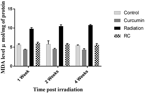 |
Figure 1 Effects of irradiation pre- and post-treatment with curcumin on MDA levels (µmol/mg of protein) at 1, 2, and 4 weeks post-irradiation. |
Skin Tissue CAT Activity
One, 2, and 4 weeks post-irradiation, CAT activity in the skin tissue samples were found to be significantly lower in the R group compared to VC (P<0.0001). Treatment with curcumin before and after irradiation significantly reversed CAT activity (P<0.0001). Treatment with curcumin also significantly increased CAT activity in the skin compared to the VC group (P<0.0001) at 1, 2, and 4 weeks. No significant differences were observed between the levels of CAT in the skin tissues of C compared with the RC group (P>0.05), as shown in Figure 2. The effects of curcumin and radiation were also time dependent (P<0.05).
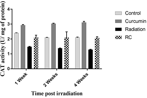 |
Figure 2 Effects of irradiation pre- and post-treatment with curcumin on CAT activity (U/mg of protein) at 1, 2, and 4 weeks post-irradiation. |
Skin Tissue SOD Activity
One, 2, and 4 weeks post-irradiation, SOD activity in the skin tissue samples was found to be significantly lower in the R group compared to VC. Treatment with curcumin before and after irradiation reversed SOD activity (P<0.0001). Treatment with curcumin also significantly increased SOD activity in the skin compared to the VC group (P<0.05). There was a significant difference between the levels of SOD in the skin tissues of curcumin compared with the RC group (P<0.01), as shown in Figure 3. The effects of curcumin and radiation were not time dependent (P>0.05).
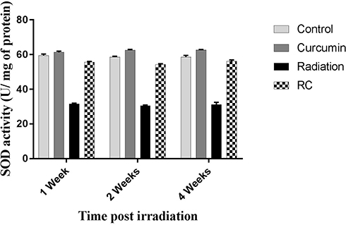 |
Figure 3 Effects of irradiation pre- and post-treatment with curcumin on SOD activity (U/mg of protein) at 1, 2, and 4 weeks post-irradiation. |
GSH-Px Activity in Skin Tissue
One, 2, and 4 weeks post-irradiation, GSH-Px activity in the skin tissue samples were found to be significantly lower in the R group compared to VC (P<0.0001). Treatment with curcumin before and after irradiation significantly reversed CAT activity (P<0.0001). Significant differences were observed between the levels of CAT in the skin tissues of VC compared with the RC group (P<0.05), as shown in Figure 4. The effects of curcumin as well as radiation were not time dependent (P>0.05).
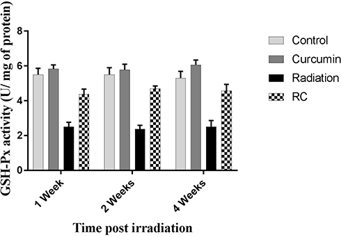 |
Figure 4 Effects of irradiation pre- and post-treatment with curcumin on GSH-Px activity (U/mg of protein) at 1, 2, and 4 weeks post-irradiation. |
Discussion
Findings from biochemical evaluations showed that exposure to radiation led to significant damage to skin tissues. Furthermore, comparison between the radiation and control groups showed a significant increase in MDA production in the radiation group. This is in agreement with previous findings which showed that radiation-induced oxidative damage increases MDA levels of irradiated tissues.9,10 Irradiation of the skin tissues also led to a significant reduction in SOD and CAT activities as well as GSH levels when compared to the control group. These results show the effect of ionizing radiation in upsetting the antioxidant balance in biological systems due to the production of excess free radicals.11 Antioxidant imbalance has been shown to induce detrimental effects on healthy tissues.12
Proliferating cells such as those of the skin are highly radiosensitive and are mostly affected by irradiation.4,13 Irradiation of the skin could damage proliferating stem cells as well as further development of ischemic and fibrous tissue via reduction of resident and recruited stem cells in the vascularized tissue.14 Radiation-induced oxidative damage gives rise to changes in lipid bilayer fluidity and permeability. Thus, ROS are responsible for structural and functional damage to membrane lipids. Furthermore, cell membrane permeability and variations in tissue ionic contents are hampered.15,16 Results from the present study show that the administration of curcumin before and after irradiation prevented radiation-induced oxidative damage to the skin as well as increased antioxidant system. These findings are in agreement with previous studies which reported the ability of curcumin to inhibit the development of lipid peroxidation and neutralize ROS.17,18 It has also been shown to attenuate the expressions of several inflammatory mediators including nuclear factor κB (NF-κB), cyclooxygenase-2 (COX-2), inducible nitric oxide synthase (iNOS), lipoxygenases, inflammatory cytokines (IL-1, IL-6, IL-8), chemokines, etc.19
To ensure that the therapeutic effects of radiotherapy are fully maximized, it is imperative that side-effects to normal tissues are greatly minimized. Thus, in recent decades, several investigations have been conducted on the use of radioprotectors to counter the deleterious effects of ionizing radiation on normal tissues. An ideal radioprotector should protect healthy tissues without sparing tumor cells. Radioprotectors can come in either natural or chemical forms. Natural radioprotectors such as melatonin, curcumin, sheng-mai-san, dan-hong, metformin, etc. have been reported to protect against ionizing-radiation toxicities to several organs.5,20 Furthermore, they are easily available, cheap, and less toxic. These are major advantages over chemical radioprotectors.
While curcumin has been investigated for its radioprotective effects, studies have also shown that curcumin has anti-cancer abilities.21,23 Thus, curcumin could be a double-edged sword in cancer therapy. Hence, based on these interesting findings in addition to its low toxicity and sensitization of tumor cells, there is growing interest for clinical applications of curcumin in radiotherapy, especially in its ability to protect the skin which is inevitably exposed to ionizing radiation.
Conclusion
Findings from the present study have shown that curcumin has the potential to protect against radiotherapy-induced oxidative damage to the skin via restoration of the antioxidant balance of irradiated skin tissues. We recommend future clinical studies to further evaluate its clinical efficacy.
Disclosure
The authors report no conflicts of interest in this work.
References
1. Ringborg U, Bergqvist D, Brorsson B, et al. The Swedish council on technology assessment in health care (SBU) systematic overview of radiotherapy for cancer including a prospective survey of radiotherapy practice in Sweden 2001–summary and conclusions. Acta Oncol. 2003;42(5–6):357–365. doi:10.1080/02841860310010826
2. Porock D, Nikoletti S, Kristjanson L. Management of radiation skin reactions: literature review and clinical application. Plast Surg Nurs. 1999;19(4):185–192.
3. Ryan JL. Ionizing radiation: the good, the bad, and the ugly. J Invest Dermatol. 2012;132(3 Pt 2):985–993. doi:10.1038/jid.2011.411
4. Najafi M, Motevaseli E, Shirazi A, et al. Mechanisms of inflammatory responses to radiation and normal tissues toxicity: clinical implications. Int J Radiat Biol. 2018;94(4):335–356. doi:10.1080/09553002.2018.1440092
5. Yahyapour R, Shabeeb D, Cheki M, et al. Radiation protection and mitigation by natural antioxidants and flavonoids: implications to radiotherapy and radiation disasters. Curr Mol Pharmacol. 2018;11(4):285–304. doi:10.2174/1874467211666180619125653
6. Cheng AL, Hsu CH, Lin JK, et al. Phase I clinical trial of curcumin, a chemopreventive agent, in patients with high-risk or pre-malignant lesions. Anticancer Res. 2001;21(4b):2895–2900.
7. Kolivand S, Amini P, Saffar H, et al. Evaluating the radioprotective effect of curcumin on rat’s heart tissues. Curr Radiopharm. 2019;12(1):23–28. doi:10.2174/1874471011666180831101459
8. Jourdan MM, Lopez A, Olasz EB, et al. Laminin 332 deposition is diminished in irradiated skin in an animal model of combined radiation and wound skin injury. Radiat Res. 2011;176(5):636–648.
9. Jeong BK, Song JH, Jeong H, et al. Effect of alpha-lipoic acid on radiation-induced small intestine injury in mice. Oncotarget. 2016;7(12):15105–15117. doi:10.18632/oncotarget.7874
10. Cagin YF, Parlakpinar H, Polat A, et al. The protective effects of apocynin on ionizing radiation-induced intestinal damage in rats. Drug Dev Ind Pharm. 2016;42(2):317–324. doi:10.3109/03639045.2015.1052080
11. Cihan YB, Ozturk A, Gokalp SS. Protective role of royal jelly against radiation-induced oxidative stress in rats. Int J Hematol Oncol. 2013;28(4):079–87. doi:10.4999/uhod.11016
12. Kaya H, Delibas N, Serteser M, Ulukaya E, Ozkaya O. The effect of melatonin on lipid peroxidation during radiotherapy in female rats. Strahlenther Onkol. 1999;175(6):285–288. doi:10.1007/BF02743581
13. Prise KM, Saran A. Concise review: stem cell effects in radiation risk. Stem Cells. 2011;29(9):1315–1321. doi:10.1002/stem.690
14. Schonmeyr BH, Wong AK, Soares M, Fernandez J, Clavin N, Mehrara BJ. Ionizing radiation of mesenchymal stem cells results in diminution of the precursor pool and limits potential for multilineage differentiation. Plast Reconstr Surg. 2008;122(1):64–76. doi:10.1097/PRS.0b013e31817743cd
15. Pandey B, Mishra K. In vitro studies on radiation induced membrane oxidative damage in apoptotic death of mouse thymocytes. Int J Low Radiat. 2003;1(1):113–119. doi:10.1504/IJLR.2003.003478
16. Tabarraei Y, Bakhshandeh Z, Behrouzkia Z, et al. Short-term changes in histopathological markers of irradiated rat’s lung: preliminary study. Res J Pharm Biol Chem Sci. 2014;5(3):307–315.
17. Jha NS, Mishra S, Jha SK, Surolia A. Antioxidant activity and electrochemical elucidation of the enigmatic redox behavior of curcumin and its structurally modified analogues. Electrochim Acta. 2015;151:574–583. doi:10.1016/j.electacta.2014.11.026
18. Al-Rubaei ZM, Mohammad TU, Ali LK. Effects of local curcumin on oxidative stress and total antioxidant capacity in vivo study. Pak J Biol Sci. 2014;17(12):1237–1241. doi:10.3923/pjbs.2014.1237.1241
19. Cho YJ, Yi CO, Jeon BT, et al. Curcumin attenuates radiation-induced inflammation and fibrosis in rat lungs. Korean J Physiol Pharmacol. 2013;17(4):267–274. doi:10.4196/kjpp.2013.17.4.267
20. Musa AE, Shabeeb D. Radiation-induced heart diseases: protective effects of natural products. Medicina (Kaunas). 2019;55(5):126. doi:10.3390/medicina55050126
21. Fan Z, Yao J, Li Y, Hu X, Shao H, Tian X. Anti-inflammatory and antioxidant effects of curcumin on acute lung injury in a rodent model of intestinal ischemia reperfusion by inhibiting the pathway of NF-Kb. Int J Clin Exp Pathol. 2015;8(4):3451–3459.
22. Jordan BC, Mock CD, Thilagavathi R, Selvam C. Molecular mechanisms of curcumin and its semisynthetic analogues in prostate cancer prevention and treatment. Life Sci. 2016;152:135–144. doi:10.1016/j.lfs.2016.03.036
23. Wilken R, Veena MS, Wang MB, Srivatsan ES. Curcumin: a review of anti-cancer properties and therapeutic activity in head and neck squamous cell carcinoma. Mol Cancer. 2011;10(1):12. doi:10.1186/1476-4598-10-12
 © 2020 The Author(s). This work is published and licensed by Dove Medical Press Limited. The full terms of this license are available at https://www.dovepress.com/terms.php and incorporate the Creative Commons Attribution - Non Commercial (unported, v3.0) License.
By accessing the work you hereby accept the Terms. Non-commercial uses of the work are permitted without any further permission from Dove Medical Press Limited, provided the work is properly attributed. For permission for commercial use of this work, please see paragraphs 4.2 and 5 of our Terms.
© 2020 The Author(s). This work is published and licensed by Dove Medical Press Limited. The full terms of this license are available at https://www.dovepress.com/terms.php and incorporate the Creative Commons Attribution - Non Commercial (unported, v3.0) License.
By accessing the work you hereby accept the Terms. Non-commercial uses of the work are permitted without any further permission from Dove Medical Press Limited, provided the work is properly attributed. For permission for commercial use of this work, please see paragraphs 4.2 and 5 of our Terms.