")
Back to Journals » Vascular Health and Risk Management » Volume 12
Critical evaluation of the MitraClip system in the management of mitral regurgitation
Authors Deuschl F, Schofer N, Lubos E, Blankenberg S, Schäfer U
Received 3 June 2015
Accepted for publication 17 September 2015
Published 11 January 2016 Volume 2016:12 Pages 1—8
DOI https://doi.org/10.2147/VHRM.S65185
Checked for plagiarism Yes
Review by Single anonymous peer review
Peer reviewer comments 3
Editor who approved publication: Dr Daniel Duprez
Florian Deuschl* Niklas Schofer* Edith Lubos, Stefan Blankenberg, Ulrich Schäfer
Department of General and Interventional Cardiology, University Heart Center Hamburg, Hamburg, Germany
*These authors contributed equally to this work
Abstract: The MitraClip (MC) system is a device for percutaneous, transseptal edge-to-edge reconstruction of the mitral valve (MV) in patients with severe mitral regurgitation (MR) not eligible for surgery. Recently, a number of studies have underlined the therapeutic benefit of the MC system for patients with extreme and high risk for MV surgery suffering from either degenerative or functional MR. The MC procedure shows negligible intraprocedural mortality, low periprocedural complication rates, and a significant reduction in MR, as well as an improvement in functional capacity and most importantly quality of life. Presently, the MC system has become an additional interventional tool in the concert of surgical methods. It hereby enlarges the spectrum of MV repair for the Heart Team. Lately, many reviews focused on the MC system. The current review describes the developments in the treatment of MR with the MC system.
Keywords: MitraClip, mitral regurgitation, percutaneous mitral valve repair
Introduction
With increasing age, the prevalence of mitral regurgitation (MR) is rapidly growing.1 Aging of the world’s population challenges health care professionals worldwide to develop new and less invasive treatment options for the elderly.
The MitraClip (MC) system is by now an established interventional therapy for severe MR in a very selective group of patients with high or extreme risk for conventional surgery (class IIB, evidence class C, recommendation, European Guidelines)2 – namely the elderly, patients with multiple comorbidities, and/or patients with severely reduced ejection fraction.
Mitral valve repair (MVR) – despite the absence of randomized trials – is the gold standard for the treatment of severe MR. If repair is not suitable, mitral valve replacement (MVRx) is the only surgical option.3 In young and low surgical risk patients, results are excellent; the mortality rate for MVR is low with 1.4%, and for MVRx with 1.6%. However, in octogenerians and patients with high surgical risk, 30-day mortality has been shown to be substantially higher with 11.0% for MVR and 18.9% for MVRx. For the latter, there is a great need for a less invasive interventional therapy. The MC device (Abbott Laboratories, Abbott Park, IL, USA) is a transvenous, transseptal, edge-to-edge repair system for patients with high surgical risk for the treatment of severe functional (FMR) and degenerative MR (DMR). In 2005, the first results of the Endovascular Valve Edge-to-Edge Repair Study (EVEREST) I trial were published.5 At present, over 22,000 patients (as of April 2015, according to the manufacturer Abbott Laboratories) have been treated worldwide (Table 1).
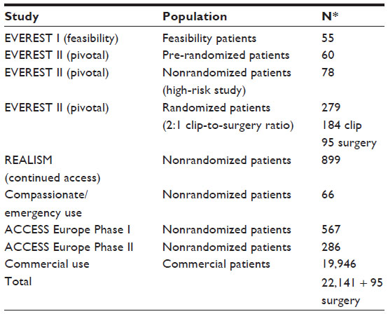 | Table 1 MitraClip clinical trials and commercial use with corresponding number of patients |
In the USA, the commercial use of the MC system started in 2014 with a steadily growing number and holds a current market share of 20%–25% (Figure 1). Globally, the etiology of MR is 2/3 FMR and 1/3 DMR (Figure 2).
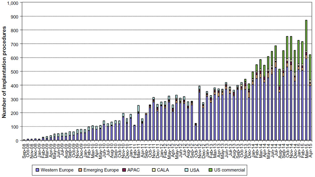 | Figure 1 Worldwide experience with the MitraClip procedure from September 2008 until April 2015. |
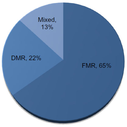 | Figure 2 Etiology of mitral regurgitation according to the manufacturer Abbott Laboratories (as of April 30, 2015). |
European guidelines recommend MC treatment for both FMR and DMR,2 whereas the MC system is approved only for DMR in the USA. After 10 years of clinical experience with percutaneous MVR, this review focuses on the results of recent registries and clinical studies, such as EVEREST II,6–10 ACCESS-EU,11,12 GRASP,13,14 TRAMI,15–17 and one meta-analysis.18
The MitraClip: eligibility criteria and procedure
To guarantee safe positioning of the clip, anatomical eligibility criteria are recommended. A coaptation length of ≥2 mm, a coaptation depth of <11 mm, and in the case of degenerative disease, a flail gap of <10 mm and a flail width of <15 mm are favorable.19 The MC system consists of a steerable guide catheter that is introduced transfemoral, and through echocardiographic guiding transseptal into the left atrium (Figure 3). The clip delivery system can be introduced through the guide catheter. Once the delivery system is completely introduced into the guide, the operator can move the MC in all dimensions under echocardiographic guidance and fluoroscopic confirmation. In the area of the MR, the clip is guided directly above the leaflets by 3D-echo, and the orientation of the clip arms should be perpendicular to the line of coaptation.
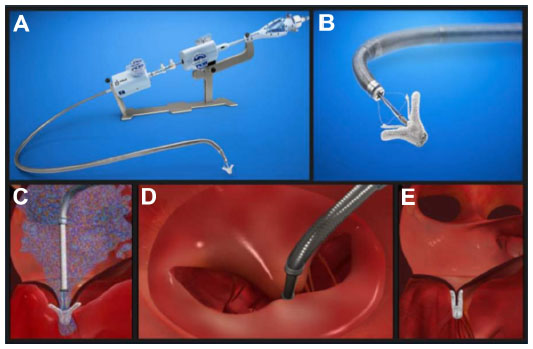 | Figure 3 The MitraClip system. |
The MC is lowered through the valve into the left ventricle to load the leaflets on the clip arms. The grippers fix the leaflets to the clip arms, and then the arms are closed and the MC can be released from the delivery system (Figure 3).
Echocardiography is the most important guiding modality for all steps of the procedure, especially during clip positioning, whereas fluoroscopy is only essential for wiring, transseptal puncture, and control of coaxial alignment of the clip to the line of coaptation during transvalvular maneuvering. Further use of fluoroscopy as a second tool of visualization is optional and operator-dependent. Fluoroscopy duration is approximately 25 minutes. Contrast agents are not needed.12
Conventional management of MR in comparison to the MC system
Standard of care for severe MR is surgical treatment, preferably mitral valve (MV) reconstruction before MVRx.20 So far, the EVEREST II trial is the only randomized controlled trial comparing MV surgery (MVS) and MC.10 Of note, patients in this study were eligible for surgery in contrast to the majority of MC studies. In this study, MC shows a clear inferiority regarding acute efficacy in MR reduction as well as an inferiority in the composite end point of primary efficacy (freedom of death, MVS or reoperation, and MR grade 3+ or 4+) at 1-year follow-up in the intent-to-treat analysis (73% for MVS vs 55% for MC, P=0.007). Following MC, 20% of the patients had to be reoperated compared to 2.2% after MVS (4-year follow-up: 24.8% and 5.5%, respectively). More patients had MR grade 2+ after MC therapy. However, patients showed less symptomatic heart failure according to the New York Heart Association (NYHA) class when compared to those who underwent MVS. Interestingly, at 4-year follow-up the composite end point of freedom from death, surgery, or MR ≥ grade 3+ showed a rate of 39.8% in the MC group and 53.4% after MVS, a difference that was no longer statistically significant (P=0.07). The interventional and surgical long-term results proved to be stable at 4 years with 25% vs 5.5% (MC vs MVS) of the patients requiring surgery for MV dysfunction. Remarkably, if a good result after 6 months was found with the MC, the likelihood of recurrent MR was low, and there was no evidence of late device-related complications.10
A meta-analysis of 21 studies with 6,463 patients compared the outcome of MVS (n=3,265) and MC (n=3,198) demonstrating similar high rates of procedural success (MVS 98% vs MC 96%).21 However, while MVS was superior with respect to 30-day technical failure rate (0.6% vs 3.2%, P=0.002), outcome for MC was superior to MVS in the pooled key safety analysis at 30 days (mortality: 3.3%, 95% confidence interval [CI] 2.6–4.2 vs 16.2%, 95% CI 13.0–20.0; stroke: 1.1%, 95% CI 0.6–1.6 vs 4.5%, 95% CI 3.6–5.3; bleeding: 4.2%, 95% CI 3.0–7.0 vs 59.0%, 95% CI 50.0–67.0; prolonged mechanical ventilation: 1.7%, 95% CI 1.1–2.2 vs 36.3%, 95% CI 33.1–40.0). These results were shown despite a higher surgical risk profile in the patient group treated with MC (eg, mean left ventricular ejection fraction [LVEF] of 38% vs 52% [MC vs MVS]).21 One-year mortality of MC patients was 13%. Since there is no long-term data available for MVS, results are not comparable. A further limitation of this meta-analysis is the fact that most of these studies on MC therapy were derived from registries or retrospectively analyzed case series; they are not randomized or controlled, especially for the patient population characteristics.
MC therapy has to be compared to the best medical therapy in heart failure patients with severe MR. This group of patients have a 1-year mortality of 20%, a 5-year mortality of 50%, and a high rate of recurrent hospitalization for heart failure.22 Trials prospectively comparing best medical treatment with interventional therapy are ongoing. The currently enrolling studies COAPT (ClinicalTrials.gov registration number: NCT01626079) and RESHAPE-HF (ClinicalTrials.gov registration number: NCT01772108) will address this important question in the near future.
Mortality and safety
The largest meta-analysis comprising 2,980 patients from 16 studies (12 European and four North American) demonstrated a very low intra-procedural mortality of only 0.1%. Nevertheless, 30-day mortality increased to 4.2%, and all-cause mortality during a mean follow-up of 310 days was 15.8%.18 Thirty-day mortality ranged between 0.9%14 and 4.7%12 in most clinical trials. Recent trials report a high 1-year mortality ranging between 12% and 18.2%.12,14,20 Significant comorbidities expressed by a high logistic European System for Cardiac Operative Risk Evaluation (EuroSCORE) I of 23.4%±1.5% most likely accounts for the high mortality rate during the first year.18 In the GRASP registry, 55% of deaths during the first year were attributed to noncardiac reasons.14
Complications
In the meta-analysis, the most relevant procedure-associated complication was major bleeding (requiring transfusion) with 9.7% followed by stroke/transient ischemic attack (1.3%), chordal rupture (0.8%), pericardial tamponade (0.7%), and myocardial infarction (0.4%).18 The ACCESS registry reports even lower rates of stroke (0.7%) and bleeding complications (3.8%).12 In summary, complication rates associated with MC are low (Table 2), particularly when compared to those associated with transcatheter aortic valve replacement. Here, arterial puncture and calcification of vessels and the aortic annulus increase the risk of transient ischemic attack as well as stroke, ranging from 4.0% to 6.7%,23 and of major vascular complications, ranging from 8.2%24 to 16.2%.23
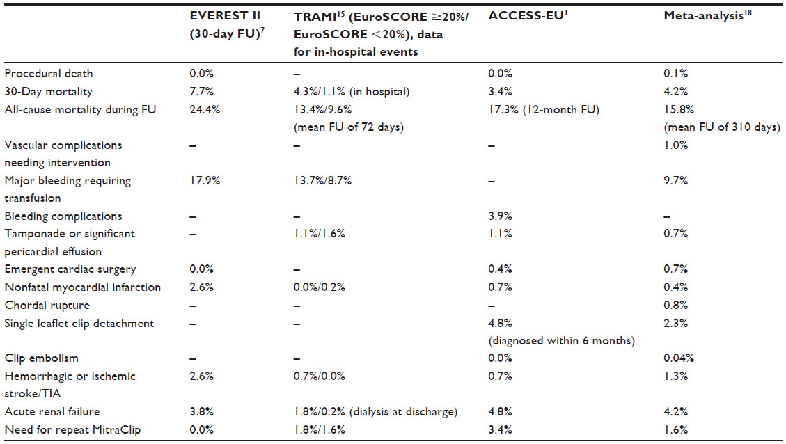 | Table 2 Complications of percutaneous mitral valve repair |
Heart failure and MC
MR in combination with congestive heart failure has a very poor prognosis.22,25 Reverse left ventricular (LV) remodeling and improvements of symptoms after MVS in patients with advanced LV dysfunction have been reported in several studies.26,27 Thirty-day mortality ranges between 8% and 9%.26,28 In this regard, MC is a promising, minimally invasive percutaneous treatment technique.29 In a study, 108 patients with predominantly FMR and LV dysfunction (mean LVEF 28%±11% with LVEF <40% in 88% of patients) demonstrated an impressively low 30-day mortality of only 1.8%.30 Most importantly, there are accumulating data that the MC appears to be not only safe but also efficacious in patients with FMR.30 In this regard, the ACCESS-EU registry showed a similar clinical improvement in patients with an LVEF ≤30% vs >30%. In addition, there is some evidence of reverse remodeling after successful treatment with the MC.31
There is an ongoing discussion whether a severely depressed LV function might be susceptible to further acute reduction in an LVEF induced by surgical correction of MR. Interestingly, the occurrence of the so-called afterload mismatch was frequently found (26%) in a study comprising 73 patients with FMR and severely reduced LVEF (27%±9%).32 Comparison of patients with vs without afterload mismatch after MC revealed that LV end-diastolic diameter (71±8 mm vs 67±7 mm, P=0.02) and LV end-systolic diameter (57±9 mm vs 53±7 mm, P=0.04) were larger, pulmonary pressure was higher (49±10 mmHg vs 40±10 mmHg, P=0.04), and right ventricular dysfunction was more prevalent (68% vs 31%, P=0.049). The authors suggested that afterload mismatch was the consequence of an abrupt increase in LV end-systolic wall stress after MR correction on a preexisting status of absent or reduced contractile reserve. Fortunately, the observed hemodynamic deterioration in patients was a transient phenomenon and did not translate into an adverse outcome at 12 months (1-year survival: 81.2% vs 75.2%, P=0.44). This study observed no difference in the need for inotropes between patients with and without afterload mismatch in the early postoperative time period.32
The previously mentioned study comprising only patients with FMR and reduced LVEF reported that 57.7% of patients required inotropic support on the intensive care unit and 13% of patients were transiently bridged with intra-aortic balloon counter pulsation (IABP) underlining the incidence of a transient window of aggravated heart failure immediately after intervention.30
Procedural success, long-term outcome, and predictors of procedural success
Acute procedural success is defined as a reduction in MR to ≤grade 2+. A recent meta-analysis showed acute procedural success in 91.4% of the patients. Persistent MR reduction was found in 85.3% of the patients at 30-days follow-up and in 86.9% at a mean follow-up of 310 days (ranging from 80 days to 4 years).18 In only 3% of the patients, the therapy failed, and no clip could be implanted. A single-center study in 108 patients with predominantly FMR and LV dysfunction (mean LVEF 28%±11% with 88% having an LVEF <40%) showed a procedural success rate of 99%.30
The MC procedure is associated with a low mortality rate. However, it is of great importance to better define predictors of adverse clinical outcome. Only few studies have addressed this topic. In a study of selected FMR patients with severe LV dysfunction, univariate analysis demonstrated an adverse outcome for pre-interventional logistic EuroSCORE I ≥20% (hazards ratio [HR] 4.4, 95% CI 1.8–9.5, P=0.01) and pre-interventional proBNP >1,600 pg/mL (HR 21.2, 95% CI 2.5–38, P=0.01), a need for post-interventional IABP treatment (HR 3.8, 95% CI 1.2–13.5, P=0.02), and peri-interventional occurrence of acute kidney injury (HR 4.1, 95% CI 2–16, P=0.01).30 These findings are in line with results of a single-center study (65% FMR, 35% DMR) analyzing predictors of midterm clinical and survival outcome (all-cause mortality or hospitalization): NYHA IV at baseline (HR 2.4, 95% CI 1.4–4.3, P=0.002) and glomerular filtration rate <60 mL/min/1.73 m2 (HR 2.05, 95% CI 1.1–4.0, P=0.03), Sociey of Thoracic Surgeons score >12% (HR 2.20, 95% CI 1.3–3.8, P=0.004), and failure of procedural success (HR 2.66, 95% CI 1.4–5.0, P=0.002).33
In a study of 300 MC patients (68% FMR, 32% DMR), regurgitant orifice area ≥70.8 mm2 and trans-mitral pressure gradient ≥4 mmHg in combination with an MV orifice area ≤3.0 cm2 (assessed by echocardiography) were defined as predictors of increased risk for procedural failure.34
Quality of life
Functional capacity according to the NYHA class showed a relief of symptoms (86% in the NYHA class I and II) 1 year after treatment.30 The meta-analysis showed an improved functional capacity according to the NYHA (class I and II) in 76.6% of the patients.18 Moreover, the analysis demonstrated an improvement in LVEF, 6-minute walk distance, and quality of life.18 The gain in 6-minute walk distance (260.6±13.6 m at baseline vs 359.8±24.9 m at follow-up) was larger than the gain being described after cardiac resynchronization therapy.23 Similar data were reported for FMR patients with severe LV dysfunction (328.7±80.1 m; mean improvement 108 m). Most interestingly, there were clear signs of LV reverse remodeling with an increase in LVEF (27%±9.8% to 34.7%±10.4%, P=0.02 at 1-year follow-up) and with a decrease in LV end-diastolic volume as well as in LV end-systolic volume.30
Conclusion
For patients with severe MR and high surgical risk, the treatment with the MC has meanwhile evolved as the therapy of choice in Europe (FMR and DMR) and North America (DMR). There is a class IIb (level of evidence C) recommendation in the European guidelines for interventional mitral device therapy in severe symptomatic FMR as well as DMR in all patients with high surgical risk. Additionally, life expectancy has to exceed 1 year, and the Heart Team (cardiologist, cardiac surgeon) should mutually agree that patients are ineligible for surgery.2 Additionally, MR pathology has to meet special criteria for intervention by defined echocardiographic parameters.6,35 North American 2014 guidelines for the management of valvular heart disease20 strictly follow the results of the EVEREST I and II trials and recommend interventional mitral device therapy only for DMR.6,7,10 In conclusion, the MC system has evolved as the most important transcatheter MVR therapy till date. Intraprocedural mortality is low, adverse events are seldom, and short- as well as long-term results are satisfactory.
The number of case reports and smaller case series, where the MC device was used as a bailout strategy apart from current guideline recommendations, is rapidly growing. These reports show that with increasing device experience, the anatomical criteria for the applicability of MC implantation broaden. Current guideline-based indications might therefore underestimate the potential of MC therapy.
- In the case of severe coaptation failure, two MCs were implanted via a double-guide approach with two simultaneously introduced clip delivery systems. The first clip was initially used to improve coaptation between the posterior and anterior leaflet for the second clip to be positioned for principal MR treatment. Once a successful grasp was performed with the second clip, the first clip was reopened and optimized (“mitral titration technique”).36
- The MC was used after MVR to reduce residual severe MR.37,38
- The MC was implanted in a series of patients with left ventricular outflow tract (LVOT) obstruction with subsequent MR due to systolic anterior movement of the anterior leaflet in hypertropic obstructive cardiomyopathy patients. Not only MR but also subvalvular LVOT gradient was successfully reduced.39,40
- The MC was used as primary rescue therapy for patients with severe MR in cardiogenic shock and/or critically ill.41–43
- The MC was implanted in a trileaflet MV.44
These cases demonstrate the potential of the device and might inspire reconsideration of the use of MC in future trials. Due to the remarkably low peri-interventional risk, the MC might be considered as a bailout therapy for treatment of severe MR even in critically ill patients.
However, new devices for the therapy of MR are entering the market, ultimately broadening the spectrum of patients being treated by catheter-based techniques in the near future. In line with surgical reconstruction, a combined use of different interventional devices, for example, MC and annuloplasty device, may be the future of interventional MV therapy.
Disclosure
F Deuschl received speakers honoraria and traveling support from Abbott Laboratories. U Schaefer received research funding, speakers honoraria, and traveling support from Abbott Laboratories, and is a faculty member at Crossroads Abbott Laboratories in Brussels. E Lubos received research funding, speakers honoraria, and traveling support from Abbott Laboratories. The authors report no other conflicts of interest in this work.
References
Nkomo VT, Gardin JM, Skelton TN, Gottdiener JS, Scott CG, Enriquez-Sarano M. Burden of valvular heart diseases: a population-based study. Lancet. 2006;368(9540):1005–1011. doi:10.1016/S0140-6736(06)69208-8. | |
Vahanian A, Alfieri O, Andreotti F, et al. Guidelines on the management of valvular heart disease (version 2012). Eur Heart J. 2012;33(19):2451–2496. doi:10.1093/eurheartj/ehs109. | |
Bonow RO, Carabello BA, Chatterjee K, et al. 2008 focused update incorporated into the ACC/AHA 2006 guidelines for the management of patients with valvular heart disease: a report of the American College of Cardiology/American Heart Association Task Force on Practice Guidelines (Writing Committee to revise the 1998 guidelines for the management of patients with valvular heart disease): endorsed by the Society of Cardiovascular Anesthesiologists, Society for Cardiovascular Angiography and Interventions, and Society of Thoracic Surgeons Circulation. 2008;118(15):e523–e661. doi:10.1161/CIRCULATIONAHA.108.190748. | |
Chikwe J, Goldstone AB, Passage J, et al. A propensity score-adjusted retrospective comparison of early and mid-term results of mitral valve repair versus replacement in octogenarians. Eur Heart J. 2011;32(5):618–626. doi:10.1093/eurheartj/ehq331. | |
Feldman T, Wasserman HS, Herrmann HC, et al. Percutaneous mitral valve repair using the edge-to-edge technique: six-month results of the EVEREST phase I clinical trial. J Am Coll Cardiol. 2005;46(11):2134–2140. doi:10.1016/j.jacc.2005.07.065. | |
Feldman T, Foster E, Glower DD, et al. Percutaneous repair or surgery for mitral regurgitation. N Engl J Med. 2011;364(15):1395–1406. doi:10.1097/01.SA.0000410146.91957. d6. | |
Whitlow PL, Feldman T, Pedersen WR, et al. Acute and 12-month results with catheter-based mitral valve leaflet repair: The EVEREST II (Endovascular Valve Edge-to-Edge Repair) High Risk Study. J Am Coll Cardiol. 2012;59(2):130–139. doi:10.1016/j.jacc.2011.08.067. | |
Lim DS, Reynolds MR, Feldman T, et al. Improved functional status and quality of life in prohibitive surgical risk patients with degenerative mitral regurgitation after transcatheter mitral valve repair. J Am Coll Cardiol. 2014;64(2):182–192. doi:10.1016/j.jacc.2013.10.021. | |
Glower DD, Kar S, Trento A, et al. Percutaneous mitral valve repair for mitral regurgitation in high-risk patients: results of the EVEREST II study. J Am Coll Cardiol. 2014;64(2):172–181. doi:10.1016/j.jacc.2013.12.062. | |
Mauri L, Foster E, Glower DD, et al. 4-Year results of a randomized controlled trial of percutaneous repair versus surgery for mitral regurgitation. J Am Coll Cardiol. 2013;62(4):317–328. doi:10.1016/j.jacc.2013.04.030. | |
Reichenspurner H, Schillinger W, Baldus S, et al. Clinical outcomes through 12 months in patients with degenerative mitral regurgitation treated with the mitraclip® device in the ACCESS-EUrope phase I trial. Eur J Cardiothoracic Surg. 2013;44(4):280–288. doi:10.1093/ejcts/ezt321. | |
Maisano F, Franzen O, Baldus S, et al. Percutaneous mitral valve interventions in the real world: early and 1-year results from the ACCESS-EU, a prospective, multicenter, nonrandomized post-approval study of the Mitraclip therapy in Europe. J Am Coll Cardiol. 2013;62(12):1052–1061. doi:10.1016/j.jacc.2013.02.094. | |
Attizzani GF, Ohno Y, Capodanno D, et al. Gender-related clinical and echocardiographic outcomes at 30-day and 12-month follow up after MitraClip implantation in the GRASP registry. Catheter Cardiovasc Interv. 2015;85(5):889–897. doi:10.1002/ccd.25715. | |
Grasso C, Capodanno D, Scandura S, et al. One- and twelve-month safety and efficacy outcomes of patients undergoing edge-to-edge percutaneous mitral valve repair (from the GRASP registry). Am J Cardiol. 2013;111(10):1482–1487. doi:10.1016/j.amjcard. 2013.01.300. | |
Wiebe J, Franke J, Lubos E, et al. Percutaneous mitral valve repair with the mitraclip system according to the predicted risk by the logistic EuroSCORE: preliminary results from the German Transcatheter Mitral Valve Interventions (TRAMI) registry. Catheter Cardiovasc Interv. 2014;598(800):591–598. doi:10.1002/ccd.25493. | |
Schillinger W, Hünlich M, Baldus S, et al. Acute outcomes after MitraClip® therapy in highly aged patients: results from the German TRAnscatheter Mitral valve Interventions (TRAMI) registry. EuroIntervention. 2013;9(1):84–90. doi:10.4244/EIJV9I1A13. | |
Baldus S, Schillinger W, Franzen O, et al. Mitra Clip therapy in daily clinical practice: initial results from the German transcatheter mitral valve interventions (TRAMI) registry. Eur J Heart Fail. 2012; 14(9):1050–1055. doi:10.1093/eurjhf/hfs079. | |
Vakil K, Roukoz H, Sarraf M, et al. Safety and efficacy of the MitraClip® system for severe mitral regurgitation: a systematic review. Catheter Cardiovasc Interv. 2014;84(1):129–136. doi:10.1002/ccd.25347. | |
Feldman T, Kar S, Rinaldi M, et al. Percutaneous mitral repair with the MitraClip system. Safety and midterm durability in the initial EVEREST (Endovascular Valve Edge-to-Edge REpair Study) cohort. J Am Coll Cardiol. 2009;54(8):686–694. doi:10.1016/j.jacc.2009.03.077. | |
Nishimura RA, Otto CM, Bonow RO, et al. 2014 AHA/ACC guideline for the management of patients with valvular heart disease: executive summary: a report of the American College of Cardiology/American Heart Association Task Force on Practice Guidelines. Circulation. 2014;129(23):e650. doi:10.1161/CIR.0000000000000029. | |
Philip F, Athappan G, Tuzcu EM, Svensson LG, Kapadia SR. MitraClip for severe symptomatic mitral regurgitation in patients at high surgical risk: a comprehensive systematic review. Catheter Cardiovasc Interv. 2014;84(4):581–590. doi:10.1002/ccd.25564. | |
Goel SS, Bajaj N, Aggarwal B, et al. Prevalence and outcomes of unoperated patients with severe symptomatic mitral regurgitation and heart failure: comprehensive analysis to determine the potential role of mitraclip for this unmet need. J Am Coll Cardiol. 2014;63(2):185–186. doi:10.1016/j.jacc.2013.08.723. | |
Linde C, Gold MR, Abraham WT, et al. Long-term impact of cardiac resynchronization therapy in mild heart failure: 5-year results from the resynchronization reverses remodeling in systolic left ventricular dysfunction (REVERSE) study. Eur Heart J. 2013;34(33):2592–2599. doi:10.1093/eurheartj/eht160. | |
Popma JJ, Adams DH, Reardon MJ, et al. Transcatheter aortic valve replacement using a self-expanding bioprosthesis in patients with severe aortic stenosis at extreme risk for surgery. J Am Coll Cardiol. 2014;63(19):1972–1981. doi:10.1016/j.jacc.2014.02.556. | |
Rossi A, Dini FL, Faggiano P, et al. Independent prognostic value of functional mitral regurgitation in patients with heart failure. A quantitative analysis of 1256 patients with ischaemic and non-ischaemic dilated cardiomyopathy. Heart. 2011;97(20):1675–1680. doi:10.1136/hrt.2011.225789. | |
De Bonis M, Taramasso M, Grimaldi A, et al. The GeoForm annuloplasty ring for the surgical treatment of functional mitral regurgitation in advanced dilated cardiomyopathy. Eur J Cardiothoracic Surg. 2011;40(2):488–495. doi:10.1016/j.ejcts.2010.11.048. | |
De Bonis M, Lapenna E, Verzini A, et al. Recurrence of mitral regurgitation parallels the absence of left ventricular reverse remodeling after mitral repair in advanced dilated cardiomyopathy. Ann Thorac Surg. 2008;85(3):932–939. doi:10.1016/j.athoracsur.2007.11.021. | |
Braun J, Bax JJ, Versteegh MIM, et al. Preoperative left ventricular dimensions predict reverse remodeling following restrictive mitral annuloplasty in ischemic mitral regurgitation. Eur J Cardiothoracic Surg. 2005;27(5):847–853. doi:10.1016/j.ejcts.2004.12.031. | |
De Bonis M, Lapenna E, La Canna G, et al. Mitral valve repair for functional mitral regurgitation in end-stage dilated cardiomyopathy: role of the “edge-to-edge” technique. Circulation. 2005;112(9 Suppl):1402–1408. doi:10.1161/CIRCULATIONAHA.104.525188. | |
Taramasso M, Maisano F, Latib A, et al. Clinical outcomes of MitraClip for the treatment of functional mitral regurgitation. EuroIntervention. 2014;10(6):746–752. | |
Grayburn PA, Foster E, Sangli C, et al. Relationship between the magnitude of reduction in mitral regurgitation severity and left ventricular and left atrial reverse remodeling after mitraclip therapy. Circulation. 2013;128(15):1667–1674. doi:10.1161/CIRCULATIONAHA.112.001039. | |
Melisurgo G, Ajello S, Pappalardo F, et al. Afterload mismatch after mitraclip insertion for functional mitral regurgitation. Am J Cardiol. 2014;113(11):1844–1850. doi:10.1016/j.amjcard.2014.03.015. | |
Puls M, Tichelbäcker T, Bleckmann A, et al. Failure of acute procedural success predicts adverse outcome after percutaneous edge-to-edge mitral valve repair with MitraClip. EuroIntervention. 2014;9(12):1407–1417. doi:10.4244/EIJV9I12A238. | |
Lubos E, Schlüter M, Vettorazzi E, et al. MitraClip therapy in surgical high-risk patients: identification of echocardiographic variables affecting acute procedural outcome. JACC Cardiovasc Interv. 2014; 7(4):394–402. doi:10.1016/j.jcin.2013.12.198. | |
Armstrong EJ, Rogers JH, Swan CH, et al. Echocardiographic predictors of single versus dual MitraClip device implantation and long-term reduction of mitral regurgitation after percutaneous repair. Catheter Cardiovasc Interv. 2013;82(4):673–679. doi:10.1002/ccd.24645. | |
Schaefer U, Frerker C, Kreidel F. Simultaneous double clipping delivery guide strategy for treatment of severe coaptation failure in functional mitral regurgitation. Heart Lung Circ. 2015;24(1):98–102. doi:10.1016/j.hlc.2014.09.008. | |
Grasso C, Attizzani GF, Ohno Y, et al. Catheter-based edge-to-edge mitral valve repair after percutaneous mitral valve annuloplasty failure. JACC Cardiovasc Interv. 2014;7:e85–86. Epub 2014 Jun 18. | |
Grasso C, Ohno Y, Attizzani GF, et al. Percutaneous mitral 547 valve repair with the MitraClip system for severe mitral 548 regurgitation in patients with surgical mitral valve repair 549 failure. J Am Coll Cardiol. 2014;63:836–838. | |
Schäfer U, Kreidel F, Frerker C. MitraClip implantation as a new treatment strategy against systolic anterior motion-induced outflow tract obstruction in hypertrophic obstructive cardiomyopathy. Heart Lung Circ. 2014;23(5):e131–e135. doi:10.1016/j.hlc.2014.01.007. | |
Schäfer U, Frerker C, Thielsen T, et al. Targeting systolic anterior motion and left ventricular outflow tract obstruction in hypertrophic obstructed cardiomyopathy with a MitraClip. EuroIntervention. 2014. doi:10.4244/EIJY14M08_13. | |
Pleger ST, Chorianopoulos E, Krumsdorf U, Katus HA. Percutaneous edge-to-edge repair of mitral regurgitation as a bail-out strategy in critically ill patients. J Invasive Cardiol. 2013;25(2):69–72. | |
Zuern CS, Schreieck J, Weig HJ, Gawaz M, May AE. Percutaneous mitral valve repair using the MitraClip in acute cardiogenic shock. Clin Res Cardiol. 2011;100(8):719–721. doi:10.1007/s00392-011-0324-1. | |
Couture P, Cloutier-Gill L-A, Ducharme A, Bonan R, Asgar AW. MitraClip intervention as rescue therapy in cardiogenic shock: one-year follow-up. Can J Cardiol. 2014;30(9):1108.e15–1108.e16. doi:10.1016/j.cjca.2014.03.045. | |
Freixa X, Hayami D, Basmadjian AN, Asgar AW. MitraClip repair of a “trileaflet” regurgitant mitral valve. EuroIntervention. 2015;11(3):355. doi:10.4244/EIJY14M07_05. |
 © 2016 The Author(s). This work is published and licensed by Dove Medical Press Limited. The full terms of this license are available at https://www.dovepress.com/terms.php and incorporate the Creative Commons Attribution - Non Commercial (unported, v3.0) License.
By accessing the work you hereby accept the Terms. Non-commercial uses of the work are permitted without any further permission from Dove Medical Press Limited, provided the work is properly attributed. For permission for commercial use of this work, please see paragraphs 4.2 and 5 of our Terms.
© 2016 The Author(s). This work is published and licensed by Dove Medical Press Limited. The full terms of this license are available at https://www.dovepress.com/terms.php and incorporate the Creative Commons Attribution - Non Commercial (unported, v3.0) License.
By accessing the work you hereby accept the Terms. Non-commercial uses of the work are permitted without any further permission from Dove Medical Press Limited, provided the work is properly attributed. For permission for commercial use of this work, please see paragraphs 4.2 and 5 of our Terms.