")
Back to Journals » Veterinary Medicine: Research and Reports » Volume 14
Combined Adjuvant Formulations Enhanced an Immune Response of Trivalent Foot and Mouth Disease Vaccine in Cattle
Authors Ayele G, Getachew B, Bari FD, Bayissa B, Muluneh A, Abayneh T, Gelaye E , Edao BM
Received 26 December 2022
Accepted for publication 7 April 2023
Published 15 April 2023 Volume 2023:14 Pages 47—62
DOI https://doi.org/10.2147/VMRR.S397590
Checked for plagiarism Yes
Review by Single anonymous peer review
Peer reviewer comments 2
Editor who approved publication: Professor Young Lyoo
Getu Ayele,1 Belayneh Getachew,1 Fufa Dawo Bari,2 Berecha Bayissa,1 Ayelech Muluneh,3 Takele Abayneh,1 Esayas Gelaye,1 Bedaso Mammo Edao2
1Research and Development Directorate, National Veterinary Institute, Bishoftu, Oromia, Ethiopia; 2Department of Microbiology, Immunology and Vet Public Health, College of Veterinary Medicine, Addis Ababa University, Bishoftu, Oromia, Ethiopia; 3Department of Microbiology, Animal Health Institute, Sebeta, Oromia, Ethiopia
Correspondence: Bedaso Mammo Edao, Email [email protected]
Introduction: Foot-and-mouth disease is globally one of the most economically important viral diseases of cloven-hoofed animals that can be controlled by different strategies, where vaccination plays an important role. Selection of adjuvant added to vaccine preparation is crucial in ensuring the protective effect of the vaccine. Aluminum hydroxide gel mixed with saponin (AS) is widely used adjuvant, with its suboptimal immune response in FMD vaccine. The present study was undertaken to evaluate different ingredients of adjuvants for inactivated trivalent (A, O and SAT 2) FMD vaccine and to demonstrate the effect of booster dose in cattle.
Methods: Cattle were grouped into five; four experimental and one control, with six animals in each group and immunized with trivalent vaccine with various formulations of adjuvants. Immune response was measured using Solid Phase Competitive Enzyme Linked Immune Sorbent Assay (SPCE).
Results: The antibody level in cattle immunised with a vaccine formulation containing a mixture of aluminum hydroxide gel and saponin (AS) were significantly lower than AS boosted group for the three serotypes (p< 0.05, t-test), which directs the need for booster dose. Whereas the antibody response in the AS + oil group was higher followed by oil alone. The AS preparation with a booster dose has shown better immune response compared to the group without.
Conclusion: The findings of this study could suggest that oil based and AS with oil could replace the conventional aluminum hydroxide gel and saponin adjuvants in FMD vaccine preparations. Challenge test was not successful indicating the need for further research on the virus infectivity.
Keywords: adjuvant, foot and mouth disease, immune response, serotype
Introduction
Ethiopia is one of the countries which has the largest livestock population in Africa.1 Despite this, the products and productivity of livestock are still very less due to the impacts of economically important diseases of animals.2 Among these, foot-and-mouth disease (FMD) is a highly contagious viral disease of cloven-hoofed animals affecting cattle, pigs, sheep, and goats.3 The disease is characterized by fever and the presence of vesicular lesion or sores on the tongue, lips, teats, snouts, in the oral cavity and between the hoofs. It is characterized by high morbidity, low mortality, severe production and weight losses in adult animals. However, it causes high mortality in young animals.4
The FMD virus (FMDV) is a member of the genus Aphthovirus, which belongs to the family Picornaviridae. The virus genome is a positive single stranded RNA, which is about 8.5 Kb, encodes structural proteins including viral proteins (VP) 1, VP2, VP3, VP4 and nonstructural proteins.5 To date, there are seven serotypes which are known to cause FMD. These serotypes are identified as Southern African Territories (SAT) 1, SAT 2, SAT 3, A, O, C, and Asia 1 with different genotypes/topotypes, lineages and variants.5,6 In Africa, six of the seven serotypes of FMD viruses except Asia 1 have been reported in the continent.7–9 Among these three serotypes, SAT 1, SAT 2 and SAT 3 are unique in Africa.9–11
In Ethiopia, the disease is endemic and the report of Ministry of Agriculture showed that, the highest incidence of outbreak was occurred from 1997 to 2006 throughout the country. According to the report, the maximum outbreak was recorded in the central part of Ethiopia.12 Many sero-epidemiological surveys conducted in different parts of the country have showed a prevalence result of up to ~26%. Among the seven serotypes of FMDV that are immunologically different, five of them (O, A, SAT 1, SAT 2 and C) were reported to be present in Ethiopia.8,13–15 The FMDV serotype O is the most prevalent (73.3%) followed by serotype A (19.5%), SAT 2 (4.1%) and SAT 1 (1.8%). After 1983, serotype C has not been reported in Ethiopia.8,13,16
The endemic presence of the FMDV is highly challenging due to its impacts on trade market and productivity of livestock. Thus, different measures are required to eradicate and control the disease, including vaccination strategies and control of animal movement.17 However, the vaccine requires continuous improvement to overcome the limitations of currently existing FMD vaccines and to increase its protective ability.18 This can be achieved by the selection of proper antigen and adjuvants mixed with the vaccine.19 An ideal adjuvant is the one which can stimulate specific components of the humoral immune response early, that can promote production of high antibody titer with prolonged duration of immunity. It should also stimulate the cell-based immune response.20
In Ethiopia, considering the wide prevalence of the disease, the National Veterinary Institute (NVI) is producing inactivated trivalent (for serotype O, A and SAT 2) vaccine. The vaccine is produced only for cattle and adjuvanted with aluminum hydroxide gel and saponin.21 Aluminum hydroxide gel and saponin and oil adjuvant vaccines are the most commonly used adjuvant in inactivated FMD vaccine to control the disease. The expected lifespan of livestock, such as cattle is 18 to 22 years on average. Thus, approximately 24–44 vaccinations are needed in these animals’ lifetimes to maintain their health. Therefore, a cheap and effective vaccine is required to fight the disease in livestock especially in low- and middle-income countries like Ethiopia. The adjuvant preparation containing aluminum hydroxide gel and saponin has an advantage in that the antigen is released slowly as it is absorbed into the gel. However, vaccine containing aluminum hydroxide gel and saponin as an adjuvant has several limitations, including the induction of short-lived immune responses and the consequent need for frequent vaccination. In contrast, the oil-adjuvanted FMD vaccines are more effective in providing long-lasting protection than a standard aluminum hydroxide gel and saponin based vaccines.22,23 Since 1970, adjuvants used in FMD vaccine have been transformed from gel to oil, thereby overcoming the problem of poor immunization.9 Comparative studies of aluminum hydroxide gel and saponin with oil-based adjuvant have been conducted and revealed that oil adjuvant showed better immune response than aluminum hydroxide gel and saponin.24 Providing early protection against the disease is another advantage of oil-based adjuvant.25 Furthermore, study conducted on a combination of aluminum hydroxide gel and saponin and oil induced better immunity.26 Despite the documented advantages of oil based and combined adjuvants in enhancing immune response, there is limited scientific evidence on adjuvant evaluation and selection of proper adjuvants in Ethiopia. Therefore, the present study was carried out to evaluate immune response induced by different formulations of adjuvants for inactivated trivalent FMD vaccines in cattle, assess immune response elicited with and without booster vaccine, and test the potency of formulated vaccines by challenging the experimental animals with the live virus.
Materials and Methods
Description of the Study Area
The study was conducted from November 2020 to June 2021 in the National Veterinary Institute, Bishoftu, Ethiopia. All experimental works were conducted at NVI. The antibody response against FMDV was measured using ELISA at the Animal Health Institute (AHI), Sebeta, Ethiopia.
Experimental Animals
The animals used in this experimental study were supplied by the National Veterinary Institute. They were zebu breed male cattle aged three to four years old, apparently healthy or free from any clinical signs. Prior to vaccination, all cattle were ear tagged and the identification number was given. All the study animals used for this experimental study were bled and tested for the presence of anti-FMDV nonstructural protein (NSP) antibodies using the FMD nonstructural enzyme-linked immunosorbent assay (NS-ELISA) (IDEXX Switzerland). Only cattle with a negative result with the FMD NS-ELISA were used for the study. They were randomly allocated into five groups with six cattle in four groups (6 cattle/group) and five in control/non-vaccinated groups and a total of 29 cattle were used. Throughout the study period, they were kept separately and cared for at the animal rooms of NVI with adequate provision of feed and water. The cattle in each vaccine group were vaccinated subcutaneously (SC) with inactivated trivalent (serotype A, O and SAT 2) FMD vaccine formulated with different adjuvants in the middle of the cervical area and blood samples were collected at 0, 7, 14, 21, 28 and 42 days after vaccination for serology testing. Generally, these cattle were used in the evaluation of humoral immune response induced because of vaccine formulated with different adjuvants and booster dose.
Experimental Design
A longitudinal study was applied to evaluate the immune response of the experimental animals. Twenty-nine (29) male cattle were grouped into five groups as follows to evaluate the immune response elicited by different formulations of adjuvants as well as to evaluate the effect of booster dose. The first group was vaccinated with the inactivated trivalent FMD vaccine adjuvanted with the conventional aluminum hydroxide gel and saponin (AS) (without boosting). The second group was vaccinated similarly as the first group with the AS adjuvanted vaccine and the booster dose was given for this group at 14 days post vaccination. The third group was vaccinated with mineral oil adjuvanted inactivated trivalent FMD vaccine. The fourth group was vaccinated with vaccine formulated with AS plus oil and the last group was left as control/non-vaccinated. In this experimental study, every vaccine formulates with a different adjuvant was equally likely applied to each group and every experimental calf within the group. Allocation of adjuvants to each calf was on a random basis to distribute experimental error equally for all groups. At the end of the experiment, the humoral immune response of each group was measured by using solid-phase competitive ELISA (SPCE) and the result of antibody response of each calf within each group was recorded.
Cell Culture
Cryopreserved suspension of baby hamster kidney cell line (BHK-21) was provided by the Pan African Veterinary Vaccine Center (PANVAC) and used for vaccine preparation and virus titration according to the method described by Hassan.27
Vaccine Preparation
The FMDV serotype A/21/ETH/2008, O/ETH/38/2005, SAT 2/ETH/2009 locally isolated antigen was used for vaccine preparation using BHK-21 cell line. Maintenance media (Hank’s media) was also used for cultivation and inoculum preparation as described by Kallel et al.28 Before inoculation of the virus into the grown cell, the inoculum was prepared by diluting FMDV adapted to BHK-21 cell in 3 passages in 1:100 dilutions in Hank’s media and antibiotic were added in the inoculum. For sterility test, a little amount of inoculum was inoculated into bacteriological media to identify the presence of any type of bacteria. Consequently, the confluent BHK-21 monolayer cell was allowed to be infected with the diluted viruses on each flask for the three serotypes aseptically. The culture supernatants of the virus adapted and grown on BHK-21 cell monolayers were separately harvested after 24 hours post-infection and poured into sterile container. The sterility test was conducted on bacteriological media. The virus supernatant was then preserved aseptically at −20°C until further work.
Purification
The harvested FMDV serotype A, O and SAT 2 from infected BHK-21 cell culture were initially concentrated by addition of 50% of ammonium sulfate and stirred at low speed overnight.29 The viruses were further purified by using high-speed centrifugation at 3000 rpm for 30 minutes to remove unwanted substances or cell debris according to a method described by Nevarro del Canizo.30 The concentrated FMDV serotypes A, O and SAT 2 were re-suspended in PBS with pH of 7.2.
Sterility of Prepared Vaccine
The prepared vaccine was tested for the presence of aerobic and anaerobic bacteria, fungal and mycoplasma contaminants by culturing of vaccine sample in Thioglycolate broth, Sabouraud agar and mycoplasma medium.
Virus Titration
The three FMDV serotypes used as the vaccine strains were titrated by using 2% incomplete MEM added to 96-microtiter plate wells. Tenfold serial dilution starting with 10−1 by mixing 0.5 mL of virus in 4.5 mL of MEM. Similarly, consequent transfer of 0.5 mL from the first virus dilution to the next using aseptic and sterile pipette for each dilution and finally 0.5 mL was discarded without changing the pipette. Thus, from each virus dilution in the universal bottles (10−1 to 10−8), 100 µL/wells were dispensed into the wells of their respective rows on 96-wells microtiter plates filled with 100µL/wells of BHK-21 cells, which was prepared 48 hours prior to infection. The plates were sealed and incubated at 37°C in an atmosphere of 5% CO2 incubator for 48 hours. The cytopathic effect (CPE) was observed under a microscope after 48 hours and the infectivity titer of each virus was determined according to Spearman–Karber method.31 The titer of the virus was expressed as log10 TCID50 /mL.
Inactivation of the Virus
The viruses were then pooled in 1:1:1 ratio and inactivation was achieved by using a final concentration of 0.06% of formaldehyde according to a method described by Barteling and Cassim.32 Complete inactivation of the virus was confirmed by using BHK-21 cell culture.33 Finally, purified and inactivated FMDV serotypes were stored at +4 °C until used.
Adjuvant Preparation
Mineral Oil Adjuvant
Water in oil (W/O) emulsion or incomplete Freund’s adjuvant (IFA) used in this experimental study was prepared according to NVI protocol by mixing the following mineral oils: 3.6% Montanide 888, 5% Tween 80, and 48% PBS and 43.3% Marcol 52. The mixture was homogenized and sterilized by autoclaving at 121°C for 15 minutes. The prepared adjuvant was stored at room temperature in the dark place until used.
Saponin
Saponin (SIGMA- ALDRICH) adjuvant was prepared in 10%, filtered by 0.22 µm pore filter sheet for sterilization. The final concentration of 0.3% v/v was used to formulate with the vaccine (NVI protocol).
Aluminum Hydroxide Gel
Aluminum Hydroxide Gel
10% of aluminum hydroxide gel was prepared according to the method described by El-Sayed et al34 and autoclaved at 121°C for 15 minutes and stored at 4°C until used.
Vaccine Formulation with Adjuvants
Formulation of the vaccine with adjuvants was performed as follows: Formulation 1: inactivated trivalent FMD vaccine with aluminum hydroxide gel and saponin (AS), Formulation 2: inactivated trivalent FMD vaccine mixed with mineral oil adjuvant, Formulation 3: inactivated trivalent FMD vaccine with combination of AS and mineral oil adjuvant. In these formulations, 0.3% of saponin and 10% of aluminum hydroxide gel were used to formulate with the vaccine and oil-based adjuvant was formulated with aqueous vaccine in ratio of 50:50 according to El-Sayed et al.34
Immunization of the Animals
A total of 29 local cattle of zebu breeds, aged 3 to 4 years’ old, held at an experimental room of animal facility and quarantine department of NVI were selected for this study. The cattle were randomly grouped into five groups and the cattle in each group were vaccinated subcutaneously (SC) using individual syringes of 10mL and 21G needle for each group in the middle of the cervical area with 4 mL of one of the following vaccines. Group 1 were vaccinated with the trivalent FMD vaccine adjuvanted with conventional AS and group 2 were similarly vaccinated as group 1, but they took the booster dose on 14th day post vaccination. Group 3 were vaccinated with the vaccine formulated with the oil-based adjuvant. Group 4 were vaccinated with the vaccine containing a combination of standard AS and oil. The fifth group were left as control. After the cattle were given the first dose of 4 mL SC (at 0 day), all the group except group 2 were immunized twice at the interval of 14 days post vaccination with the same protocol and dose used for primary vaccination to boost the first immunization.
Blood and Serum Sample Collection and Processing
The blood sample were collected from each group of animals to measure the humoral immune response of vaccinated and non-vaccinated cattle on day 0, 7th, 14th, 21st, 28th, and 42nd days post vaccination. About 5 mL blood sample was collected from all experimentally immunized and control group in a clean sterile plain vacutainer tube from the jugular vein. Prior to collection, the animals were restrained in a crash and the site of collection was palpated to determine the location of the jugular vein and disinfected with 70% ethanol. The vein was pressed firmly by thumb and the needle was inserted into the engorged vein by free hand and the blood was collected in a plain vacutainer tube. The collected blood was left at room temperature for 24 hours to clot. After 24 hours, sera were harvested aseptically into clean cryovial tubes which were labeled as day 0, 7, 14, 21, 28, and 42 as well as by the tag number of the animals and stored at −20°C until testing. The serum samples were then transported to NAHDIC with ice box for ELISA assay.
Serological Assay
Solid phase competitive ELISA (SPCE): Serum samples collected from the vaccinated and non-vaccinated cattle were tested for the antibody response against the three serotypes of FMDV (A, O and SAT2). The test was carried out using solid phase competitive ELISA (SPCE) assay to detect structural proteins specific to the three FMDV serotypes (A, O, and SAT 2). The assay is applied to measure antibodies in serum samples of FMDV infected or vaccinated animals using anti-FMDV monoclonal antibodies (mAbs), specific for FMDV serotypes. In SPCE assay, the test sera were incubated with the trapped inactivated antigen, enabling the specific antibodies present in the sample to bind to the respective antigen. Then, the anti-FMDV mAb, conjugated with peroxidase is dispensed. The reaction of conjugated antibody with homologous antigen were inhibited by antibodies of positive sample previously bound to the virus, while in the case of negative sample the conjugated mAb were bound to the virus and the reaction is appreciated by color development.35
Therefore, in the present study, the SPCE kits ready to use were used to measure antibody levels elicited by FMD vaccines containing serotype A, O and SAT 2. The test was performed aseptically for each serotype according to the instructions given in the manual (IZSLER Brescia, Italy). Ninety-six wells ELISA microplates supplied pre-coated with inactivated antigen of FMDV serotypes captured by the homologous mAb were used. In the assay, 3 plates/sample coated with specific antigens of type A, O and SAT2 were used. The first two wells A1 and B1 were used for positive control (ie, 1/10 and 1/30 dilution of positive sera were added in A1 and B1 wells respectively). In the A1, the positive sera (7.5μL) and ELISA buffer (67.5μL) were mixed using mono channel micropipette and in B1, 50μL buffer was dispensed. After mixing, 25μL of the positive control serum was transferred from the dilution of 1/10 to the subsequent well B1 to obtain the dilution of 1/30 and mixed. Then, without changing the tip, 25μL was pipetted from the well B1 and discarded to maintain the final volume of 50μL/well. Accordingly, 50μL negative control was distributed in each of the four wells in positions of C1, D1, E1 and F1. The test was continued with the distribution of 45μL/well ELISA diluent buffer and 5μL/well of test sera to gain a final dilution of 1/10 and mixed using the multichannel pipette. The plates were covered and incubated at room temperature for 60 minutes to allow antigen–antibody (Ag-Ab) reaction. After incubation, 25μL/well HRPO (Horse Reddish Peroxidase)-conjugate diluted at 1/10 dilution was added to each well. Then, the plates were covered and incubated at room temperature for 60 minutes. After 1-hour incubation, the fluid was removed from the wells. The plates were filled with 200μL/well washing solution containing PBS-tween 20 diluted in distilled water (1/10 dilution) and left at room temperature for 3 minutes without removing the solution. Washing solution was removed and the washing was repeated 3 times. The last washing was left at room temperature for 5 minutes (in total of four washing cycles). Therefore, the plates were tapped firmly onto a clean absorbent towel to remove residual fluids. Fifty μL/well substrate/chromogen solution was added to all wells and the plates were allowed to incubate at room temperature for 20 minutes in a dark place. Finally, the stop solution (50μL/well) was added to stop the reaction between substrate and enzyme and the plates were gently shaken prior to reading. Immediately after the addition of the stop solution, the optical density (OD) of each well was measured at the wavelength of 450 nm using the microplate reader. The ELISA plate absorbance was converted to the percent of inhibition (PI) value. The PI of 70% or above at 1/10 dilution, was considered the cut-off and the animals were regarded as antibody positive.
The percentage of inhibition (PI) was calculated according to the formula described by OIE:36 (100 – [mean optical density of the group/mean optical density of the negative control] × 100). The percentage of inhibition implies competition between antibody present in a test sample and the HRP conjugated guinea-pig anti-FMDV antisera for the specific FMDV antigen pre-coated on the ELISA plate.36 Therefore, PI is directly proportional to the antibody level in the test sera.
Criteria for Test Validity
The spectrophotometric reading of the negative control wells was expected to give OD value of 1 or higher for FMDV serotype O and SAT2 and, whilst 0.8 or more for serotype A. The serum in the positive control wells were expected to give 90% or higher inhibition at 1/10 dilution and >50% at 1/30 dilution for serotype A, and O, whereas, 80% or higher at 1/10 dilution and >80% at 1/30 dilution for SAT2 serotype.
Challenge with Live Virus
After twenty-eight days of post vaccination, the vaccinated and control group were challenged by intradermal inoculation on the upper surface of the tongue with 0.1 mL of the virus per site according to the protocol described by OIE36 and Pacheco et al37. All the challenge experiments were carried out in a highly contained environment at NVI. Locally isolated live FMDV serotype O provided by NVI and having 106.4 TCID50 /mL infectivity titer was used as a challenge virus. After inoculation of the challenge virus, the experimental animals were restrained and carefully examined daily for any clinical signs of FMD and lesion, inside the mouth in the muzzle, on the tongue and feet for two weeks. Additionally, their rectal temperatures were monitored twice per day for the first seven consecutive days and recorded.
Data Management and Analysis
Data from SPCE ELISA result was recorded on Microsoft Excel spreadsheet and data analysis was performed using statistical software (STATA version 12.). Analysis of variance (ANOVA) was used to determine statistical significance between the adjuvant formulation with respect to their immune response. Students t-test was also used to compare pairwise comparisons of the mean OD of vaccine groups. To report the effects as statistically significant p<5% (0.05) at 95% confidence interval (CI), was used.
Result
Sterility and Safety Test Result
The sterility test of the vaccine formulations showed that the vaccines were free from aerobic and anaerobic bacteria, fungal and mycoplasma contaminants. Similarly, the assessment of the absence of CPE on BHK-21 cells was used to confirm complete virus inactivation (Table 1). Thus, the formulated vaccines were considered to be safe for animal experimentation as per the OIE requirements for vaccine preparation.33
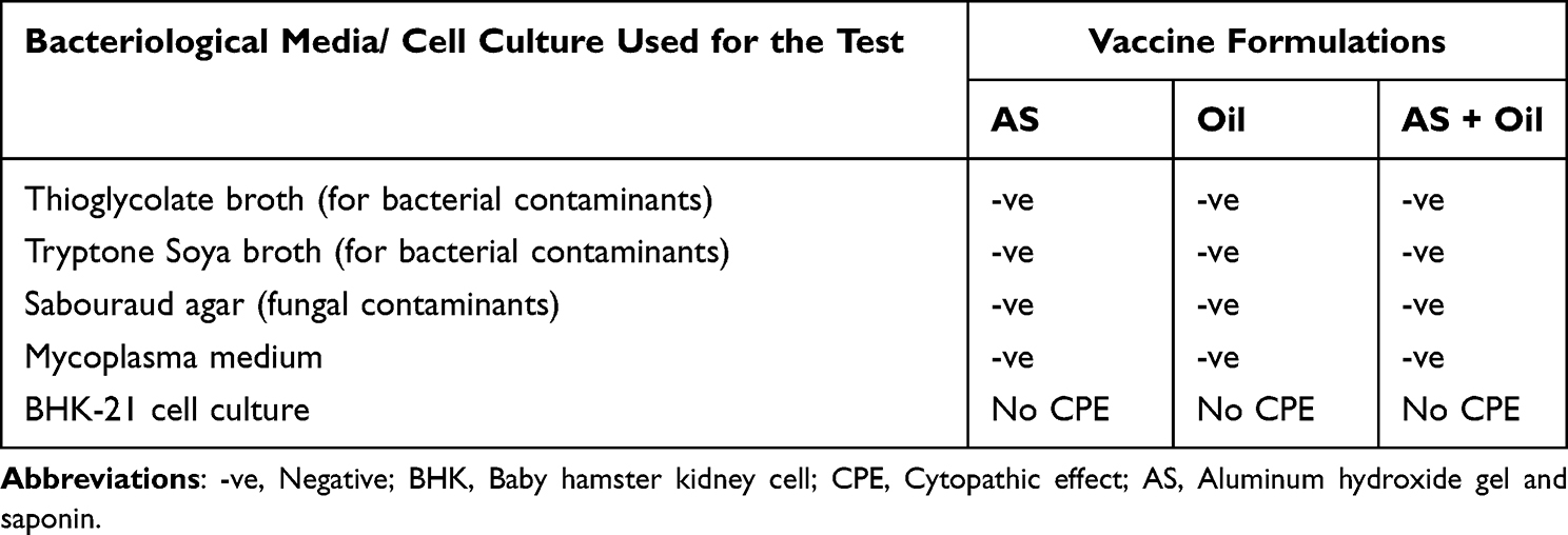 |
Table 1 Summary of Sterility and Safety Test Results of the Prepared Trivalent FMD Vaccines |
Immune Response Result for FMDV Serotype a
In experimental groups inoculated with vaccine formulations containing a mixture of aluminum hydroxide gel and saponin, aluminum hydroxide gel and saponin with a booster dose and aluminum hydroxide gel, saponin and oil, and oil alone, there was an evident increase in antibody level from day 7 to 42 post vaccinations for serotype A (Table 2, Figures 1 and 2). On day 42 after vaccination, the percent of inhibition observed was highest in the experimental group where a mixture of AS plus oil was used as an adjuvant (PI=99.26%) followed by groups inoculated with the vaccine formulation containing oil alone (PI=97.76%) and AS with a booster dose (PI=94.79%). The experimental group inoculated with the formulation, which contains AS alone had the lowest immune response (PI=89.57%) (Table 3). There was a significant difference in humoral immune response in the experimental group vaccinated with the formulation containing AS plus oil (p<0.05) as evidenced by the mean OD value and highest percentage of inhibition compared to other groups.
 |
Table 2 Mean Optical Density (OD) of Antibody Response of Cattle Against FMDV Serotype A |
 |
Table 3 Mean Percentage Inhibition of Antibody Response of Cattle Against FMDV Serotype A as Measured by SPCE |
 |
Figure 1 Line graphs displaying antibody response (OD value) of individual cattle in each experimental group using Solid Phase Competitive ELISA (SPCE) (serotype (A). Control (non-vaccinated animals), Oil (animals vaccinated with oil adjuvanted vaccine), AS (animals vaccinated with aluminum hydroxide gel and saponin adjuvanted vaccine), AS boost (animals vaccinated with aluminum hydroxide gel and saponin adjuvanted vaccine and boosted), AS + Oil (animals vaccinated with Aluminum hydroxide gel, saponin and oil adjuvanted vaccine). *Booster vaccination was given on 14th day. |
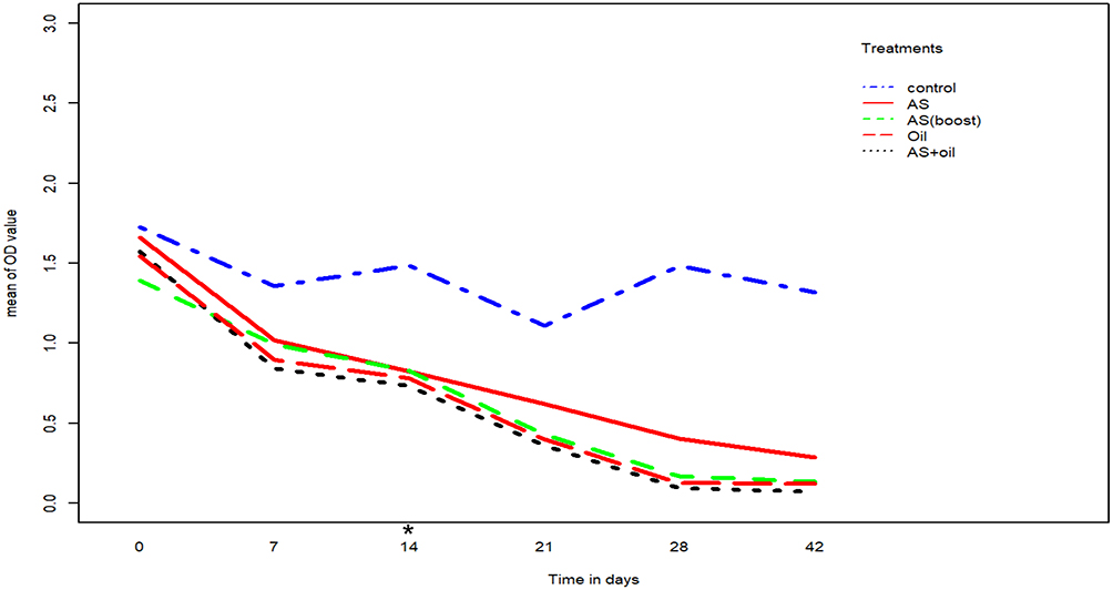 |
Figure 2 Line graph showing the mean antibody response of all experimental groups against serotype A at 0, 7, 14, 21, 28 and 42 days post vaccination using Solid Phase Competitive Elisa (SPCE). The blue dotted line represents control groups; the red line represents groups vaccinated with aluminum hydroxide gel and saponin adjuvanted vaccine (AS); the green dotted line represents groups vaccinated with aluminum hydroxide gel and saponin adjuvanted vaccine and boosted (AS boosted); the red dotted line represents groups vaccinated with oil adjuvanted vaccine (Oil); and the black dotted line represents groups vaccinated with aluminum hydroxide gel, saponin and oil (AS + Oil). *Booster vaccination was given on 14th day. |
Immune Response Result for FMDV Serotype O
In all the four experimental groups, the results revealed that the mean antibody response for FMDV serotype O increased considerably during the post-vaccination period (Table 4, Figures 3 and 4). Significantly higher antibody titers were recorded from day 0 to reach its maximum value at day 42 in experimental groups inoculated with formulations containing AS and mineral oil (1.58, 0.04), and oil alone (1.57, 0.05), (p<0.05). However, the titers were significantly lower in experimental groups inoculated with vaccine preparation adjuvanted with AS (1.62, 0.19) followed by groups inoculated with AS with a booster dose (1.44, 0.12) at days 0 and 42, respectively, compared to other groups (p<0.05). The maximum percent of inhibition at 42 days post vaccination was recorded in experimental groups inoculated with vaccine formulations containing AS plus mineral oil (P=98.47%) and mineral oil alone (P=98.08%). The percent of inhibition in experimental group’s inoculated with vaccine formulations containing AS with a booster dose was 95.4%, whereas this was 92.72% without a booster (Table 5).
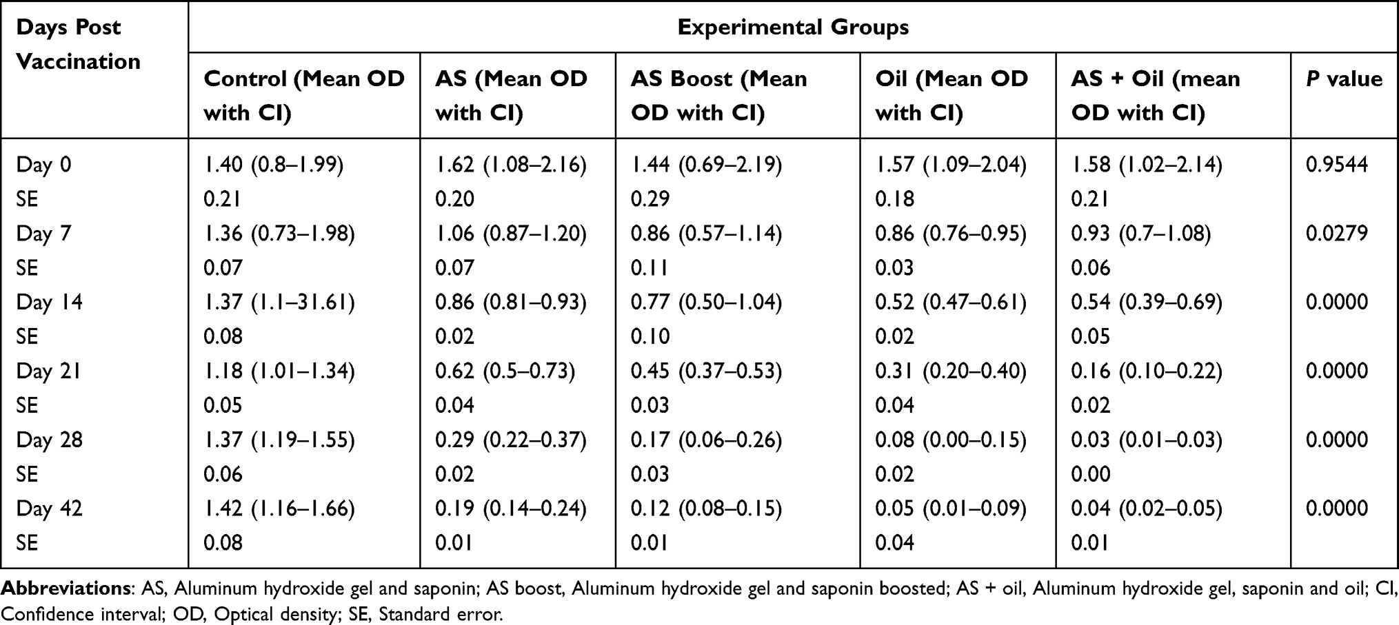 |
Table 4 Mean Optical Density (OD) of Antibody Response of Cattle Against FMDV Serotype O |
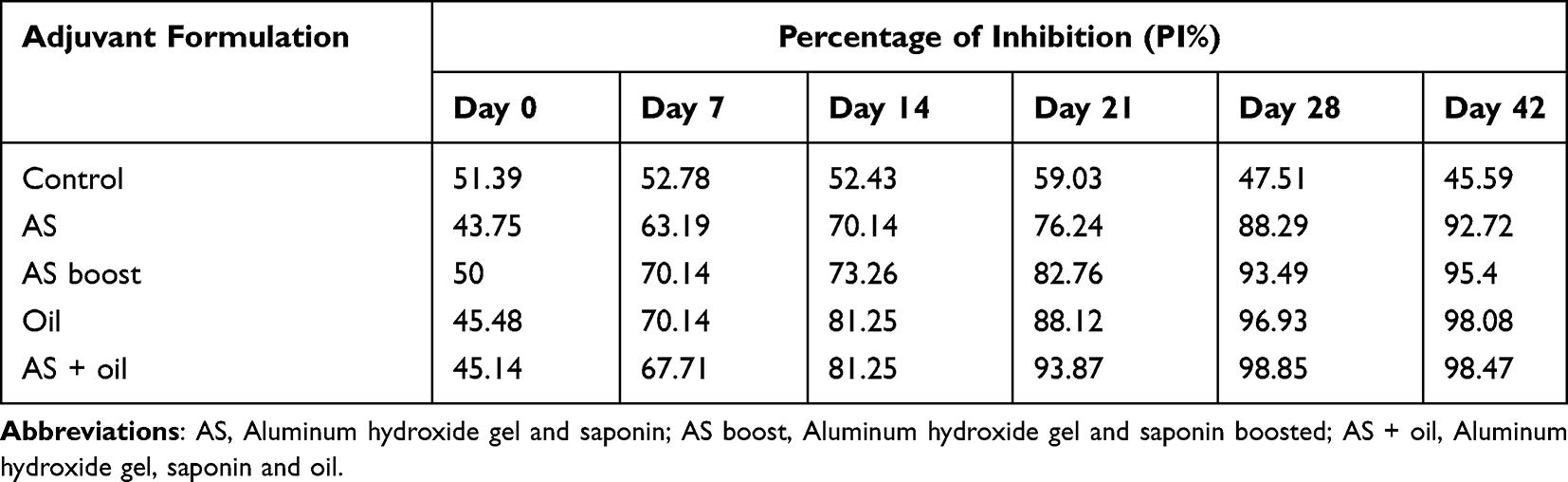 |
Table 5 Mean Percentage Inhibition of Antibody Response of Cattle Against FMDV Serotype O as Measured by SPCE |
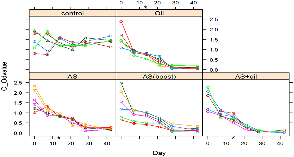 |
Figure 3 Line graphs displaying the antibody response (OD value) of individual animals in each group using Solid Phase Competitive ELISA (SPCE) (serotype (O). Control (non-vaccinated animals), Oil (animals vaccinated with oil adjuvanted vaccine), AS (animals vaccinated with aluminum hydroxide gel and saponin adjuvanted vaccine), AS boost (animals vaccinated with aluminum hydroxide gel and saponin adjuvanted vaccine and boosted), AS + Oil (animals vaccinated with Aluminum hydroxide gel, saponin and oil adjuvanted vaccine). *Booster vaccination was given on 14th day. |
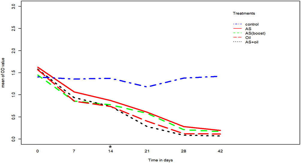 |
Figure 4 Line graph showing the mean antibody response of the groups against serotype O at 0, 7, 14, 21, 28 and 42 days post vaccination using Solid Phase Competitive ELISA (SPCE). The blue dotted line represents control groups; the red line represents groups vaccinated with aluminum hydroxide gel and saponin adjuvanted vaccine (AS); the green dotted line represents groups vaccinated with aluminum hydroxide gel and saponin adjuvanted vaccine and boosted (AS boosted); the red dotted line represents groups vaccinated with oil adjuvanted vaccine (Oil); and the black dotted line represents groups vaccinated with aluminum hydroxide gel, saponin and oil (AS+ Oil). *Booster vaccination was given on 14th day. |
Immune Response Result for FMDV Serotype SAT 2
In a similar pattern to immune responses recorded for the other two serotypes (Serotype A and Serotype O), the highest mean antibody level and percent of inhibition were observed in groups inoculated with vaccine formulation containing a mixture of AS plus oil, and oil alone (Tables 6 and 7, Figures 5 and 6). Whereas, the results showed that groups immunized with the vaccine prepared with AS induced less antibody response and percent of inhibition followed by groups vaccinated with AS with booster dose.
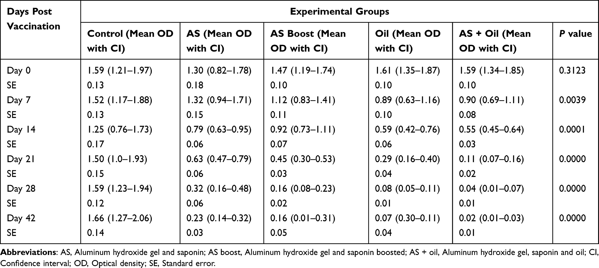 |
Table 6 Mean Optical Density (OD) of Antibody Response of Cattle Against FMDV Serotype SAT 2 |
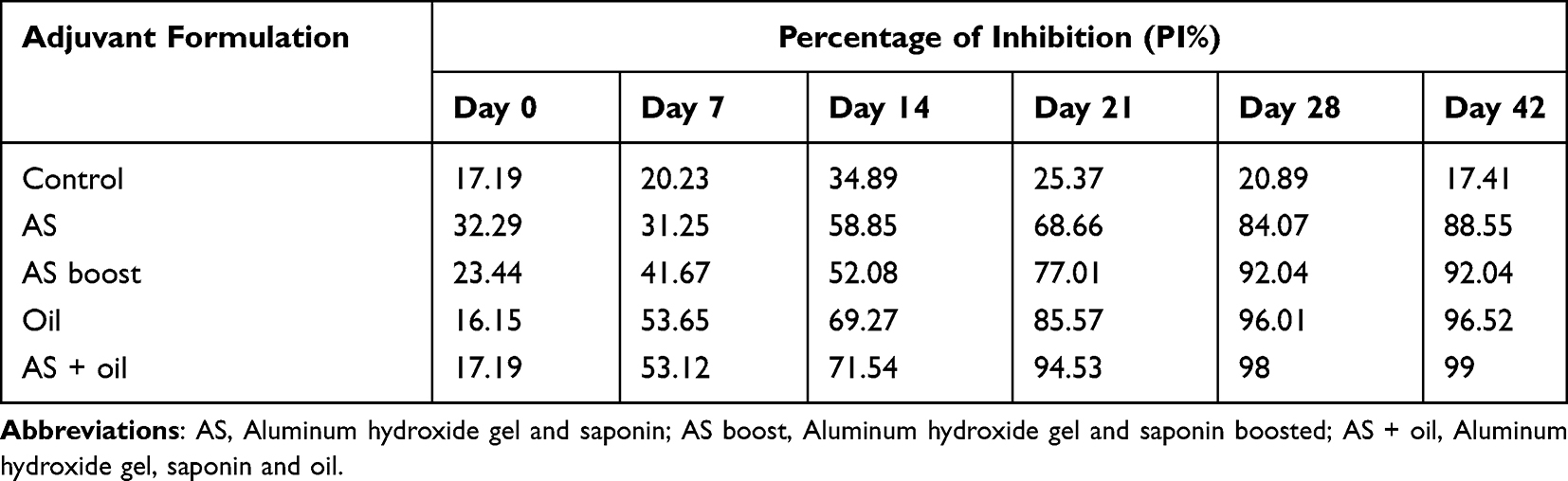 |
Table 7 Mean Percentage Inhibition of Antibody Response of Cattle Against FMDV Serotype SAT 2 as Measured by SPCE |
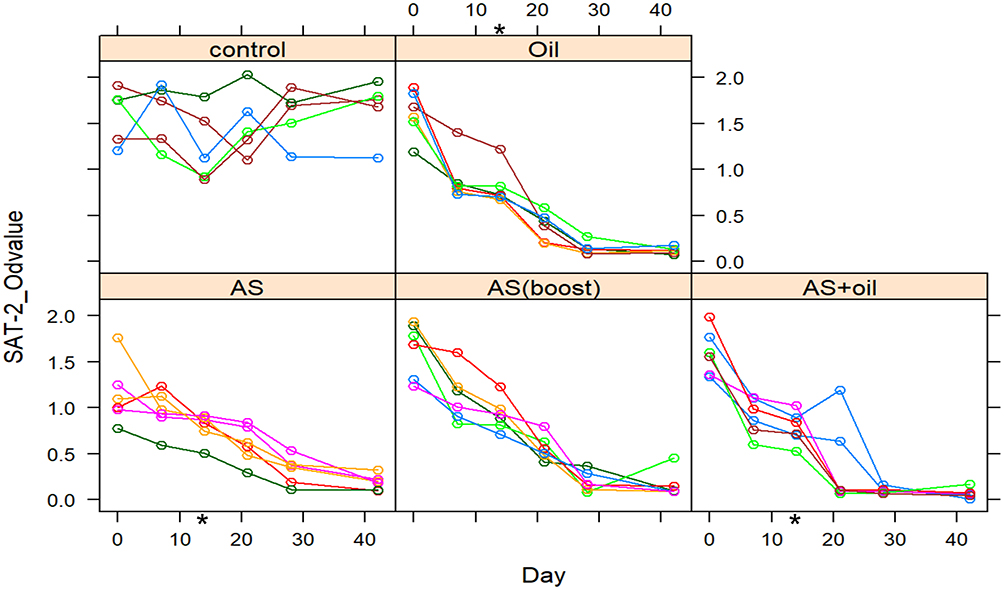 |
Figure 5 Line graphs displaying the antibody response of individual animals in each group using Solid Phase Competitive ELISA (SPCE) (serotype SAT 2). Control (non-vaccinated animals), Oil (animals vaccinated with oil adjuvanted vaccine), AS (animals vaccinated with aluminum hydroxide gel and saponin adjuvanted vaccine), AS boost (animals vaccinated with aluminum hydroxide gel and saponin adjuvanted vaccine and boosted), AS + Oil (animals vaccinated with Aluminum hydroxide gel, saponin and oil adjuvanted vaccine). *Booster vaccination was given on 14th day. |
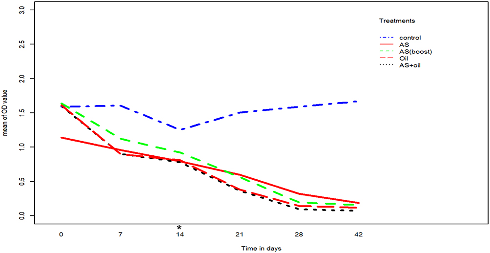 |
Figure 6 Line graph showing the mean antibody response of the experimental groups against serotype SAT 2 at 0, 7, 14, 21, 28 and 42 days post vaccination using Solid Phase Competitive ELISA (SPCE). The blue dotted line represents control groups; the red line represents groups vaccinated with aluminum hydroxide gel and saponin adjuvanted vaccine (AS); the green dotted line represents groups vaccinated with aluminum hydroxide gel and saponin adjuvanted vaccine and boosted (AS boosted); the red dotted line represents groups vaccinated with oil adjuvanted vaccine (Oil); and the black dotted line represents groups vaccinated with aluminum hydroxide gel, saponin and oil (AS + Oil). *Booster vaccination was given on 14th day. |
The Challenge Study
This study which involved injecting experimental animals’ upper tongues intradermally with 106.4 TCID50/mL of the homologous FMDV serotype O at 28 days post vaccination, failed and needs further investigation. None of the experimental animals including the control groups showed clinical disease or lesions suggestive of FMD.
Discussion
Foot-and-Mouth Disease (FMD) is an acute, febrile, and highly contagious vesicular disease of cloven-hoofed animals caused by FMDV. It is one of the most devastating diseases of livestock that causes significant economic losses following an outbreak.38 Control of the disease particularly in endemic areas could be achieved through vaccination with the use of quality vaccine prepared with locally circulating serotypes and effective adjutants.39 Adjuvants have been used for more than 70 years even when the mode of their action had not been understood.40 The use of adjuvants in inactivated FMD vaccine formulation plays a vital role in improving the development of rapid and long-lasting protective immunity. Thus, the findings of the present study are imperative in contributing valuable information in the understanding of the effective formulations of FMD vaccines and selection of adjuvants used in the eradication of the disease.
Overall, in all cattle vaccinated with various experimental vaccine formulations SPCE result indicated that there was a decrease in the OD value of a conjugate antibody during the post-vaccination period (Tables 2–7 and Figures 1–6), which is because of the competition between antibodies in the test sera and enzyme conjugated antibody for the same antigen. The decrease in the OD value is indirectly proportional to the antibody level in the test serum. This in turn indicates an increase in the antibody titers and the percentage of inhibition as the antibodies available in the test sera impede binding of conjugated antibody (Tables 2, 4 and 6).
Evaluation of antibody responses to the serotype A, O and SAT 2, AS, oil-based, and combination of AS with oil adjuvanted vaccines revealed considerable variation both in the rapidity of development and magnitude of humoral immune response (Figures 1, 3 and 5). The variation in immune response is related primarily to difference in formulation and booster injection to experimental animals.
In groups vaccinated with AS formulations in all the three serotypes used in this study, the booster dose has shown a statistically significant difference in mean antibody response compared to groups without the booster dose (p<0.05). This is in agreement with Cloete et al21 and Patil et al,41 who described the poor immune response elicited by inactivated FMD vaccine formulated with aluminum hydroxide gel and saponin necessitate relatively frequent re-vaccinations to provide protective immunity. Lyons et al38 also suggested that among factors that limit an effective performance of the inactivated FMD vaccine, the requirement for repeat boosting. Also, similar results obtained by Cokcaliskan et al42 in cattle suggested that better immune response in cattle could be due to the effect of booster dose.
The antibody response between AS boosted and oil-based group were not statistically significant for the three serotypes until day 21 post vaccination (p>0.05). As described by Garcon et al,43 this may be due to the reason that oil emulsions adjuvant function as delivery systems by the formation of depot effect that traps antigen at the injection site and by slowing the release of antigen to induce a long-lasting immune response. However, the mean antibody response reached to the maximum in aluminum hydroxide gel and saponin boosted group for serotypes A, O and SAT2 on days 28 and 42 were significantly lower than in groups with oil alone (p<0.05). These results were concordant with those reported by Patil et al,44 who found that oil adjuvant elicited a better immune response than aluminum hydroxide gel and saponin vaccine.
The use of the combined formulation of AS and mineral oil as an adjuvant is not common. In the present study, a mixture of AS and mineral oil adjuvant for inactivated trivalent foot and mouth disease vaccine was evaluated. The ELISA results and statistical analysis revealed that experimental group that had mixed adjuvant showed relatively a higher post-vaccination antibody response than those that received the vaccine from the three serotypes of FMDV with a single adjuvant suggesting that the combination of AS and mineral oil have enhancing effect on the immune response. This was partly in agreement with Garcon et al,43 in that FMD vaccine prepared with mixing aluminum hydroxide gel and oil-based adjuvant produced a better humoral immune response than oil alone. A study conducted in mice by Park et al,45 showed that combinations of aluminum hydroxide gel and oil-based adjuvant revealed the highest protection rate. Moreover, the results also partially corresponded to previous work conducted by Park et al,9 who reported a combination of oil with a gel enhanced the immune response in experimental animals and concluded that oil improves the effects of gel of adjuvant.
On the other hand, a synergy between oil and saponin for immune response has been reported in some studies. Cokcaliskan et al3 observed a strong antibody response in cattle vaccinated with FMD vaccine formulated with combined oil and saponin adjuvants. The study conducted by Bazid et al46 in guinea pigs and cattle with monovalent FMDV serotype O, also reported that oil-based FMD vaccines containing saponin induced higher antibody level than vaccine without saponin. These findings are partially corroborating with the finding of the present study that enhanced antibody response was obtained by the use of aluminum hydroxide gel and saponin in combination with mineral oil adjuvant in a FMD vaccine compared to other adjuvants.
Various combination adjuvants consisting of a variety of molecules act synergistically by activating a variety of immune mechanisms. These “combination adjuvants” significantly improve vaccine efficacy by modulating, enhancing, or extending the immune response and at the same time reducing the amount of antigen.47 Even though the cost-benefit analysis of adjuvant combinations is not conducted in this study, the results of AS+ oil combinations in this study indicate a potential alternative to the conventional combination in FMD vaccinations in cattle.
In this experimental study, the virus prepared for the challenge test did not develop any typical clinical signs and lesions of FMD in the control group. This is in agreement with the finding of Cloete et al,22 who reported that the control cattle did not develop any clinical disease. This might be due to the serial passage of the virus used for vaccine production (laboratory adapted virus) which could lead to losing its infectivity. It also suggests that in order to increase the virus’s infectivity the virus need to be re-adapted to animal virulence by passage in cattle and lab animals.22
Conclusion
This study demonstrated that cattle vaccinated with a single injection of vaccine formulations containing aluminum hydroxide gel and saponin as an adjuvant had a lower antibody response compared to cattle boosted at day 14 with the same vaccine formulation. These observations confirmed that the booster dose is necessary to enhance the immune response of aluminum hydroxide gel and saponin adjuvanted vaccine against FMD. Besides, trivalent FMD vaccine prepared with oil and mixture of aluminum hydroxide gel, saponin and oil adjuvant vaccine elicited better immune response than the other adjuvant formulation. Thus, our data strongly suggest that oil-based and aluminum hydroxide gel and saponin with oil adjuvanted vaccine could replace the aluminum hydroxide gel and saponin in FMD vaccine. Since the combination of three adjuvants in the FMD vaccine is the first of its kind, future experimental research on the evaluation of these adjuvants, including duration of protection and biological characterization of viral infectivity is highly recommended.
Abbreviations
ARERC, Animal Research Ethics Review Committee; AS, Aluminum hydroxide gel-Saponin; BHK, Baby Hamster Kidney; ELISA, Enzyme-Linked Immune Sorbent Assay; FAO, Food and Agriculture Organization; FMD, Foot and Mouth Disease; FMDV, Foot and Mouth Disease Virus; NAHDIC, National Animal Health Diagnostic and Investigation center; NVI, National Veterinary Institute; O/W, Oil in Water; OIE, Office International des Epizooties; PANVAC, Pan African Veterinary Vaccine Center; PBS, Phosphate Buffer Saline; SAT, Southern African Territory; SPCE, Solid Phase Competitive Elisa; TCID, Tissue Culture Infectivity Dose.
Data Sharing Statement
The datasets used and/or analysed during the current study are available from the corresponding author on reasonable request and with permission of IRB and/or other responsible bodies in Ethiopia.
Ethical Approval
The experimental animals were treated according to the protocol of ethical guidelines of the Animal Welfare Committee of Addis Ababa University College of Veterinary Medicine and Agriculture (AAU-CVM). They were kept and cared in the animal facility of NVI and used for the experiment after approval from the Animal Research Ethics Review Committee (ARERC) of Addis Ababa University College of Veterinary Medicine and Agriculture (AAU-CVM) (Ref No: VM/ERC/27/06/13/2021).
Acknowledgment
The authors would like to thank the National Veterinary Institute and the National Animal Health Institute (AHI) for supporting the study.
The study was the first author’s master’s thesis presented to Addis Ababa University, College of Veterinary Medicine, and Agriculture, and it can be found online in the Addis Ababa University Institutional thesis repository. It was the research project funded by the National Veterinary Institute, and all co-authors contributed significantly to the research work.
Funding
This study was financially supported by the National Veterinary Institute (NVI), Bishoftu, Ethiopia. The funding organization had no any role in the design of the study and collection, analysis, and interpretation of data and in writing the manuscript.
Disclosure
The authors declare that there are no conflicts of interest regarding the publication of this paper.
References
1. Asresie A, Zemedu L, Adigrat E. The contribution of livestock sector in Ethiopian economy. Rev Adv Life Sci Technol. 2015;29:1.
2. Sulayeman M, Dawo F, Mammo B, Gizaw D, Shegu D. Isolation, molecular characterization and sero-prevalence study of foot-and-mouth disease virus circulating in central Ethiopia. BMC Vet Res. 2018;14(1):110. doi:10.1186/s12917-018-1429-9
3. Çokçalışkan C, Türkoğlu T, Sareyyüpoğlu B, et al. QS-21 enhances the early antibody response to oil adjuvant foot-and-mouth disease vaccine in cattle. Clin Exp Vaccine Res. 2016;5(2):138–147. doi:10.7774/cevr.2016.5.2.138
4. Knight-Jones TJD, Rushton J. The economic impacts of foot and mouth disease: what are they, how big are they and where do they occur? Prev Vet Med. 2013;112:161–173. doi:10.1016/j.prevetmed.2013.07.013
5. Jamal SM, Belsham GJ. Foot-and-mouth disease: past, present and future. Vet Res. 2013;44(1):1–14. doi:10.1186/1297-9716-44-116
6. Tadesse B. Review on the evaluation of efficacy of foot and mouth disease vaccine. Rep Opinion. 2018;10(2):55–56.
7. Maree FF, Kasanga CJ, Scott KA, et al. Challenges and prospects for the control of foot-and-mouth disease: an African perspective. Vet Med. 2014;5:119. doi:10.2147/VMRR.S62607
8. Ayelet G, Mahapatra M, Gelaye E, et al. Genetic characterization of foot-and-mouth disease viruses, Ethiopia, 1981–2007. Emerg Infect Dis. 2009;15(9):1409. doi:10.3201/eid1509.090091
9. Park ME, Lee SY, Kim RH, et al. Enhanced immune responses of foot-and-mouth disease vaccine using new oil/gel adjuvant mixtures in pigs and goats. Vaccine. 2014;32(40):5221–5227. doi:10.1016/j.vaccine.2014.07.040
10. Bastos ADS, Sangare O. Geographic distribution of SAT-2 type foot-and-mouth disease virus genotypes in Africa.
11. Brito BP, Rodriguez LL, Hammond JM, Pinto J, Perez AM. Review of the global distribution of foot-and-mouth disease virus from 2007 to 2014. Transbound Emerg Dis. 2015;64:316–332. doi:10.1111/tbed.12373
12. Ayelet G, Gelaye E, Guitian J, Sahle M, Knowles NJ, Mahapatra M. The Status of Foot and Mouth Disease (FMD) in Ethiopia. Italy: The Global control of FMD; 2008.
13. Yirgalem M, Dawo F, Gizaw D, Mamo B, Bilata T, Shegu D. Phylogenetic and sequence variability analyses of Vp1 Protein of foot and mouth disease viruses in cattle in Amhara Region of Ethiopia; 2020.
14. Megersa B, Beyene B, Abunna F, Regassa A, Amenu K, Rufael T. Risk factors for foot and mouth disease seroprevalence in indigenous cattle in Southern Ethiopia: the effect of production system. Trop Anim Health Prod. 2009;41(6):891–898. doi:10.1007/s11250-008-9276-5
15. Yahya M, Hailemariam Z, Amare LB, Rufael T. ‘Seroprevalence of foot and mouth disease in traditionally managed cattle in East and West Hararghe zones, Ethiopia. Rev Elev Med Vet Pays Trop. 2013;66(1):1.
16. Jemberu WT, Mourits M, Rushton J, Hogeveen H. Cost-benefit analysis of foot and mouth disease control in Ethiopia. Prev Vet Med. 2016;132:67–82. doi:10.1016/j.prevetmed.2016.08.008
17. Arzt J, Belsham GJ, Lohse L, Bøtner A, Stenfeldt C. Transmission of foot-and-mouth disease from persistently infected carrier cattle to naive cattle via transfer of oropharyngeal fluid. Msphere. 2018;3(5). doi:10.1128/mSphere.00365-18
18. Xiao C, Rajput ZI, Liu D, Hu S. Enhancement of serological immune responses to foot-and-mouth disease vaccine by a supplement made of extract of Cochin Chinamomordica seeds. Clin Vaccine Immunol. 2007;14(12):1634–1639. doi:10.1128/CVI.00339-07
19. Ibrahim EES, Gamal WM, Hassan AI, Mahdy SED, Hegazy AZ, Abdel-Atty MM. Comparative study on the immune potentiator effect of ISA 201, ISA 61, ISA 50, ISA 206 used in trivalent foot and mouth disease vaccine. Vet World. 2015;8(10):1189. doi:10.14202/vetworld.2015.1189-1198
20. Park JH. Requirements for improved vaccines against foot-and-mouth disease epidemics via transfer of oropharyngeal fluid. Clin Exp Vaccine Res. 2013;2(1):8. doi:10.7774/cevr.2013.2.1.8
21. Ashenafi B Costs and Benefits of Foot and Mouth Disease Vaccination Practices in Commercial Dairy Farms in Central Ethiopia [MSc thesis]. Wageningen University; 2012.
22. Cloete M, Dungu B, Van Staden LI, Ismail-Cassim N, Vosloo W. Evaluation of different adjuvants for foot-and-mouth disease vaccine containing all the SAT serotypes. Onderstepoort J Vet Res. 2008;75(1):17–31. doi:10.4102/ojvr.v75i1.84
23. Choi JH, You SH, Ko MK, et al. Improved immune responses and safety of foot-and-mouth disease vaccine containing immunostimulating components in pigs. J Vet Sci. 2020;21(5). doi:10.4142/jvs.2020.21.e74
24. Selim A, Abouzeid N, Aggour AM, Sobhy NM. Comparative study for immune efficacy of two different adjuvants bivalent FMD vaccines in sheep. J Am Sci. 2010;6:1292–1298.
25. Lyer AV, Ghosh S, Singh SN, Deshmukh RA. Evaluation of three ‘ready to formulate’ oil adjuvants for foot-and-mouth disease vaccine production. Vaccine. 2000;19(9–10):1097–1105. doi:10.1016/S0264-410X(00)00337-6
26. Gamaledin WM, EhabElsayed AI, Hassanin AA, Mohamed WS. Performance of aluminium hydroxide gel and ISA oils adjuvanted foot and mouth disease vaccines. ARC J Anim Vet Sci. 2019;5(3):1–9.
27. Hassan AI. Effect of different culture systems on the production of foot and mouth disease trivalent vaccine. Vet World. 2016;9(1):32. doi:10.14202/vetworld.2016.32-37
28. Kallel H, Jouini A, Majoul S, Rourou S. Evaluation of various serum and animal protein free media for the production of a veterinary rabies vaccine in BHK-21 cells. J Biotechnol. 2002;95(3):195–204. doi:10.1016/S0168-1656(02)00009-3
29. Kim H, Kim AY, Kim JS, et al. Determination of the optimal method for the concentration and purification of 146S particles for foot-and-mouth disease vaccine production. J Virol Methods. 2019;269:26–29. doi:10.1016/j.jviromet.2019.04.009
30. Nazzarodel Cañizo AAN, Mazza M, Bellinzoni R, Cascone O. Foot and mouth disease virus concentration and purification by affinity chromatography. Appl Biochem Biotechnol. 1997;61(3):399–409. doi:10.1007/BF02787811
31. Karber G. Beitrage zur kille kiven behand–lung pharmakologis reihenver-suche naunyn schiede berg. Naunyn-Schmiedebergs Arch Pharm. 1931;126:280–283.
32. Barteling SJ, Cassim NI. Very fast (and safe) inactivation of foot-and-mouth disease virus and enteroviruses by a combination of binary ethyleneimine and formaldehyde. Dev Biol. 2004;119:449–455.
33. OIE PB. NB: Version Adapted by the World Assembly of Delegates of the Office International Des Epizooties. Paris, France: OIE PB; 2009.
34. El-Sayed EI, Mossad WG, Hassanin AI, W. S. Immunomodulating efficacy of different adjuvants in formulation of Foot and Mouth disease vaccine relative to its immunogenicity. J Appl Vet Sci. 2020;5(3):14–30. doi:10.21608/javs.2020.98318
35. Pinto J. Foot-and-Mouth Disease Situation Food and Agriculture Organization of the United Nations Monthly Report. Roma, Italy: FAO; 2017.
36. OIE. Foot and mouth disease (infection with foot and mouth disease virus) (NB: version adopted in May 2017): OIE terrestrial manual; 2018.
37. Pacheco JM, Brum MC, Moraes MP, Golde WT, Grubman MJ. Rapid protection of cattle from direct challenge with foot-and-mouth disease virus (FMDV) by a single inoculation with an adenovirus-vectored FMDV subunit vaccine. Virology. 2005;337(2):205–209. doi:10.1016/j.virol.2005.04.014
38. Rodriguez LL, Grubman MJ. Foot and mouth disease virus vaccines. Vaccine. 2009;27:90–96. doi:10.1016/j.vaccine.2009.08.039
39. Lyons NA, Lyoo YS, King DP, Paton DJ. Challenges of generating and maintaining protective vaccine-induced immune responses for foot-and-mouth disease virus in pigs. Front Vet Sci. 2016;3:102. doi:10.3389/fvets.2016.00102
40. Macedo LB, Lobato ZIP, Fialho SL, Viott ADM, Guedes RMC, Silva-Cunha A. Evaluation of different adjuvants formulations for bluetongue vaccine. Braz Arch Biol Technol. 2013;56(6):932–941. doi:10.1590/S1516-89132013005000002
41. Patil PK, Bayry J, Ramakrishna C, et al. Immune responses of sheep to quadrivalent double emulsion foot-and-mouth disease vaccines: rate of development of immunity and variations among other ruminants. J Clin Microbiol. 2012;40(11):4367. doi:10.1128/JCM.40.11.4367-4371.2002
42. Çokçalışkan C, Türkoğlu T, Uzunlu E, et al. Influence of vaccine potency and booster administration of foot-and-mouth disease vaccines on the antibody response in cattle with maternal antibodies. J Vet Sci. 2017;18(1):315. doi:10.4142/jvs.2017.18.S1.315
43. Garçon N, Leroux-Roels G, Cheng WF. Vaccine adjuvants. Perspect Vaccinol. 2011;1(1):89–113. doi:10.1016/j.pervac.2011.05.004
44. Patil PK, Bayry J, Ramakrishna C, Hugar B, Misra LD, Natarajan C. Immune responses of goats against foot-and-mouth disease quadrivalent vaccine: comparison of double oil emulsion and aluminium hydroxide gel vaccines in eliciting immunity. Vaccine. 2002;20(21–22):2781–2789. doi:10.1016/S0264-410X(02)00184-6
45. Park ME, Lee SY, Kim RH, et al. Altered adjuvant of foot-and-mouth disease vaccine improves immune response and protection from virus challenge. Trials Vaccinol. 2016;5:97–104. doi:10.1016/j.trivac.2016.04.006
46. Bazid AHI, El-Alfy HA, El-Didamony G, Elfeil WK, El-Sayed MM, Fawzy M. Adjuvant effect of saponin in an oil-based monovalent (serotype O) foot-and-mouth disease virus vaccine on the antibody response in Guinea pigs and cattle. Arch Virol. 2021;2021:1–8.
47. Levast B, Awate S, Babiuk L, Mutwiri G, Gerdts V, van Drunen Littel-van den Hurk S. Vaccine potentiation by combination adjuvants. Vaccines. 2014;2(2):297–322.
 © 2023 The Author(s). This work is published and licensed by Dove Medical Press Limited. The full terms of this license are available at https://www.dovepress.com/terms.php and incorporate the Creative Commons Attribution - Non Commercial (unported, v3.0) License.
By accessing the work you hereby accept the Terms. Non-commercial uses of the work are permitted without any further permission from Dove Medical Press Limited, provided the work is properly attributed. For permission for commercial use of this work, please see paragraphs 4.2 and 5 of our Terms.
© 2023 The Author(s). This work is published and licensed by Dove Medical Press Limited. The full terms of this license are available at https://www.dovepress.com/terms.php and incorporate the Creative Commons Attribution - Non Commercial (unported, v3.0) License.
By accessing the work you hereby accept the Terms. Non-commercial uses of the work are permitted without any further permission from Dove Medical Press Limited, provided the work is properly attributed. For permission for commercial use of this work, please see paragraphs 4.2 and 5 of our Terms.