")
Back to Journals » International Journal of General Medicine » Volume 15
Circular RNAs Hsa_circ_101555 and Hsa_circ_008068 as Diagnostic Biomarkers for Early-Stage Lung Adenocarcinoma
Authors Lian X, Cao D, Hu X, Mo W, Yao X, Mo J, Wang H
Received 28 March 2022
Accepted for publication 31 May 2022
Published 9 June 2022 Volume 2022:15 Pages 5579—5589
DOI https://doi.org/10.2147/IJGM.S367999
Checked for plagiarism Yes
Review by Single anonymous peer review
Peer reviewer comments 2
Editor who approved publication: Dr Scott Fraser
Xue Lian,1,2 Dakui Cao,2 Xun Hu,3 Weiqiang Mo,2 Xiujuan Yao,4 Juanfen Mo,5 Haiqin Wang1,2
1Department of Respiratory Medicine, Bengbu Medical College, Bengbu, Anhui, 233000, People’s Republic of China; 2Department of Respiratory Medicine, The Second Affiliated Hospital of Jiaxing University, Jiaxing, Zhejiang, 314000, People’s Republic of China; 3Department of Thoracic Surgery, The Second Affiliated Hospital of Jiaxing University, Jiaxing, Zhejiang, 314000, People’s Republic of China; 4Department of Pathology, The Second Affiliated Hospital of Jiaxing University, Jiaxing, Zhejiang, 314000, People’s Republic of China; 5The Key Laboratory, The Second Affiliated Hospital of Jiaxing University, Jiaxing, Zhejiang, 314000, People’s Republic of China
Correspondence: Haiqin Wang, Department of Respiratory Medicine, The Second Hospital of Jiaxing, 1518 Huanchen North Road, Jiaxing, Zhejiang, 314000, People’s Republic of China, Email [email protected]
Background: Lung adenocarcinoma (LUAD) is a life-threatening disease worldwide with a high mortality rate. The early diagnosis of LUAD is crucial for improving subsequent treatment and prognosis. However, biomarkers for early detection remain a clinical challenge in LUAD. Here, we aimed to develop circular RNAs (circRNAs) in circulating plasma from LUAD patients as valuable diagnostic biomarkers in LUAD.
Methods: CircRNA expression was determined by circRNA microarray in three pairs of LUAD tumour tissues and patient-matched normal lung tissues. Hsa_circ_101555 and hsa_circ_008068 were selected as potential biomarkers in LUAD tissues and plasma by RT–PCR, respectively. The diagnostic value was analysed by the area under the curve (AUC) and the receiver operating characteristic (ROC) test.
Results: Our results showed that 6261 circRNAs were upregulated and 7238 circRNAs were downregulated in LUAD tumour tissues compared with patient-matched normal lung tissues. Hsa_circ_101555 and hsa_circ_008068 were filtered as biomarkers for early-stage LUAD. Q-PCR results showed that hsa_circ_101555 and hsa_circ_008068 were significantly upregulated in both LUAD cancer tissues and circulating plasma. Hsa_circ_101555 and hsa_circ_008068 were positively associated with tumour differentiation, tumour size and CEA (P< 0.05). The ROC analysis showed that hsa_circ_101555 and hsa_circ_008068 had a better diagnostic potential compared to the traditional biomarkers (CEA, SCC, CYFRA21-1) in the detection of early-stage LUAD.
Conclusion: The circular RNAs hsa_circ_101555 and hsa_circ_008068 could serve as novel diagnostic biomarkers for early-stage LUAD.
Keywords: circRNAs, lung adenocarcinoma, diagnostic biomarker, circ101555
Introduction
Lung adenocarcinoma (LUAD) is one of the most commonly diagnosed lung cancers and is the main cause of cancer-related death worldwide, accounting for more than 50% of non-small-cell lung cancers, and the percentage continues to increase.1,2 LUAD is characterized by its poor clinical prognosis and high morbidity and severely affects the quality of life of patients.3 Despite substantial efforts in the treatment of lung cancer, the 5-year survival rates remain lower than 17%.4 If diagnosis and treatment are started in the early stage, the prognosis of lung cancer is notably improved. Therefore, early diagnostic biomarkers are required to improve the prevention, treatment, and survival rates of LUAD.
Currently, lung tissue biopsy is still the gold standard technique for accurately diagnosing LUAD, but specimens are often difficult to obtain.5 As a complement, low-dose computed tomography (LDCT) screening is a noninvasive method for the early detection of LUAD.6 Nevertheless, the percentage of subjects with pulmonary nodules and the rate of false-positive diagnosis are both relatively high.7 In the past decade, liquid biopsy has been an adjunctive test to help evaluate the malignancy potential and showed no radiation exposure compared with LDCT.8 Biomarkers are detected in body fluids and include exosomes, long noncoding RNAs (lncRNAs), microRNAs (miRNAs), circulating tumour nucleic acids (ctDNA), and circulating tumour cells (CTCs).9 However, the early diagnosis of LUAD has a low sensitivity in liquid biopsy. Hence, a noninvasive and effective liquid biopsy for detecting early-stage LUAD is urgently needed.
Circular RNAs (circRNAs) are a novel class of endogenous noncoding RNAs that are widely expressed in eukaryotic cells.10 They are primarily expressed in exons or introns, generating exonic circRNAs or intronic circRNAs.11 CircRNAs were detected as miRNA sponges that compete with endogenous RNAs to affect gene expression, thereby regulating their downstream target genes.12 The transcripts of circRNAs are considered stable and highly conserved, with no free 5′ cap or 3′ tail in their structure.13 In recent years, increasing evidence suggests that circRNAs participate in various biological processes implicated in human malignant tumours, such as proliferation, migration, invasion, and apoptosis.14–16 For instance, hsa_circ_0072309 was downregulated in breast cancer and acted as a miR-492 sponge to promote cell proliferation, migration, and invasion.17 Liang et al indicated that circ β-catenin was increased in liver cancer tissues and affected liver cancer cell growth via the Wnt/β-catenin signalling pathway.18 In lung cancer, hsa_circ_0007580 was verified to promote lung cancer cell proliferation and invasion by sponging miR-545-3p to inhibit protein kinase Ca (PKCA) by activating the p38/MAPK signalling pathway.19 Furthermore, circ_0074027 plays an oncogenic role in lung cancer by sponging miRNA-2467-3p to upregulate bromodomain-containing protein 4 (BRD4) and activate the MAPK signalling pathway.20 These characteristics of circRNA demonstrate that dysregulation of circRNAs plays critical roles in cancer initiation and development.
In the present study, the differential expression profiles of tissue circRNAs in LUAD were first investigated by circRNA microarray and identified hsa_circ_101555 and hsa_circ_008068 as significantly upregulated in lung tissues. Subsequently, the target circRNAs were further validated by reverse transcription-quantitative polymerase chain reaction (RT–qPCR) to verify the diagnostic efficacy of circRNAs from tissues and plasma in LUAD patients. CircRNAs may be developed to serve as new early diagnostic biomarker profiles in LUAD.
Methods
Subjects
In this study, a total of fourteen paired LUAD tumour tissues and matched non cancer tissues were obtained from patients who underwent surgery and were diagnosed with stage I LUAD at the Second Affiliated Hospital of Jiaxing University between January 2020 and July 2021. Thirty plasma samples were collected from stage I LUAD patients. Thirty plasma samples of healthy controls were enrolled from the physical examination centre at the Second Affiliated Hospital of Jiaxing University. This study was approved by the ethics committees of the Second Affiliated Hospital, Jiaxing University (Ethics number: jxey-2020SW019), which accorded with the ethical standards formulated in the Helsinki Declaration. All patients offering clinical materials provided written informed consent and signed informed consent forms. The clinical characteristics of LUAD patients are presented in Table 1.
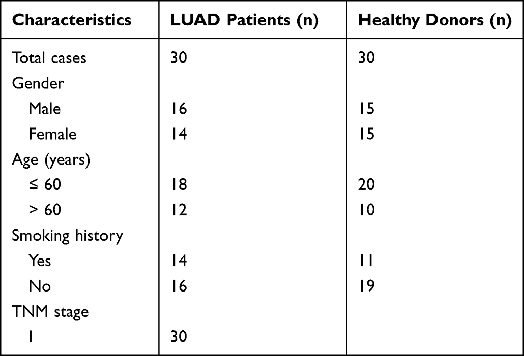 |
Table 1 Clinical Characteristics of LUAD Patients |
Microarrays
We detected the expression profile of circRNAs in six tissue samples, including three paired LUAD cancer tissues and patient-matched normal lung tissues. Microarray analysis was performed by KangChen Biotech, Shanghai. Total RNA from each sample was quantified using a NanoDrop ND-2000. The sample preparation and microarray hybridization were performed based on Arraystar’s standard protocols. Briefly, total RNA was digested with RNase R (Epicentre, Inc.) to remove linear RNAs and enrich circRNAs. Then, the enriched circRNAs were amplified and transcribed into fluorescent circRNAs utilizing a random priming method (Arraystar Super RNA Labelling Kit; Arraystar). The labelled circRNAs were hybridized onto an Arraystar Human circRNA Array V2 (8x15K, Arraystar). After washing the slides, the arrays were scanned by an Agilent Scanner G2505C.
Quantile normalization and subsequent data processing were performed using the R software limma package. Differentially expressed circRNAs with statistical significance between the two groups were identified through volcano plot filtering. Differentially expressed circRNAs between two samples were identified through fold change filtering. Hierarchical clustering was performed to show the distinguishable circRNA expression pattern among samples.
Quantitative Real-Time Reverse Transcription PCR
Tissue RNA was extracted by TRIzol reagent (Invitrogen, USA). Plasma RNA was extracted using RNA extract reagent according to the manufacturer’s protocol (HiPure Serum/Plasma RNA Kit, Magen). A PrimeScriptTM RT reagent kit (RR047A, Takara) was used to reverse transcribe mRNA into cDNA. Then, TB Green fast q-PCR mix (RR820A, Takara) was applied to perform RT-qPCR using an ABI StepOne Plus system according to the manufacturer’s instructions. The reaction mixture was incubated at 95 ℃ for 2 min, followed by 40 cycles at 95 ℃ for 30s, 57 ℃ for 30s and 72 ℃ for 30s. β-Actin was used as the internal control to calculate the relative mRNA levels of target genes using the 2-ΔΔCT method. The relative expression levels of circRNAs were calculated using the 2−ΔΔCT method. β-Actin was selected as the housekeeping gene and normalized as the reference.
Statistical Analysis
Statistical calculations were performed using GraphPad Prism Software 7.0 (GraphPad, USA). The Wilcoxon matched-pairs signed rank test was used to compare the expression levels of circRNAs between LUAD tumour tissues and patient-matched normal lung tissues. t-tests were used for the analysis of differential hsa_circ_101555 and hsa_circ_008068 expression between the plasma of LUAD patients and healthy donors. Correlations were analysed using Spearman’s rank correlation test. The receiver operating characteristic (ROC) curve and area under the ROC curve (AUC) were used to evaluate the diagnostic performance of the selected circRNAs. A P value < 0.05 was considered statistically significant.
Results
Differentially Expressed circRNAs by Microarray in LUAD Cancer Tissues
A circRNA expression microarray was performed to determine circRNA expression in three pairs of LUAD tumour tissues and patient-matched normal lung tissues. We detected a genome-wide analysis of circRNAs using the Arraystar Human CircRNA Microarray. In total, 6261 circRNAs were identified as upregulated and 7238 circRNAs were downregulated with FC ≥ 1.5 and P values ≤ 0.05 in LUAD tumour tissues compared to adjacent normal tissues. Hierarchical clustering was used to illustrate the differentially expressed circRNAs from the microarray analysis (Figure 1A). Volcano plots and scatter plots showed the variations in circRNA expression between LUAD tumour tissues and adjacent normal lung tissues (Figure 1B and C). Types and counts of differentially expressed circRNAs were regulated by quantile normalization (Figure 1D). In the circRNA microarray, 18 upregulated and 10 downregulated circRNAs were significantly differentially expressed between the tumour and normal tissues (Table 2). These data indicated that the expression of circRNAs in LUAD tumour tissues was different from that in adjacent normal tissues.
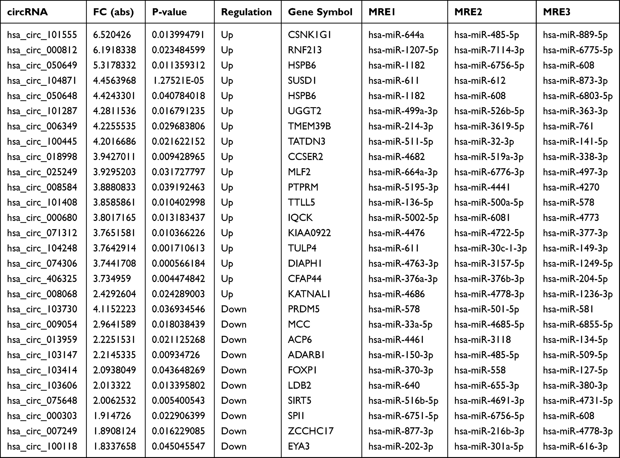 |
Table 2 Differentially Expressed circRNAs Between LUAD Tumour Tissues and Patient-Matched Normal Lung Tissues |
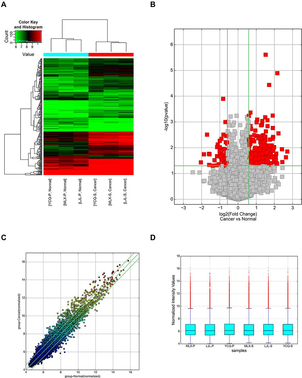 |
Figure 1 Differential expression levels of circRNAs in the tumour tissues and normal tissues of LUAD patients. (A) Hierarchical clustering of circRNA microarray analysis from three paired LUAD tumour tissues and matched adjacent normal tissues showed the differential expression of circRNAs (FC ≥ 1.5 and P values ≤ 0.05). (B) Volcano plots were constructed to determine the distribution of circRNAs using the fold change and P values. The left red points represent the differentially downregulated circRNAs, and significantly upregulated circRNAs are shown by right red points. (C) A scatter plot was used to assess variations in circRNA expression levels between LUAD tumour tissues and adjacent normal tissues. The values of the X and Y axes in the scatter plot are the normalized signal values of the samples (log2 scaled). The circRNAs above the top green line and below the bottom green line were changed by more than 1.5-fold between the two compared groups. (D) The box plot shows that the circRNAs were regulated by quantile normalization. |
Hsa_circ_101555 and Hsa_circ_008068 Were Highly Expressed in Tumour Tissues and Plasma of LUAD Patients
We chose 2 upregulated circRNAs (hsa_circ_101555 and hsa_circ_008068) with a higher fold change as the candidate circRNAs. Hsa_circ_101555 and hsa_circ_008068 have not been reported for the diagnosis of early-stage LUAD. Therefore, the relative expression of hsa_circ_101555 and hsa_circ_008068 were further measured in fourteen LUAD tumour tissues and patient-matched normal lung tissues by q-PCR experiments. As shown in the results, hsa_circ_101555 was significantly increased in LUAD tumour tissues compared to patient-matched normal tissues (P < 0.05, Figure 2A). Similarly, hsa_circ_008068 was also obviously upregulated in LUAD tumour tissues compared to adjacent normal tissues (P < 0.05, Figure 2B). Furthermore, we detected the two circRNAs in the plasma of thirty LUAD patients and thirty healthy donors. The results showed that hsa_circ_101555 and hsa_circ_008068 were both significantly upregulated between the plasma of LUAD patients and healthy donors (P < 0.05, Figure 2C and D).
 |
Figure 2 Hsa_circ_101555 and hsa_circ_008068 expressed in the tissues and plasma of LUAD patients. (A) The mRNA of hsa_circ_101555 in the tissues of LUAD patients compared with matched adjacent normal tissues. (B) The mRNA of hsa_circ_008068 in the tissues of LUAD patients compared with matched adjacent normal tissues. (C) Hsa_circ_101555 expressed in the plasma of LUAD patients compared with healthy donors. (D) Hsa_circ_008068 expressed in the plasma of LUAD patients compared with healthy donors. Differences between groups were analysed using a t-test. *P < 0.05, **P < 0.01, ***P < 0.001, significantly different from the values in the normal tissues and healthy donors. |
Correlation Between the Two circRNAs and Tumour Indicators in LUAD Patients
To further explore the relationship between the expression of circRNAs and traditional tumour indicators in LUAD patients, we collected and analysed the patients’ clinical data. As shown in Table 3, hsa_circ_101555 and hsa_circ_008068 were significantly associated with tumour differentiation (high and moderate, poor) and tumour size (≤ 3 cm, > 3 cm) in LUAD patients (P < 0.05). However, no significant correlations were found between the circRNAs and tumour location (P > 0.05). Furthermore, the serum levels of CEA were positively associated with the plasma levels of hsa_circ_101555 and hsa_circ_008068 in LUAD patients (P < 0.05, Figure 3A and B). Nevertheless, the serum levels of SCC were not significantly correlated with the plasma levels of these two circRNAs in LUAD patients (P > 0.05, Figure 3C and D). The serum levels of CYFRA21-1 were positively associated with circulating hsa_circ_008068 in LUAD patients (P < 0.05, Figure 3F), whereas there was no correlation in hsa_circ_101555 (P > 0.05, Figure 3E).
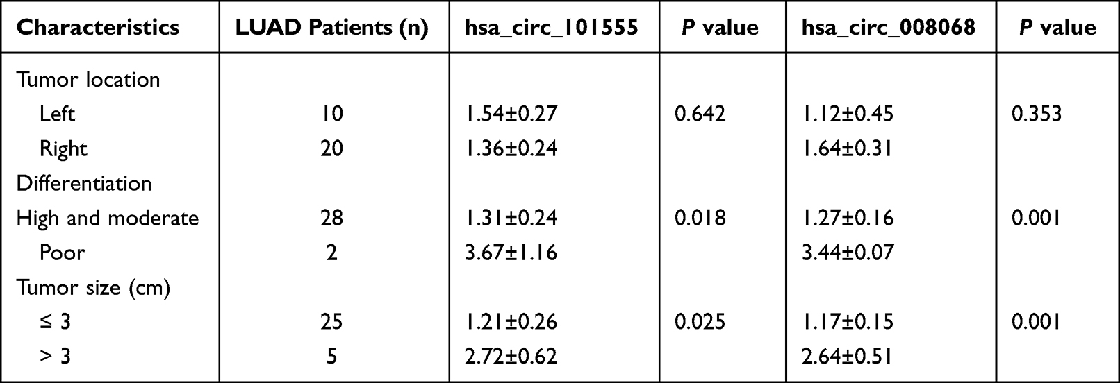 |
Table 3 Correlation Between the Expression of Plasma circRNAs and Clinical Characteristics in LUAD Patients |
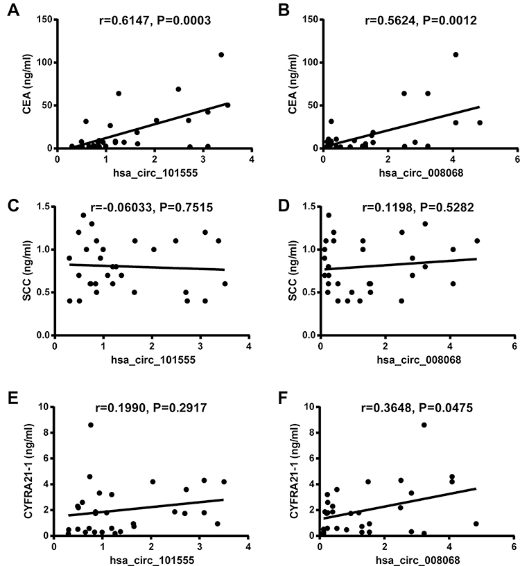 |
Figure 3 Traditional tumour marker correlations with hsa_circ_101555 and hsa_circ_008068 in the plasma of early-stage LUAD patients. (A) Correlation between hsa_circ_101555 and CEA in LUAD patients. (B) Correlation between hsa_circ_008068 and CEA in LUAD patients. (C) Correlation between hsa_circ_101555 and SCC in LUAD patients. (D) Correlation between hsa_circ_008068 and SCC in LUAD patients. (E) Correlation between hsa_circ_101555 and CYFRA21-1 in LUAD patients. (F) Correlation between hsa_circ_008068 and CYFRA21-1 in LUAD patients. Correlation analysis was performed using the Spearman’s rank correlation test. |
Hsa_circ_101555 and Hsa_circ_008068 Had a High Diagnostic Value for Early-Stage LUAD Patients
To confirm the potential value of hsa_circ_101555 and hsa_circ_008068 as tumour biomarkers for early diagnosis in LUAD patients, we analyzed the ROC curves of the two circRNAs and three traditional biomarkers (CEA, SCC, CYFRA21-1) in the plasma of LUAD patients. Our data showed that the AUC value of hsa_circ_101555 was 0.708 (95% CI = 0.578 to 0.838, sensitivity = 76.67% and specificity = 60.00%, Figure 4A). The AUC value of hsa_circ_008068 was 0.624 (95% CI = 0.482 to 0.767, sensitivity = 63.33% and specificity = 53.33%, Figure 4B). The AUC value of CEA was 0.599 (Figure 4C). The AUC value of SCC was 0.506 (Figure 4D). The AUC value of CYFRA21-1 was 0.531 (Figure 4E). Moreover, we combined circRNAs and three tumor markers (CYFR21-1, NSE and CA72-4) to make a combined diagnostic model. The AUC value of hsa_circ_101555 and three tumor markers was 0.753 (Figure 4F). The AUC value of hsa_circ_008068 and three tumor markers was 0.704 (Figure 4G). Specific values are shown in Table 4.
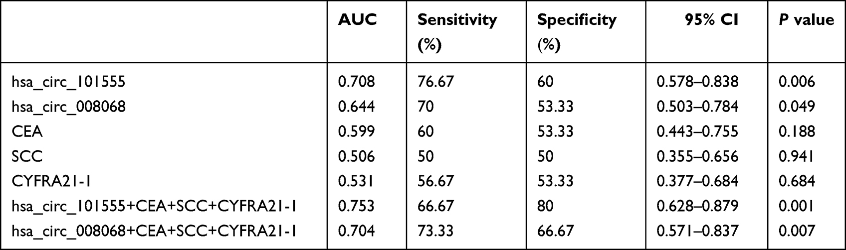 |
Table 4 The Diagnostic Efficacy of circRNAs and Three Traditional Biomarkers for the Detection of LUAD |
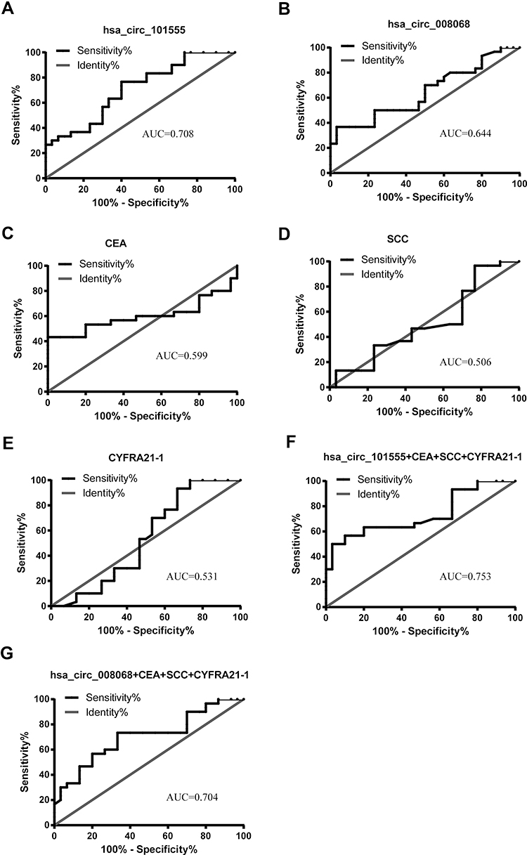 |
Figure 4 ROC curve analyses of circRNAs and three traditional biomarkers for the detection of early-stage LUAD. (A) ROC analysis for the detection of LUAD using hsa_circ_101555. (B) ROC analysis for the detection of LUAD using hsa_circ_008068. (C) ROC analysis for the detection of LUAD using CEA. (D) ROC analysis for the detection of LUAD using SCC. (E) ROC analysis for the detection of LUAD using CYFRA21-1. (F) ROC analysis for the detection of LUAD using hsa_circ_101555 combined three tumour biomarkers. (G) ROC analysis for the detection of LUAD using hsa_circ_008068 combined three tumour biomarkers. The AUC values are given on the graphs. |
Discussion
LUAD is the most common histological subtype of lung cancer and is prone to cancer recurrence and metastasis.4 The early diagnosis of LUAD is the most effective way to alleviate cancer progression and reduce deaths.5 However, detecting early-stage LUAD still presents a difficult challenge. In this study, we analysed the expression of circRNAs in three pairs of LUAD cancer tissues and adjacent normal lung tissues using a circRNA microarray. We identified 6261 circRNAs that were upregulated and 7238 circRNAs that were downregulated in tumour tissues compared with patient-matched normal tissues in LUAD patients. Meanwhile, our findings demonstrated that hsa_circ_101555 and hsa_circ_008068 were more highly expressed in LUAD tumour tissues and plasmas than in normal tissues and healthy plasmas, respectively. Therefore, hsa_circ_101555 and hsa_circ_008068 were filtered as potential screening/diagnostic LUAD markers.
High-throughput sequencing technology has detected many circRNAs in human peripheral blood, tissues, and exosomes.21 Previous studies revealed that abnormal circRNA expression is associated with tumour progression and may be a potential biomarker for malignant tumours. Recently, hsa_circ_002059 was reported to be downregulated in gastric cancer tissues and plasma and may be a potential clinical diagnostic marker for diagnosing gastric cancer.22 Similarly, hsa_circ_0001649 was significantly downregulated in hepatocellular carcinoma and was more sensitive and specific than the traditional marker α-fetoprotein (AFP) in hepatocellular carcinoma.22 In lung adenocarcinoma patients, hsa_circ_0013958 was confirmed to be markedly increased in tumour tissues and plasma, which was found to be positively associated with lymph node metastasis and TNM stages.23 Nevertheless, few circRNAs have been detected as diagnostic markers in LUAD, and many functions and molecular mechanisms of circRNAs remain unclear. Our results showed that hsa_circ_101555 and hsa_circ_008068 were highly expressed in lung tissues and plasma from LUAD patients. At present, there are no reports of hsa_circ_101555 and hsa_circ_008068 in LUAD patients. In hepatocellular carcinoma, circular RNA hsa_circ_101555 has been reported to promote tumour cell proliferation and migration by sponging miR-145-5p and regulating CDCA3 expression.24 Another study showed that hsa_circ_101555 promoted colorectal cancer progression as a competing endogenous RNA of miR-597-5p.25 Hence, we speculated that hsa_circ_101555 has similar functions in LUAD patients, but the precise mechanisms need further elucidation. Moreover, hsa_circ_008068 has not been reported in various diseases. Our results indicated that hsa_circ_008068 was significantly increased in tumour tissues and plasmas of LUAD patients compared with healthy donors. Moreover, we observed that the dysregulated expression of hsa_circ_101555 and hsa_circ_008068 in LUAD patients was overexpressed, but the molecular mechanisms need to be addressed with further research.
Traditional clinical indicators, such as tumour differentiation, tumour size, tumour location, CEA, CYFRA21-1, and SCC, are used to facilitate differential diagnoses and determine prognosis in lung cancer.26 Elevated levels of indicators are significantly associated with lung cancer diagnosis and progression. In our study, hsa_circ_101555 and hsa_circ_008068 were positively associated with CEA in LUAD patients. We consider that hsa_circ_101555 and hsa_circ_008068 also provide useful biochemical parameters in the screening, diagnosis, and prognosis of LUAD. Moreover, our correlation analysis identified that circRNAs were irrelevant to CYFRA21-1 and SCC, which are considered to reflect squamous cell cancers. In addition, traditional tumour biomarkers have a low sensitivity for early diagnoses due to the low tumour burden released into peripheral circulation in early-stage lung cancer.27 We also observed the diagnostic value of these two circRNAs compared with CEA, CYFRA21-1, and SCC. The ROC curves showed that the AUC values of hsa_circ_101555 and hsa_circ_008068 were excellent compared with those of traditional tumour markers for the diagnosis of early LUAD. We consider that circRNAs could be detection markers in early-stage LUAD. Currently, combined diagnosis had more excellent diagnostic value than single marker. This prompted us that the use of multiple highly sensitivity and specificity circRNAs combined diagnosis model may provide better value in detection of early-stage LUAD. Nonetheless, our study was limited to a small sample size but may serve as a novel marker in this area.
In summary, the present study identified abnormal circRNAs between tumour tissues and patient-matched normal lung tissues from LUAD patients. Hsa_circ_101555 and hsa_circ_008068 were confirmed to be potential targets in LUAD patients by RT–PCR. Thereafter, the two circRNAs were found to be positively associated with CEA and CYFRA21-1. The results demonstrated that hsa_circ_101555 and hsa_circ_008068 were potential early diagnostic markers for LUAD.
Acknowledgments
This work was supported by Health Bureau of Zhejiang Province, Zhejiang, China (2020PY028).
Disclosure
The authors report no conflicts of interest to declare in this work.
References
1. Siegel RL, Miller KD, Fuchs HE, Jemal A. Cancer statistics, 2021. CA Cancer J Clin. 2021;71(1):7–33. doi:10.3322/caac.21654
2. Tavernari D, Battistello E, Dheilly E, et al. Nongenetic evolution drives lung adenocarcinoma spatial heterogeneity and progression. Cancer Discov. 2021;11(6):1490–1507. doi:10.1158/2159-8290.CD-20-1274
3. Sun GZ, Zhao TW. Lung adenocarcinoma pathology stages related gene identification. Math Biosci Eng. 2019;17:737–746. doi:10.3934/mbe.2020038
4. Hirsch FR, Scagliotti GV, Mulshine JL, et al. Lung cancer: current therapies and new targeted treatments. Lancet. 2017;389:299–311. doi:10.1016/S0140-6736(16)30958-8
5. Collins LG, Haines C, Perkel R, Enck RE. Lung cancer: diagnosis and management. Am Fam Physician. 2007;75:56–63.
6. Veronesi G, Baldwin DR, Henschke CI, et al. Recommendations for Implementing Lung Cancer Screening with Low-Dose Computed Tomography in Europe. Cancers (Basel). 2022;12(6):0. doi:10.3390/cancers12061672
7. Rulli KS, Matthews E. Using low-dose computed tomography for early detection of lung cancer. Radiol Technol. 2020;92:23–31.
8. Pisapia P, Malapelle U, Troncone G. Liquid biopsy and lung cancer. Acta Cytol. 2019;63(6):489–496. doi:10.1159/000492710
9. Alix-Panabieres C, Pantel K. Liquid biopsy: from discovery to clinical application. Cancer Discov. 2021;11(4):858–873. doi:10.1158/2159-8290.CD-20-1311
10. Patop IL, Wust S, Kadener S. Past, present, and future of circRNAs. EMBO J. 2019;38:e100836. doi:10.15252/embj.2018100836
11. Zang J, Lu D, Xu A. The interaction of circRNAs and RNA binding proteins: an important part of circRNA maintenance and function. J Neurosci Res. 2020;98(1):87–97. doi:10.1002/jnr.24356
12. Conn SJ, Pillman KA, Toubia J, et al. The RNA binding protein quaking regulates formation of circRNAs. Cell. 2015;160(6):1125–1134. doi:10.1016/j.cell.2015.02.014
13. Tao M, Zheng M, Xu Y, Ma S, Zhang W, Ju S. CircRNAs and their regulatory roles in cancers. Mol Med. 2021;27:94. doi:10.1186/s10020-021-00359-3
14. Patop IL, Kadener S. circRNAs in Cancer. Curr Opin Genet Dev. 2018;48:121–127. doi:10.1016/j.gde.2017.11.007
15. Zhang Q, Wang W, Zhou Q, et al. Roles of circRNAs in the tumour microenvironment. Mol Cancer. 2020;19(1):14. doi:10.1186/s12943-019-1125-9
16. Kristensen LS, Jakobsen T, Hager H, Kjems J. The emerging roles of circRNAs in cancer and oncology. Nat Rev Clin Oncol. 2022;19(3):188–206. doi:10.1038/s41571-021-00585-y
17. Yan L, Zheng M, Wang H. Circular RNA hsa_circ_0072309 inhibits proliferation and invasion of breast cancer cells via targeting miR-492. Cancer Manag Res. 2019;11:1033–1041. doi:10.2147/CMAR.S186857
18. Liang WC, Wong CW, Liang PP, et al. Translation of the circular RNA circbeta-catenin promotes liver cancer cell growth through activation of the Wnt pathway. Genome Biol. 2019;20:84. doi:10.1186/s13059-019-1685-4
19. Chen S, Lu S, Yao Y, et al. Downregulation of hsa_circ_0007580 inhibits non-small cell lung cancer tumorigenesis by reducing miR-545-3p sponging. Aging. 2020;12(14):14329–14340. doi:10.18632/aging.103472
20. Gao P, Wang Z, Hu Z, Jiao X, Yao Y. Circular RNA circ_0074027 indicates a poor prognosis for NSCLC patients and modulates cell proliferation, apoptosis, and invasion via miR-185-3p mediated BRD4/MADD activation. J Cell Biochem. 2020;121(3):2632–2642. doi:10.1002/jcb.29484
21. Gao Y, Shang S, Guo S, et al.. lnc2Cancer 3.0: an updated resource for experimentally supported lncRNA/circRNA cancer associations and web tools based on RNA-seq and scRNA-seq data. Nucleic Acids Res. 2021;49(D1):D1251–D1258. doi:10.1093/nar/gkaa1006
22. Li P, Chen S, Chen H, et al. Using circular RNA as a novel type of biomarker in the screening of gastric cancer. Clin Chim Acta. 2015;444:132–136. doi:10.1016/j.cca.2015.02.018
23. Zhu X, Wang X, Wei S, et al. hsa_circ_0013958: a circular RNA and potential novel biomarker for lung adenocarcinoma. FEBS J. 2017;284(14):2170–2182. doi:10.1111/febs.14132
24. Gu X, Zhang J, Ran Y, et al. Circular RNA hsa_circ_101555 promotes hepatocellular carcinoma cell proliferation and migration by sponging miR-145-5p and regulating CDCA3 expression. Cell Death Dis. 2021;12(4):356. doi:10.1038/s41419-021-03626-7
25. Chen Z, Ren R, Wan D, et al. Hsa_circ_101555 functions as a competing endogenous RNA of miR-597-5p to promote colorectal cancer progression. Oncogene. 2019;38(32):6017–6034. doi:10.1038/s41388-019-0857-8
26. Sears CR, Mazzone PJ. Biomarkers in Lung Cancer. Clin Chest Med. 2020;41(1):115–127. doi:10.1016/j.ccm.2019.10.004
27. McLellan R, Marshall H, Dent A, Bowman RV, Yang IA, Fong KM. Diagnosis and treatment of early lung cancer. Aust J Gen Pract. 2020;49(8):508–512. doi:10.31128/AJGP-11-19-5148
 © 2022 The Author(s). This work is published and licensed by Dove Medical Press Limited. The full terms of this license are available at https://www.dovepress.com/terms.php and incorporate the Creative Commons Attribution - Non Commercial (unported, v3.0) License.
By accessing the work you hereby accept the Terms. Non-commercial uses of the work are permitted without any further permission from Dove Medical Press Limited, provided the work is properly attributed. For permission for commercial use of this work, please see paragraphs 4.2 and 5 of our Terms.
© 2022 The Author(s). This work is published and licensed by Dove Medical Press Limited. The full terms of this license are available at https://www.dovepress.com/terms.php and incorporate the Creative Commons Attribution - Non Commercial (unported, v3.0) License.
By accessing the work you hereby accept the Terms. Non-commercial uses of the work are permitted without any further permission from Dove Medical Press Limited, provided the work is properly attributed. For permission for commercial use of this work, please see paragraphs 4.2 and 5 of our Terms.