")
Back to Journals » Medical Devices: Evidence and Research » Volume 11
Changes in cerebral oxygenation based on intraoperative ventilation strategy
Authors Dewhirst E, Walia H, Samora WP, Beebe AC, Klamar JE, Tobias JD
Received 28 November 2017
Accepted for publication 7 May 2018
Published 25 July 2018 Volume 2018:11 Pages 253—258
DOI https://doi.org/10.2147/MDER.S158262
Checked for plagiarism Yes
Review by Single anonymous peer review
Peer reviewer comments 2
Editor who approved publication: Dr Scott Fraser
Elisabeth Dewhirst,1 Hina Walia,1 Walter P Samora,2 Allan C Beebe,2 Jan E Klamar,2 Joseph D Tobias1,3
1Department of Anesthesiology and Pain Medicine, Nationwide Children’s Hospital, Columbus, OH, USA; 2Department of Orthopedic Surgery, Nationwide Children’s Hospital, The Ohio State University, Columbus, OH, USA; 3Department of Anesthesiology and Pain Medicine, The Ohio State University, Columbus, OH, USA
Introduction: Cerebral oxygenation can be monitored clinically by cerebral oximetry (regional oxygen saturation, rSO2) using near-infrared spectroscopy (NIRS). Changes in rSO2 have been shown to precede changes in pulse oximetry, providing an early detection of clinical deterioration. Cerebral oximetry values may be affected by various factors, including changes in ventilation. The aim of this study was to evaluate the changes in rSO2 during intraoperative changes in mechanical ventilation.
Patients and methods: Following the approval of the institutional review board (IRB), tissue and cerebral oxygenation were monitored intraoperatively using NIRS. Prior to anesthetic induction, the NIRS monitor was placed on the forehead and over the deltoid muscle to obtain baseline values. NIRS measurements were recorded each minute over a 5-min period during general anesthesia at four phases of ventilation: 1) normocarbia (35–40 mmHg) with a low fraction of inspired oxygen (FiO2) of 0.3; 2) hypocarbia (25–30 mmHg) and low FiO2 of 0.3; 3) hypocarbia and a high FiO2 of 0.6; and 4) normocarbia and a high FiO2. NIRS measurements during each phase were compared with sequential phases using paired t-tests.
Results: The study cohort included 30 adolescents. Baseline cerebral and tissue oxygenation were 81% ± 9% and 87% ± 5%, respectively. During phase 1, cerebral rSO2 was 83% ± 8%, which decreased to 79% ± 8% in phase 2 (hypocarbia and low FiO2). Cerebral oxygenation partially recovered during phase 3 (81% ± 9%) with the increase in FiO2 and then returned to baseline during phase 4 (83% ± 8%). Each sequential change (e.g., phase 1 to phase 2) in cerebral oxygenation was statistically significant (p < 0.01). Tissue oxygenation remained at 87%–88% throughout the study.
Conclusion: Cerebral oxygenation declined slightly during general anesthesia with the transition from normocarbia to hypocarbic conditions. The rSO2 decrease related to hypocarbia was easily reversed with a return to baseline values by the administration of supplemental oxygen (60% vs. 30%).
Keywords: pediatric, cerebral oxygenation, near infrared spectroscopy (nirs), intraoperative, ventilation
Introduction
To optimize intraoperative care, it is imperative to maintain adequate cardiac output and delivery of oxygen to the tissues. Intraoperative care includes monitoring of blood pressure (BP) and systemic oxygenation (pulse oximetry); recently, interest has been placed on measuring end-organ tissue oxygenation. Near-infrared spectroscopy (NIRS) monitors are used to measure tissue oxygenation, and are becoming more commonly used in anesthesia practice. The NIRS monitor consists of a noninvasive adhesive sensor with a laser light source and two photodetectors. Using optical technology based on the relative absorption of infrared light by different hemoglobin species, the monitor generates a measurement of regional oxygen saturation (rSO2).1 The device is typically applied to the forehead and used to measure cerebral oxygenation, but can be used to measure regional tissue saturation elsewhere in the body. Data in both the adult and pediatric literature suggest that monitoring and maintaining cerebral oxygenation may improve perioperative neurological outcomes.2,3 The NIRS monitor is being used increasingly in the operating room and intensive care unit (ICU) to monitor cerebral perfusion and guide management in various clinical scenarios, which are most established during cardiac surgery. A decrease in cerebral oxygenation correlates with events of clinical deterioration such as arrhythmia, hypotension, and hypoxia.4 Changes in cerebral oxygenation have been shown to precede those in pulse oximetry, providing an early detection of clinical deterioration.5
In the adult population, alterations in inspired oxygen concentration and expired carbon dioxide have been shown to influence cerebral oxygenation.6 Various patient, surgical, and anesthetic factors may influence intraoperative ventilation choices (minute ventilation and the fraction of inspired oxygen [FiO2]) and the resultant arterial partial pressure of oxygen (PaO2) and carbon dioxide (PaCO2).7–11 Examples include laparoscopic surgery, intrathoracic surgery, airway surgery, patient positioning, and comorbid conditions.8,9 Furthermore, various factors including a decrease in the metabolic rate related to general anesthesia may result in inadvertent hyperventilation during routine intraoperative care.9 Understanding how ventilation affects cerebral oxygenation is of clinical importance, especially when faced with other physiological changes such as anemia and decreased cardiac output that may impact tissue and cerebral oxygen delivery. The relationship between ventilation and cerebral rSO2 has not been examined in the pediatric population. The aim of the current study was to evaluate the changes in cerebral and tissue rSO2 during intraoperative changes in mechanical ventilation parameters.
Patients and methods
This study was reviewed and approved by the institutional review board (IRB) of Nationwide Children’s Hospital. As approved by the IRB, prior to study enrollment, verbal consent from a parent and assent from the patient were obtained. This study was registered with ClinicalTrials.gov (NCT02651103). Enrolled patients were children undergoing a major surgical procedure requiring placement of an arterial cannula. Prior to anesthetic induction, the NIRS monitor was placed on the forehead and over the deltoid muscle, in addition to standard American Society of Anesthesiologist (ASA) monitors. Baseline values for cerebral and tissue oxygenation were obtained using NIRS. After the induction of general anesthesia and placement of an endotracheal tube, an arterial cannula was placed and NIRS values were again measured at the following four phases of ventilation with variation of the inspired oxygen concentration (FiO2) and end-tidal carbon dioxide (ETCO2) value:
- Normocarbia (ETCO2 = 35–40 mmHg) and low FiO2 (0.3)
- Hypocarbia (ETCO2 = 25–30 mmHg) and low FiO2 (0.3)
- Hypocarbia (ETCO2 = 25–30 mmHg) and high FiO2 (0.6)
- Normocarbia (ETCO2 = 35–40 mmHg) and high FiO2 (0.6)
Following the changes in mechanical ventilation for each phase of the study, an equilibration period of 15 min was allowed prior to data collection. To ensure the accuracy of the ETCO2 value, arterial blood gas was obtained prior to the start of the study and the ETCO2 was validated using the PaCO2 measurement. At baseline and during each phase of ventilation, NIRS measurements were recorded every minute over a 5-min period. Heart rate (HR) and BP were recorded at each of the sampling intervals, and hemodynamic variables were maintained constant by adjustments in the continuous infusion of remifentanil. Depth of anesthesia was maintained at a constant value with the administration of inhaled desflurane which was titrated to maintain the bispectral index (BIS) at 50–60.
Statistical analyses
The average of the available NIRS measurements within each ventilation phase was obtained and used to compare sequential phases with paired t-tests. Sequential changes in these outcomes were evaluated using paired t-tests. Normal distribution of all the study outcomes at baseline was checked using the Shapiro–Wilk test, and tests of statistical significance at subsequent time points were robust to using nonparametric signed-rank tests. A planned sample size of 30 patients was selected to be comparable with previous studies in the adult population and to be consistent with study feasibility and projected patient enrollment. Due to the exploratory nature of this study in a pediatric population, no a priori power analysis was performed. Data analysis was performed using Stata/IC 13.1 (StataCorp LP, College Station, TX, USA), and two-tailed p < 0.05 was considered statistically significant.
Results
The study cohort included 30 patients (13 males and 17 females, age 15 ± 3 years, and weight 55 ± 15 kg). Cohort characteristics are summarized in Table 1, and the distribution of the study outcomes is shown as a box plot in Figure 1. The peripheral oxygen saturation by pulse oximetry remained at 99%–100% throughout the study. The baseline cerebral and tissue rSO2, prior to anesthetic induction while breathing room air, were 81% ± 9% and 87% ± 5%, respectively. During phase 1, cerebral rSO2 was 83% ± 8%, which decreased to 79% ± 8% in phase 2 (hypocarbia and low FiO2). Cerebral oxygenation recovered, returning to baseline values, during phase 3 (81% ± 9%) and phase 4 (83% ± 8%). Each sequential change (e.g., phase 1 to phase 2) in cerebral oxygenation was statistically significant (p < 0.01; Table 2). Tissue oxygenation increased from 87% ± 8% in phase 1 to 88% ± 7% in phase 2 (p = 0.002); remained at 88% ± 7% in phase 3; and decreased to 87% ± 8% in phase 4 (p = 0.023; Table 2). Changes in HR and BP over the course of the study are summarized in Table 3. There were no statistically significant sequential changes in HR, systolic BP, or diastolic BP over the first three study phases. When comparing phase 4 with phase 3, slight decreases were noted in HR and BP.
 | Table 1 Study cohort characteristics Abbreviation: ASA, American Society of Anesthesiologist. |
 | Figure 1 Box plot of rSO2, HR, SBP, DBP, and mean arterial pressure (MAP) over all the study phases. Notes: Box center indicates the median, box outline indicates the IQR, box whiskers indicate farthest values within 1.5 IQR of the nearest quartile, and scatter points indicate outlier values. rSO2, regional oxygen saturation. Abbreviations: BP, blood pressure; DBP, diastolic BP; HR, heart rate; IQR, interquartile range; MAP, mean arterial pressure; SBP, systolic BP. |
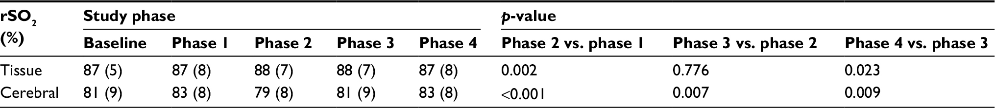 | Table 2 Cerebral and tissue oxygenation during study phases Notes: rSO2, oxygenation determined by NIRS. Values for rSO2 are presented as mean and SD. Abbreviations: NIRS, near-infrared spectroscopy. |
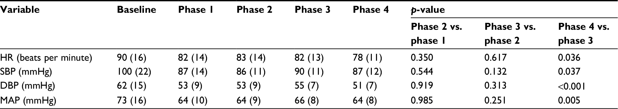 | Table 3 Hemodynamic parameters during the four study phases Note: Values for hemodynamic parameters are presented as mean and SD. Abbreviations: BP, blood pressure; DBP, diastolic BP; HR, heart rate; MAP, mean arterial pressure; SBP, systolic BP. |
Discussion
Cerebral oxygenation as measured by NIRS may provide valuable information regarding cerebral oxygen delivery and utilization in critically ill patients in both the operating room and the ICU settings. Intraoperatively, it may help guide anesthetic care, as studies in adults have shown that maintaining adequate values on NIRS may improve neurological outcomes.2,12 One of the primary regulators of cerebral blood flow (CBF) is PaCO2. Hypocarbia results in a direct effect on the cerebral vasculature with vasoconstriction and a decrease in CBF. For every 1 mmHg change in PaCO2, CBF changes by 1–2 mL/100 g per minute. As there is no impact on the cerebral metabolic rate for oxygen related to changes in PaCO2, the decrease in CBF may lead to a decrease in cerebral oxygenation. In our study, cerebral oxygenation as measured by NIRS declined during general anesthesia with the transition from normocarbia to hypocarbic conditions. Despite this decrease, no clinical impact would be expected as the starting rSO2 was high, the change with hyperventilation was small, and the resultant value remained well above the reported threshold for concern.
Our data demonstrate that, even in clinically stable patients, PaCO2 remains an important determinant of CBF and cerebral oxygenation. Changes in CBF related to alterations in intraoperative ventilation may have a greater impact on critically ill patients when other factors which impact rSO2, such as hemoglobin values and cardiac output, may be affected. We have previously noted the potential impact of the combination of anemia, hypotension, and hypocarbia on rSO2, suggesting that close attention to the control of ventilation is important during intraoperative care where inadvertent hyperventilation is commonplace.7,13
In addition, these data demonstrate that the cerebral rSO2 decrease related to hypocarbia was reversed by the administration of supplemental oxygen (60% vs. 30%). A secondary mechanism that may provide additional protection to decreases in cerebral rSO2 in the current study is the effect of anesthetic agents on cerebral oxygen needs. As general anesthetic agents (volatile anesthetic agents, propofol, barbiturates, and benzodiazepines) decrease the cerebral metabolic rate for oxygen, thereby decreasing oxygen extraction. In clinical scenarios where hyperventilation is required, such as to reduce intracranial pressure when there is impending cerebral herniation or to blunt the respiratory drive, increasing the FiO2 may be pertinent to offset any reduction in cerebral oxygenation related to decreases in CBF. Thiagarajan et al evaluated 18 adults with traumatic brain injury to determine the changes in cerebral jugular venous oxygen saturation (SjvO2) and arteriovenous oxygen content difference (AVDO2) in response to changes in PaO2 and PaCO2.15 SjvO2 decreased from 66% ± 3% to 56% ± 3% when PaCO2 decreased from 30 to 25 mmHg at a PaO2 of 100–150 mmHg. The SjvO2 values were significantly greater when the PaO2 was 200–250 mmHg (77% ± 4% and 64% ± 3%) at PaCO2 values of both 30 and 25 mmHg. The authors concluded, as we did in our study, that decreases in cerebral oxygenation (manifested as decreases in SjvO2) associated with a decrease in PaCO2, were offset by increasing the PaO2. These data are particularly relevant given the literature demonstrating episodes of cerebral ischemia and worse neurologic outcome in patients with traumatic brain injury who are exposed to hypocarbia.15
Similar results have been noted in the adult population by other investigators using various monitors of cerebral oxygenation. All of these studies have demonstrated that both alterations in inspired oxygen concentration and expired carbon dioxide impact cerebral tissue oxygenation. Tisdall et al6 measured the cerebral tissue oxygenation index (TOI) in 15 adults under hypoxic, hyperoxic, and hypo- and hypercarbic conditions. Hypoxemic and hypocarbic conditions resulted in a decrease in the TOI by 7.1% and 2.1%, respectively, while hyperoxia and hypercarbia led to increases of 2.3% and 2.6%. Picton et al10 reported a 7% decrease in rSO2 after beach chair positioning in adults maintained at an FiO2 of 0.3 and ETCO2 of 30 mmHg which was reversible by decreasing ventilation and increasing the FiO2. The same investigators reported that the rSO2 was an average of 8% higher with an FiO2 of 1.0 vs. 0.3 at an ETCO2 of 30–35 mmHg. Bouzat et al16 reported a 5% reduction in rSO2 with hyperventilation in adult patients recovering from cardiac arrest.
Our literature search revealed no previous studies in the pediatric age range that examined the effect of ventilation on cerebral oxygenation. When compared with some of the adult studies, it is not surprising that the decrease in cerebral oxygenation that we noted was smaller than that seen in the adult population, as our patients lacked comorbid conditions, such as atherosclerotic disease, which might affect CBF. When evaluating intraoperative studies, including the current study, one must also consider the impact that the anesthetic agents may have on the findings as specific anesthetic agents may affect cerebral metabolic rate for oxygenation and thereby mitigate the effects of the decrease in CBF on cerebral oxygenation.17 As the current study focused on adolescents, extrapolation of these data to younger populations is likely infeasible, and additional trials are needed in these age groups to determine the impact of changes in ventilation on cerebral rSO2.
Conclusion
No clinically significant changes in tissue oxygenation were noted related to hyperventilation, and the changes in cerebral rSO2 under the specific study conditions had no clinical impact. As PaCO2 did not affect tissue blood flow and had limited impact on cardiac output, changes in tissue oxygenation were minimal with an average value of 87% or 88% during all the study phases. The change from baseline of cerebral rSO2 was minimal, and the low value even under hypobaric conditions was well above the NIRS monitor’s low threshold value for concern. However, the impact of hypocarbia must be considered when comorbid conditions or ongoing acute issues, such as anemia or hypotension, may further impact cerebral oxygenation. In specific clinical scenarios where hyperventilation is clinically indicated, the impact on cerebral oxygenation can be mitigated by increasing the FiO2.
Acknowledgment
Preliminary results from this study were presented in abstract form in the American Society of Anesthesiologists Annual Meeting (Chicago, IL, USA; October 2016); Society for Critical Care Medicine Annual Meeting (Honolulu, HI, USA; January 2017); and the Society for Pediatric Anesthesiology Annual Meeting (Austin, TX, USA; March 2017).
Disclosure
The authors report no conflicts of interest in this work.
References
Tobias JD. Cerebral oxygenation monitoring: near infrared spectroscopy. Expert Rev Med Devices. 2006;3(2):235–243. | ||
Casati A, Fanelli G, Pietropaoli P, et al. Continuous monitoring of cerebral oxygen saturation in elderly patients undergoing major abdominal surgery minimizes brain exposure to potential hypoxia. Anesth Analg. 2005;101(3):740–747. | ||
Austin EH III, Edmonds HL Jr, Auden SM, et al. Benefit of neurophysiologic monitoring for pediatric cardiac surgery. J Thorac Cardiovasc Surg. 1997;114(5):707–715. | ||
Naguib AN, Winch P, Ro PS, Olshove V, Tobias JD. Changes in near-infrared spectroscopy and the bispectral index during tilt-table examination. Pediatr Cardiol. 2011;32(2):234–236. | ||
Tobias JD. Cerebral oximetry monitoring with near infrared spectroscopy detects alterations in oxygenation before pulse oximetry. J Intensive Care Med. 2008;23(6):384–388. | ||
Tisdall MM, Taylor C, Tachtsidis I, Leung TS, Elwell CE, Smith M. The effect on cerebral tissue oxygenation index of changes in the concentrations of inspired oxygen and end-tidal carbon dioxide in healthy adult volunteers. Anesth Analg. 2009;109(3):906–913. | ||
Rozmiarek A, Taghon T, Tobias JD. Inadvertent hyperventilation during intraoperative anesthetic care in the pediatric population. ICU Director. 2012;4:172–175. | ||
Tobias JD, Holcomb GW III, Brock JW III, Deshpande JK, Lowe S, Morgan WM III. Cardiorespiratory changes during laparoscopy in children. J Pediatr Surg. 1995;30(1):33–36. | ||
Picton P, Dering A, Alexander A, et al. Influence of ventilation strategies and anesthetic techniques on regional cerebral oximetry in the beach chair position: a prospective interventional study with a randomized comparison of two anesthetics. Anesthesiology. 2015;123(4):765–774. | ||
Picton P, Shanks A, Dorje P, Mashour GA. The influence of basic ventilation strategies on cerebral oxygenation in anesthetized patients without vascular disease. J Clin Monit Comput. 2010;24(6):421–425. | ||
Booth EA, Dukatz C, Sood BG, Wider M. Near-infrared spectroscopy monitoring of cerebral oxygen during assisted ventilation. Surg Neurol Int. 2011;2:65–68. | ||
Murkin JM, Adams SJ, Novick RJ, et al. Monitoring brain oxygen saturation during coronary bypass surgery: a randomized, prospective study. Anesth Analg. 2007;104(1):51–58. | ||
Shear T, Tobias JD. Cerebral oxygenation monitoring using near infrared spectroscopy during controlled hypotension. Paediatr Anaesth. 2005;15(6):504–508. | ||
Skippen P, Seear M, Poskitt K, et al. Effect of hyperventilation on regional cerebral blood flow in head-injured children. Crit Care Med. 1997;25(8):1402–1409. | ||
Thiagarajan A, Goverdhan PD, Chari P, Somasunderam K. The effect of hyperventilation and hyperoxia on cerebral venous oxygen saturation in patients with traumatic brain injury. Anesth Analg. 1998;87(4):850–853. | ||
Bouzat P, Suys T, Sala N, Oddo M. Effect of moderate hyperventilation and induced hypertension on cerebral tissue oxygenation after cardiac arrest and therapeutic hypothermia. Resuscitation. 2013;84(11):1540–1545. | ||
Ishiyama T, Kotoda M, Asano N, et al. Effects of hyperventilation on cerebral oxygen saturation estimated using near-infrared spectroscopy: a randomised comparison between propofol and sevoflurane anaesthesia. Eur J Anaesthesiol. 2016;33(12):929–935. |
 © 2018 The Author(s). This work is published and licensed by Dove Medical Press Limited. The full terms of this license are available at https://www.dovepress.com/terms.php and incorporate the Creative Commons Attribution - Non Commercial (unported, v3.0) License.
By accessing the work you hereby accept the Terms. Non-commercial uses of the work are permitted without any further permission from Dove Medical Press Limited, provided the work is properly attributed. For permission for commercial use of this work, please see paragraphs 4.2 and 5 of our Terms.
© 2018 The Author(s). This work is published and licensed by Dove Medical Press Limited. The full terms of this license are available at https://www.dovepress.com/terms.php and incorporate the Creative Commons Attribution - Non Commercial (unported, v3.0) License.
By accessing the work you hereby accept the Terms. Non-commercial uses of the work are permitted without any further permission from Dove Medical Press Limited, provided the work is properly attributed. For permission for commercial use of this work, please see paragraphs 4.2 and 5 of our Terms.