")
Back to Journals » International Medical Case Reports Journal » Volume 8
Bilateral simultaneous intertrochanteric fractures of femur without major trauma
Authors Aydin E, Dülgeroğlu TC , Metineren H
Received 1 March 2015
Accepted for publication 24 March 2015
Published 29 June 2015 Volume 2015:8 Pages 137—139
DOI https://doi.org/10.2147/IMCRJ.S83755
Checked for plagiarism Yes
Review by Single anonymous peer review
Peer reviewer comments 3
Editor who approved publication: Professor Ronald Prineas
Ekrem Aydin, Turan Cihan Dülgeroğlu, Hasan Metineren
Department of Orthopedics and Traumatology, Dumlupinar University School of Medicine, Kutahya, Turkey
Abstract: Bilateral simultaneous intertrochanteric femur fractures are rare and usually occur as a result of major trauma. In this paper, we present a 76-year old male patient from Turkey referred to the emergency service of our hospital with severe pain and poor mobilization in both hips. There was no history of major trauma. The patient was treated with closed proximal femoral nailing in both hips in the same operation period. At last follow-up, 6 months after surgery, the patient was able to walk with the aid of his children and resumed his previous status, after he had completed an intensive rehabilitation program.
Keywords: bilateral femur intertrochanteric fracture, PFN, elderly population
Introduction
Trochanteric femur fractures are observed frequently by orthopedic surgeons in daily practice in emergency services especially in the elderly patient population. They generally appear due to simple falls.1 Simultaneous bilateral femur intertrochanteric fractures are very rare and usually appear after major traumas or primary/secondary bone diseases.2 Although there are many case presentations in the literature about bilateral femoral neck fractures, there are very few case presentations about simultaneous bilateral femur intertrochanteric fractures.3 The proximal femur is the area where the most deficiency fractures are observed. In addition, the proximal femur is the area which is routinely subjected to maximum loading force. The deficiency fracture is a type of stress fracture caused by normal stress on abnormal bone.2 The proximal femur is a very important component of the motion system and receives high energy during movements because of carrying the whole of the body weight and due to the contractions of the muscles in this area. For this reason, the trochanteric femur fractures may occur in some cases where the bone mass is decreased and with the effects of the simple trauma and excessive muscle contractions together.
Case report
A 76-year old male from Turkey was referred to the emergency services of our hospital with severe pain and poor mobilization in both hips. In his history he had to jump over two small steps from a staircase, because his feet slipped and then the patient sat down on the step and was not able to stand up because he felt severe pain in both hips, and then he was transported to the emergency services later. The physical examination in the emergency service department revealed external rotation posture in both lower extremities, limitations in movement, sensitivity, and severe pain. After the X-ray and computed tomography, bilateral trochanteric femur fracture was detected (Figures 1 and 2). According to his medical history, even though the patient is elderly he does not have any medical comorbidities. The necessary preparations were made and the patient was taken to the operating room. Spinal anesthesia was applied, and fracture reduction was performed on the fracture table with the support of flouroscopy, and then in the same operation period, closed proximal femoral nailing was performed to both hips (Figure 3). No serious bleeding occurred, and based on this, there was no need for transfusion during and after the operation. No complications developed in the postoperative period. At last follow-up, 6 months after surgery, the patient was able to walk with his children’s assistance and resumed his previous status, after he had taken up an intensive rehabilitation program.
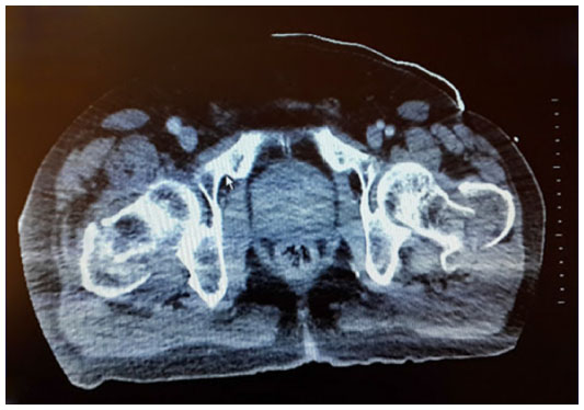 | Figure 1 Injury computed tomography image showing bilateral femur proximal fractures. |
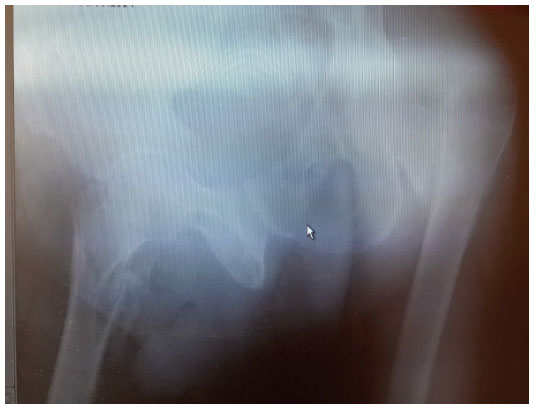 | Figure 2 Anteroposterior plain radiography of both hips showing bilateral intertrochanteric femur fractures. |
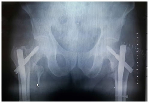 | Figure 3 Postoperative X-ray showing bilateral proximal femoral nail implementation. |
Discussion
Although femur trochanteric fractures may be observed frequently in the elderly, bilateral fractures are very rare and usually occur after serious traumas.3–5 Those which occur after simple traumas are usually in the form of femoral neck fractures.7 These kinds of patients are usually osteoporotic patients. Unilateral trochanteric fractures may occur after simple traumas.5,6,9 Bilateral fractures are very rare and occur after serious complicated traumas.8,9 Besides the effect of the direct trauma, excessive and imbalanced contractions of the muscles in the same area also have an effect in these fractures.4,5 It is known that old bones lose their elastic adaptation capacity and this facilitates the occurrence of fractures.3 It must be kept in mind that in the basis of such fractures, there will always be osteoporosis, and the operation method must be chosen according to this fact. Also, the follow-up medical support must be considered in the light of this situation.5,6 Intramedullary nails, sliding hip screws, anatomical plates, partial and total prosthesis, external fixators, and proximal femoral nails are among the materials used in the fixation of the fractures in this area.4,9 When both lower extremities are involved in the event, the fixation tool which will allow early mobilization and load of weight must be chosen. The treatment of extracapsular fracture of the hip relies on fracture pattern and stability. The sliding hip screw and plates generally are utilized for stable and displaced fracture pattern. The use of an intramedullary device is dedicated to unstable extracapsular femur intertrochanteric fracture and loss of medial cortical support of femur proximal fractures.10 Because this case is an extracapsular and unstable femur intertrochanteric fracture, we performed stable fracture fixation and preferred to apply the proximal femoral nail, which allows early mobilization and is easier tolerated by the patient. The proximal femoral nail application may be applied easily in such fractures; and since the hip abductor and rotator mechanisms receive less damage iatrogenically during the surgery, the result is a more functional hip.
Conclusion
Bilateral trochanteric fractures are very rare injuries; however, sometimes they can occur especially in patients with osteoporosis after a simple fall or jumping. In the treatment, both the patient and the surgeon may have some challenges. In choosing the surgical fixation material, it is important that the material will allow early mobilization and protect the patient from any surgical complication, thus avoiding possible complications that can stem from long-term immobilization and surgical complication. Surgeons can be sure of a successful treatment with the choice of proper fixation material and accurate planning.
Acknowledgment
Written informed consent was obtained from the patient for publication of this case report and accompanying images.
Author contributions
Turan Cihan Dülgeroğlu, Hasan Metineren, Ekrem Aydin contributed equally to this work; Ekrem Aydin was in charge of this patient’s care, including surgery and follow-up; Turan Cihan Dülgeroglu and Hasan Metineren drafted and revised the manuscript and assessed the quality of the papers. All authors contributed toward data analysis, drafting and revising the paper and agree to be accountable for all aspects of the work.
Disclosure
The authors report no conflicts of interest in this work.
References
Grisoni N, Foulk D, Sprott D, Laughlin RT. Simultaneous bilateral hip fractures in a level I trauma center. J Trauma. 2008;65(1):132–135. | |
Faraj AA. Bilateral simultaneous combined intra- and extracapsular femoral neck fracture secondary to nutritional osteomalacia: a case report. Acta Orthop Belg. 2003;69(2):201–203. | |
Rejev A. Bilateral Spontaneous İnter-Trochanteric Fractures of Proximal Femurs. Int J Surg Case Rep. 2012;5(5):246–248. | |
Copuroğlu C, Ozcan M, Dülger H, Yalnız E. Late-diagnosed Bilateral İntertrochanteric Femur Fracture During An Epileptic Seizure. Ulus Travma Acil Cerrahi Derg. 2012;18(1):92–94. | |
McGoldrick NP, Dodds MK, Green C, Synnott K. Management of Simultaneous Bilateral neck of Femur Fractures İn Elderly Patient. Geriatr Orthop Surg Rehabil. 2013;4(3):71–73. | |
Bouchoucha S, Barsaoui M, Saied W, Trifa M, Ben Khalifa S, Benghachem M. Bilateral stress fractures of the femoral neck fractures with no risk: a case report. Tunis Med. 2011;89(3):295–297. | |
Sood A, Rao C, Holloway I. Bilateral Femoral Neck Fractures in Adult Male Following minimal Trauma after Simple Mechanical Fall: A Case Report. Cases J. 2009;2(1):92. | |
Verma V, Kundu ZS, Batra A, Singh R, Sangwan SS, Gupta P. Simultaneous Bilateral Trochanteric Fractures a report of four cases. Clin J Traumatol. 2012;15(6):360–362. | |
Akçali O, Koşay C, Günal I, Alici E. Bilateral Trochanteric Fractures of The Femur İn a Patient with Chronic Renal Failure. Int Orthop. 2000; 24(3):179–180. | |
Koval KJ, Zuckerman JD. Hip Fractures a Practical Guide to Management. Springer-Verlag New York; 2000:142. |
 © 2015 The Author(s). This work is published and licensed by Dove Medical Press Limited. The full terms of this license are available at https://www.dovepress.com/terms.php and incorporate the Creative Commons Attribution - Non Commercial (unported, v3.0) License.
By accessing the work you hereby accept the Terms. Non-commercial uses of the work are permitted without any further permission from Dove Medical Press Limited, provided the work is properly attributed. For permission for commercial use of this work, please see paragraphs 4.2 and 5 of our Terms.
© 2015 The Author(s). This work is published and licensed by Dove Medical Press Limited. The full terms of this license are available at https://www.dovepress.com/terms.php and incorporate the Creative Commons Attribution - Non Commercial (unported, v3.0) License.
By accessing the work you hereby accept the Terms. Non-commercial uses of the work are permitted without any further permission from Dove Medical Press Limited, provided the work is properly attributed. For permission for commercial use of this work, please see paragraphs 4.2 and 5 of our Terms.