")
Back to Journals » International Medical Case Reports Journal » Volume 16
Bilateral Choanal Atresia in an Adolescent Female: A Rare Case Report
Authors Legesse TK, Gellaw WL, Birhanu W , Zinaye A
Received 15 January 2023
Accepted for publication 23 February 2023
Published 28 February 2023 Volume 2023:16 Pages 103—107
DOI https://doi.org/10.2147/IMCRJ.S403272
Checked for plagiarism Yes
Review by Single anonymous peer review
Peer reviewer comments 2
Editor who approved publication: Professor Ronald Prineas
Tesfaye Kebede Legesse,1 Wale L Gellaw,2 Waltengus Birhanu,2 Abenezer Zinaye1
1Department of Clinical Radiology, College of Health Sciences, Faculty of Medicine, Addis Ababa University, Addis Ababa, Ethiopia; 2Department of Otolaryngology, Head and Neck Surgery, St. Paul’s Hospital Millennium Medical College, Addis Ababa, Ethiopia
Correspondence: Abenezer Zinaye, Email [email protected]
Abstract: Choanal atresia is a rare congenital anomaly of the nasal cavities characterized by lack of patency of the posterior ends of one or both nasal cavities (choanae). It is the most common congenital anomaly of the nasal cavity. Bilateral choanal atresia accounts for a third of the cases and is almost invariably detected in the neonatal age due to respiratory distress. Detection of bilateral choanal atresia in adulthood is extremely rare and has been reported only a few times. We report a case of a teenage girl who was diagnosed with bilateral choanal atresia after presenting with longstanding snoring and intermittent nasal discharge. She was managed with bilateral transnasal endoscopic choanoplasty to restore the choanal patency.
Keywords: choanal atresia, bilateral, choanoplasty
Introduction
Choanal atresia is the lack of patency of one or both choanae.1 Although generally uncommon, it is the most common congenital anomaly of the nasal cavity. It is seen more commonly in females.2 Bilaterality is relatively common occurring in up to one third of affected patients.2 Clinical presentation of patients with choanal atresia depends on whether it is unilateral or bilateral. Unilateral choanal atresia may remain undetected well into adulthood. Bilateral choanal atresia is almost always detected in neonates and infants with signs of upper airway obstruction. Detection of bilateral choanal atresia in adults is extremely rare. As to our search, only 13 cases have been reported to date (Table 1). We present a case of bilateral choanal atresia in a 15-year-old girl who presented with longstanding snoring and intermittent nasal discharge. The diagnosis was ascertained with PNS CT and nasal endoscopy.
 |
Table 1 Previously Reported Cases of Bilateral Choanal Atresia Detected in Adulthood |
Case Report
A 15-year-old girl presented with longstanding snoring and intermittent bilateral nasal discharge. She has been breathing through her mouth for as long as she remembers. Perinatal history was not available. Initial baseline workup was unremarkable. With the suspicion of a sinonasal pathology, non-contrast CT of the paranasal sinuses (Figure 1) was done. It revealed a thickened posterior vomer measuring 7 mm, and medialized pterygoid plates resulting in narrowing of bilateral choanae. The remaining opening was seen to be completely bridged by a membranous structure. An additional incidental finding of a bipartite atlas was also noted. Nasal endoscopy (Figure 2) clearly demonstrated the atretic plate. With the diagnosis of bilateral mixed choanal atresia, transnasal endoscopic choanoplasty (Figure 3) was done on an elective basis. The atretic membranes were first perforated with suction tip. The bony component was cut with rongeur forceps. The vomer was then removed with backbiter forceps. The immediate post operative course was uneventful, and the patient was subsequently discharged. Nasal endoscopy images obtained on her 5th post-operative week (Figure 4) shows patent bilateral choanal openings. The patient also reported significant improvement of her symptoms with complete resolution of the snoring and nasal obstruction.
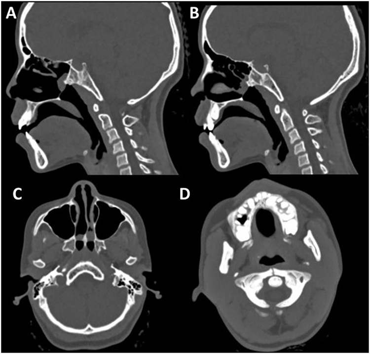 |
Figure 1 Sagittal (A and B) and axial (C) CT images of the paranasal sinuses in bone window settings demonstrate thickening of the vomer (in C), medialization of the pterygoid plates (in C) and membranous bridging of the remaining choanal opening. Minimal retained secretions are seen in the nasal cavities. A lower axial section (D) shows bipartite atlas, in which both anterior and posterior arches of C1 are unfused. |
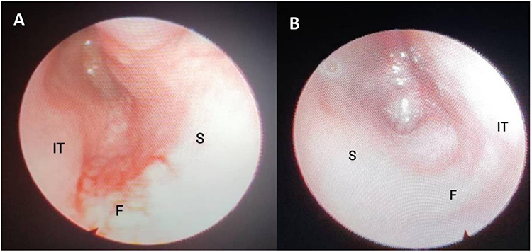 |
Figure 2 Nasal endoscopic images of the right (A) and left (B) nasal cavities demonstrate atretic plates in the posterior aspects of the nasal cavities bilaterally. Abbreviations: IT, Inferior turbinate; S, Septum; F, Floor of nasal cavity. |
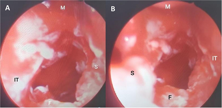 |
Figure 3 Intraoperative nasal endoscopic images show patency of the right (A) and left (B) choanae. Abbreviations: F, Floor of nasal cavity; IT, Inferior turbinate; M, Middle turbinate; S, Septum. |
 |
Figure 4 Nasal endoscopy images of the right (A) and left (B) nasal cavities 5 weeks post-op show patent choanae bilaterally. Abbreviations: F, floor of nasal cavity; L, lateral wall; S, septum; N, nasopharynx. |
Discussion
Choanal atresia is the complete blockage of the posterior nasal openings (choanae). It is thought to occur due to failure of the bucconasal membrane to degenerate during the fifth to sixth week of fetal life. It is the most common congenital nasal anomaly; it occurs in 1 in 8000 births. It is seen more commonly in females.2 Two major types have been described. The most common one is a mixed bony and membranous atresia (70% of the cases), with pure bony atresia accounting for the remaining 30% of the cases. Pure membranous atresia is another type the existence of which is questionable according to current literature.3,4
About half of patients with choanal atresia have other associated congenital malformations. Among these, CHARGE syndrome is the most commonly reported association.4 Other reported associations with choanal atresia include Treacher Collins, Pfeiffer and Crouzon syndromes.5 Cases of bilateral6 choanal atresia are more likely to be associated with other anomalies than their unilateral counterparts.7 In our case, a rare congenital variant of a bipartite (split) atlas was found.
The diagnosis of choanal atresia can be confirmed by nasal endoscopy or cross-sectional imaging. The diagnostic modality of choice in patients with suspected choanal atresia is non-contrast CT of the paranasal sinuses. In addition to making the diagnosis, CT also helps in determining the type of atresia and treatment planning.6 Suction of nasal secretions and administration of topical vasoconstrictors is recommended prior to scanning.3
Bilaterality is relatively common occurring in up to one third of affected patients.2 Clinical presentation of patients with choanal atresia depends on whether it is unilateral or bilateral. Unilateral choanal atresia may remain undetected well into adulthood; it is usually diagnosed after the patients present with unilateral discharge or obstruction. Bilateral choanal atresia on the other hand is almost always detected in neonates and infants with signs of upper airway obstruction. The typical history is that of a neonate who presents with respiratory distress and cyanosis which worsens upon feeding and is relieved with crying.8 This is explained by the fact that newborns are obligate nasal breathers; so, diagnosis of bilateral choanal atresia is considered as an emergency requiring measures such as passage of an oral airway.4 Detection of bilateral choanal atresia in adults is extremely rare. As to our search, only 13 cases have been reported to date. The age of patients previously reported range from 18 years5 to 60 years old.9 Trans-nasal endoscopic surgery was done for all patients except one patient who opted out of surgical management.10 One case of restenosis after surgery requiring repeat operation has been reported.11 Our patient underwent bilateral trans-nasal endoscopic surgery. Her post operative course is uneventful. She is currently being followed at the outpatient clinic and is doing well as of the time of writing this report.
Consent Information
Written informed consent was obtained from guardians of the patient to publish this case report. Any personal details and diagnostic images were anonymized to meet the confidentiality requirements. Institutional approval was not required to publish the case details.
Disclosure
The authors report no conflicts of interest in this work.
References
1. Cedin AC, Atallah ÁN, Andriolo RB, Cruz OL, Pignatari SN. Surgery for congenital choanal atresia. Cochrane Database Syst Rev. 2012;(2). doi:10.1002/14651858.cd008993.pub2
2. Szeremeta W, Parikh TD, Widelitz JS. Congenital Nasal Malformations. Otolaryngol Clin North Am. 2007;40(1):97–112. doi:10.1016/j.otc.2006.10.008
3. Coley BD. Caffey’s Pediatric Diagnostic Imaging E-Book. Elsevier Health Sciences; 2018. Available from: https://books.google.com.et/books?id=QZpeDwAAQBAJ.
4. Ramsden JD, Campisi P, Forte V. Choanal atresia and choanal stenosis. Otolaryngol Clin North Am. 2009;42(2):339–352. doi:10.1016/j.otc.2009.01.001
5. Anajar S, Hassnaoui J, Rouadi S, Abada R, Roubal M, Mahtar M. A rare case report of bilateral choanal atresia in an adult. Int J Surg Case Rep. 2017;37:127–129. doi:10.1016/j.ijscr.2017.05.002
6. Tinoco P, Oliveira Pereira JC, Lourenco Filho RC, et al. Bilateral choanal atresia in 34 year-old patients. Int Arch Otorhinolaryngol. 2010;14(4):481–484.
7. Burrow TA, Saal HM, De Alarcon A, Martin LJ, Cotton RT, Hopkin RJ. Characterization of congenital anomalies in individuals with choanal atresia. Arch Otolaryngol Head Neck Surg. 2009;135(6):543–547. doi:10.1001/archoto.2009.53
8. Lee WT, Koltai PJ. Nasal deformity in neonates and young children. Pediatr Clin North Am. 2003;50(2):459–467. doi:10.1016/S0031-3955(03)00036-1
9. Mengi E. Bilateral choanal atresia in a 60-year-old man: a case report and review of the literature. North Clin Istanbul. 2020;8(5):525–528. doi:10.14744/nci.2020.04557
10. Al Kindy S. Is isolated bilateral choanal atresia a real emergency? Two case reports. Saudi J Heal Sci. 2013;2(1):58. doi:10.4103/2278-0521.112633
11. El-Sawy H, Siddiq MA, Anbarasu A. Bilateral choanal atresia and paranasal sinus hypoplasia in an adult patient with hypogammaglobulinaemia. Eur Arch Oto Rhino Laryngol. 2006;263(12):1136–1138. doi:10.1007/s00405-006-0108-9
12. Panda NK, Simhadri S, Ghosh S. Bilateral choanal atresia in an adult: is it compatible with life? J Laryngol Otol. 2004;118(3):244–245. doi:10.1258/002221504322928099
13. Yasar H, Ozkul MH. Bilateral congenital choanal atresia in a 51-year-old woman. Am J Rhinol. 2016;21(6):716–718. doi:10.2500/ajr.2007.21.3100
14. Aksoy F, Demirhan H, Yildirim YS, Ozturan O. Bilateral choanal atresia in an adult - Management with mitomycin C and without stents: a case report. Cases J. 2009;2(12):3–5. doi:10.1186/1757-1626-2-9307
15. Durmaz CD, Taş V, Kocaay P, et al. Bilateral choanal atresia in an adult woman with pycnodysostosis. Congenit Anom. 2017;57(3):91–92. doi:10.1111/cga.12204
16. Verma RK, Lokesh P, Panda NK. Congenital bilateral adult choanal atresia undiagnosed until the second decade: how we did it. Allergy Rhinol. 2016;7(2):82–84. doi:10.2500/ar.2016.7.0155
17. Kars A, Bingol F, Atalay F. A rare case report: bilateral choanal atresia in an adult patient. Eur J Rhinol Allergy. 2020;3(1):26–28. doi:10.5152/ejra.2020.179
18. Sutikno B, Thaufiqurrakhman M. Transnasal endoscopic neochoanal technique: an effective procedure for bilateral choanal atresia in adult female. Int J Surg Case Rep. 2021;86(6):106338. doi:10.1016/j.ijscr.2021.106338
 © 2023 The Author(s). This work is published and licensed by Dove Medical Press Limited. The full terms of this license are available at https://www.dovepress.com/terms.php and incorporate the Creative Commons Attribution - Non Commercial (unported, v3.0) License.
By accessing the work you hereby accept the Terms. Non-commercial uses of the work are permitted without any further permission from Dove Medical Press Limited, provided the work is properly attributed. For permission for commercial use of this work, please see paragraphs 4.2 and 5 of our Terms.
© 2023 The Author(s). This work is published and licensed by Dove Medical Press Limited. The full terms of this license are available at https://www.dovepress.com/terms.php and incorporate the Creative Commons Attribution - Non Commercial (unported, v3.0) License.
By accessing the work you hereby accept the Terms. Non-commercial uses of the work are permitted without any further permission from Dove Medical Press Limited, provided the work is properly attributed. For permission for commercial use of this work, please see paragraphs 4.2 and 5 of our Terms.