")
Back to Journals » Diabetes, Metabolic Syndrome and Obesity » Volume 12
Beneficial effects of Japanese sake yeast supplement on biochemical, antioxidant, and anti-inflammatory factors in streptozotocin-induced diabetic rats
Authors Davoodi M , Karimooy FN , Budde T , Ortega-Martinez S, Moradi-Kor N
Received 22 June 2019
Accepted for publication 9 August 2019
Published 2 September 2019 Volume 2019:12 Pages 1667—1673
DOI https://doi.org/10.2147/DMSO.S220181
Checked for plagiarism Yes
Review by Single anonymous peer review
Peer reviewer comments 2
Editor who approved publication: Prof. Dr. Antonio Brunetti
Marzieh Davoodi,1 Faezeh Nemati Karimooy,2 Thomas Budde,3 Sylvia Ortega-Martinez,4 Nasroallah Moradi-Kor5
1Social Determinants of Health Research Center, Yasuj University of Medical Sciences, Yasuj, Iran; 2Department of Neuroscience, Medical School, Mashhad University of Medical Sciences, Mashhad, Iran; 3Institute of Physiology I, Westfälische Wilhelms-University, Münster, Germany; 4Department of Neurobiology, The University of Chicago, Chicago, IL, USA; 5Research Center of Physiology, Semnan University of Medical Sciences, Semnan, Iran
Correspondence: Nasroallah Moradi-Kor
Research Center of Physiology, Semnan University of Medical Sciences, Damghan Road, PO Box 35195-163, Semnan, Iran
Tel +98 233 365 4207
Email [email protected]
Background: Using chemical agents in the treatment of diabetes mellitus type 2 may have some limitations due to frequent side effects. Some novel and natural agents may be promising alternatives in this case. This study was designed to evaluate the effects of oral Japanese sake yeast supplement, as a novel agent, on biochemical antioxidant and anti-inflammatory parameters in experimentally induced diabetic rats.
Materials and methods: After inducing diabetes (55 mg/kg intraperitoneal injection of streptozotocin), 120 male adult Wistar rats were randomly divided into 5 groups and each group received 0 (control), 15, 30, or 45 mg/kg of sake yeast or was considered a nondiabetic control. Then, the serum levels of tumor necrosis factor-α, IL-6, C-reactive protein, malondialdehyde, glutathione, total antioxidant status, glucose, cholesterol, triglycerides, and insulin were evaluated and compared to baseline measures.
Results: The results showed that oral administration of sake yeast at different concentrations reduced levels of malondialdehyde, glucose, cholesterol, and triglycerides and increased levels of insulin, glutathione, and total antioxidants (P<0.05). The best responses were observed in the nondiabetic control group.
Conclusion: Sake yeast supplement may be useful as a novel agent in the treatment of diabetes.
Keywords: sake yeast, diabetes, glucose, insulin, inflammatory factors, lipid profile, antioxidants
Introduction
Diabetes mellitus is known as one of the most common metabolic diseases that is accompanied by chronic complications including nephropathy, angiopathy, retinopathy, and peripheral neuropathy.1 The diagnosis of diabetes mellitus is usually based on hyperglycemia and glucose intolerance.2 Increased blood glucose, insulin resistance, and rising inflammatory markers have been reported in patients with diabetes mellitus.3 Regarding inflammatory factors, a positive relation between levels of proinflammatory cytokines, including tumor necrosis factor-α (TNF-α) and IL-6, and insulin resistance has been found.4,5 TNF-α is known to play a role in insulin resistance and acts as a mediator between obesity and inflammatory diseases such as heart disease and type 2 diabetes.6 IL-6 and TNF-α are associated with declined glycemic control and endothelial disorder in diabetic patients.7 C-reactive protein (CRP) has also been reported to have a role in the initiation and aggravation of the classical pathways and promotion of atherosclerosis in diabetic patients.8 Imbalance between oxidant and antioxidant factors may result in oxidative stress which may increase the production of free radicals and reduces antioxidant factors.9 It is known that antioxidants are capable of removing free radicals and ROS by preventing lipid peroxidation and reducing the side effects that are caused by ROS.10 Synthetic agents have been widely used in the treatment of diabetes, but they may be confronted with major limitations due to their possible side effects.5 Therefore, the use of safe and novel agents for the treatment of diabetes is promising.
Sake yeast, Japanese rice wine, has been used in the life and culture of Japanese people for a long time. Sake is a brewed alcoholic beverage, but the brewing process is more complex in comparison with other alcoholic beverages.11 It is fermented from steamed white rice using koji and yeast. Studies have shown that sake is made up of water and ethanol,
Materials and methods
Animals
All experiments were approved by the Ethical Committee of the International Center for Intelligent Research-ICIR (ICIR-2018–185736). The experiments were conducted in accordance with the guideline of the National Institutes of Health (NIH Publication No. 85–23, revised 1996) for the care and use of laboratory animals. Wistar male rats with a weight of 200±10 g were purchased from Pastur Institute, Tehran, Iran. A total number of 120 Wistar rats were randomly divided into 5 groups and each group had 4 subgroups (n=6). Rats were hosted in well-ventilated and spacious stainless-steel cages for free movement and were fed with a standard food prepared in Javaneh Khorasan, Iran with free access to fresh water. Feed included a chow standard diet containing 3 kcal/kh energy, 20% protein, 3% fat, 4% ash, and 6% fiber. Hosting conditions included standard temperature (22±2 °C) and humidity (55±5%) and a light/dark cycle of 12 h. After inducing diabetes in rats, animals were treated with dried sake yeast powder (GSP6 as a gift from Dr Yuki Nagamori, Lion Corporation, Odawara-shi, Japan) as a food supplement for 4 weeks. The powder was dispersed in saline shortly before use and administered to animals in different doses including 0 (control), 15, 30, or 45 mg/kg by oral gavage. A nondiabetic group was also considered. The diabetic and nondiabetic control rats received saline alone and gavage solutions were refreshed daily. Body weight was measured initially and at the end of the study. The dose of sake yeast was chosen based on dose–response in the pilot study.
Pilot study
To select the optimum doses, 36 Wistar rats were deprived of food for 24 h and then were equally divided into 6 groups. Animals received the supplement in doses of 5, 10, 15, 20, 30, 45, and 60 mg/kg and were monitored for 48 h for any toxicity sign including convulsions, ataxia, hypoactivity and ventilation disorders, behavioral alteration, and related mortality. No toxicity sign was observed in this period. Finally, we selected 15, 30, and 45 mg/kg as experimental doses for further studies.
Induction of diabetes
To induce experimental diabetes, 55 mg/kg of streptozotocin (STZ; Sigma-Aldrich Co., St Louis, MO, USA) in 0.1 molar citrate buffer with pH 4.5 was intraperitoneally injected into each animal.5 After 3 days, blood samples were evaluated from each rat using a glucometer (Accu-Check; Hoffman-La Roche Ltd., Basel, Switzerland). Animals with a glucose level >250 mg/dL were considered diabetic.16
Blood sampling and blood variables
At the end of the trial and following 24 h of fasting, 60 mg/kg of sodium phenobarbital was injected to induce anesthesia. Blood samples were taken into tubes without any heparin. The samples were then centrifuged at 4000 RPM for 10 min at 4 °C and kept at −20 °C for further experiments. ELISA was applied for assessment of the levels of TNF-α and IL-6 following the instruction guide. The levels of TNF-α and IL-6 were evaluated using cytokine-specific monoclonal antibodies.5 The thiobarbituric acid reactive substances procedure was applied to evaluate the serum malondialdehyde (MDA) as suggested by Biswas et al.17 The serum concentration of glutathione (GSH) was evaluated using Ellman’s reagent (5,50-dithio-bis-2-nitrobenzoic acid).18 We used the ferric reducing antioxidant power procedure to evaluate the total antioxidant status (TAS) as reported by Budin et al.19 CRP was assessed using the CRP ELISA Kit (Thermo Fisher Scientific, Waltham, MA, USA). Commercially available kits (Pars Azmoon, Iran) were also used to examine the serum concentrations of glucose, cholesterol, triglycerides, and insulin.
Statistical analysis
Data were analyzed with GraphPad Prism statistical software (GraphPad Software, Inc., La Jolla, CA, USA). ANOVA and Dunnett’s Multiple Range Test were used to assess significance. The results are presented as mean±SD. P<0.05 was considered statistically significant.
Results
Effects of different doses of sake yeast supplementation on body weight are shown in Figure 1. As the results show, body weight was increased in nondiabetic rats (P<0.05), but was decreased in diabetic control rats and rats administered 15 and 30 mg/kg supplement (P<0.05). There was no observed significant difference between initial and final weights in rats treated with 45 mg/kg of supplement (P<0.05).
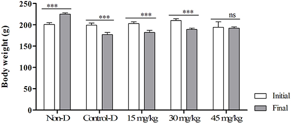 |
Figure 1 Effects of different doses of the supplement (GSP6) on body weight (g) in diabetic rats. Notes: ***Significant difference between initial and final weights at a level of 0.001; ns, nonsignificant (P>0.05). Abbreviations: Control-D, control diabetic; Non-D, nondiabetic. |
Effects of different doses of sake yeast supplementation on serum concentrations of oxidative stress and antioxidant markers are shown in Figure 2. Results indicate that oral administration of sake yeast reduced the levels of the lipid peroxidation marker MDA (Figure 2A; P<0.05) and increased the levels of the antioxidants GSH (Figure 2B) and TAS (Figure 2C) in a dose-dependent manner (P<0.05). Nondiabetic rats showed better response in comparison to other groups.
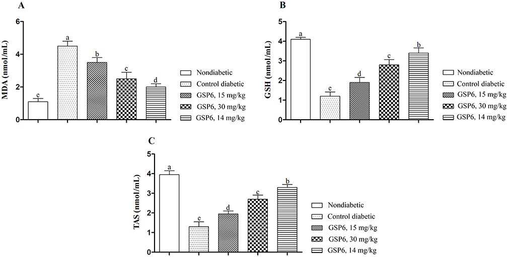 |
Figure 2 Effects of different doses of the supplement (GSP6) on the antioxidant status in diabetic rats. (A) Malondialdehyde (MDA). (B) Glutathione (GSH). (C) Total antioxidant status (TAS). Note: a–e show significant difference among groups at a level of 0.05. |
Next, the effects of experimental sake yeast treatment on levels of proinflammatory cytokines were analyzed and are shown in Figure 3. Results revealed a dose-dependent and significant decrease of serum concentrations of IL-6 (Figure 3A), TNF-α (Figure 3B), and CRP (Figure 3C) with increasing sake yeast supplementation (P<0.05). The lowest level of proinflammatory cytokines was observed by applying sake yeast at a concentration of 45 mg/kg and in the nondiabetic control (P<0.05).
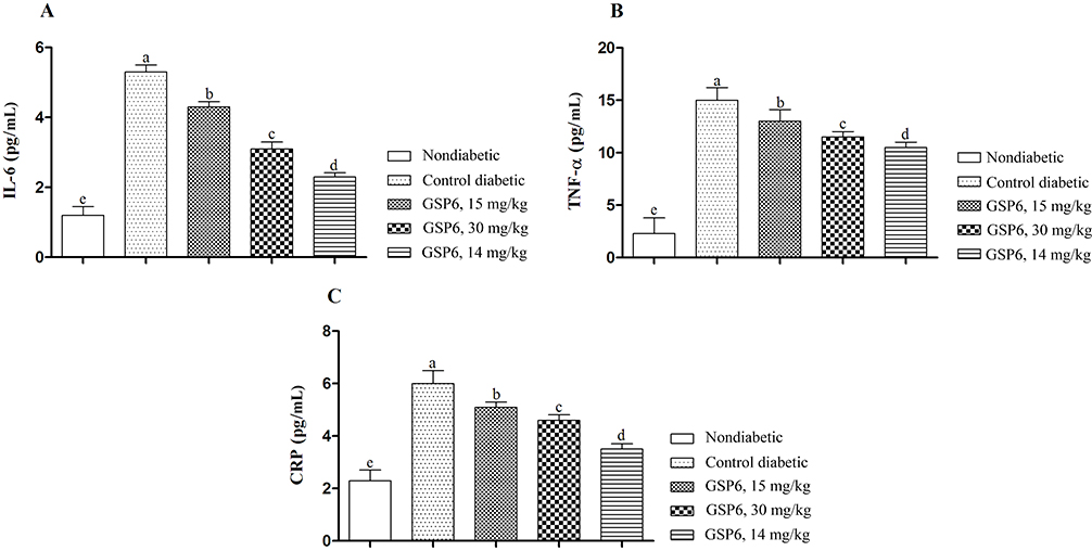 |
Figure 3 Effects of different doses of the supplement (GSP6) on proinflammatory factors in diabetic rats. (A) IL-6. (B) Tumor necrosis factor-α (TNF-α). (C) C-reactive protein (CRP). Note: a–e show significant difference among groups at a level of 0.05. |
The effects of experimental sake yeast supplementation on blood biochemical parameters are shown (Figure 4). The serum concentrations of glucose (Figure 4A), triglycerides (Figure 4C), and cholesterol (Figure 4D) were significantly decreased with increasing sake yeast dosage (P<0.05). Remarkably, the serum concentration of insulin (Figure 4B) increased in a dose-dependent manner. All treated groups showed better responses in comparison to the control group. The best responses were observed in the nondiabetic control.
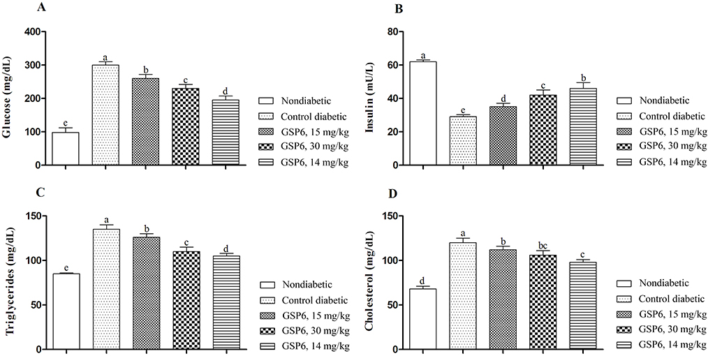 |
Figure 4 Effects of different doses of supplement (GSP6) on blood biochemical parameters in diabetic rats. (A) Glucose. (B) Insulin. (C) Triglycerides. (D) Cholesterol. Note: a-e show significant difference among groups at a level of 0.05. |
Discussion
Results showed that body weight was reduced in diabetic rats and increased in the nondiabetic group. However, animals treated with a level of 45 mg/kg did not show a significant difference (P<0.05). Reduced body weight in diabetic animals was attributed to dehydration and catabolism of fats and proteins and increased catabolic reactions which result in muscle loss and finally weight loss in diabetic rats.20 Maintaining the body weight in animals treated with 45 mg/kg of the supplement might be attributed to the control of hyperglycemia.
The results of the present study indicated an overall improvement of diabetes-related blood parameters induced by food supplementation with Japanese sake yeast. STZ-induced diabetic rats are normally characterized by reduced GSH and TAS as well as increased MDA levels, pointing to an imbalance toward peroxidation in this animal model.5 Treatment with sake yeast supplement improved GSH and TAS levels which may be due to decreased production of free radicals and increased level of antioxidants. Previous studies have reported that STZ causes diabetes by elevating sensitivity to lipid peroxidation and oxidative stress, thus pointing to mechanisms probably involved in the pathogenesis of the disease.9,21 MDA has been reported as one of the products of lipid peroxidation that is rapidly mixed with biomolecules and therefore disturbs the glucose metabolism.22 Sake yeast modulates the production of oxygen radicals which might be responsible for decreasing hyperglycemia, inflammation, and oxidative stress. Unfortunately, we could not find any study suggesting the effects of sake yeast on the antioxidant system. However, our study is the first to show that sake yeast, especially in higher doses, can increase the antioxidant factors and decrease lipid peroxidation (MDA). It could be concluded that sake yeast declines the level of MDA by increasing GSH and TAS.
Our findings indicated that sake yeast could decrease inflammatory factors in comparison to the control group. Inflammation is a basic biological process that is considered the background of many acute and chronic pathological conditions, and this action happens in response to the changes which restore tissue homeostasis by triggering different repair mechanisms. Appropriate regulation of such mechanisms is necessary to inhibit uncontrolled amplification of the initial inflammatory response which may otherwise lead the tissue repair process to collateral damage and development of diseases.23 TNF-α is known to have adverse effects including increasing adipocyte lipolysis and alterations in the insulin signaling pathway by changing tyrosine/serine phosphorylation of insulin receptor substrates.24 CRP is known as a main inflammatory factor which is regulated by IL-6, IL-1, and TNF-α and is produced in the liver in response to inflammation.25 Chang et al26 showed that diabetes mellitus can increase the level of nuclear factor-κB that is responsible for the formation of several proinflammatory cytokines. Importantly, our findings showed that supplementing with sake yeast decreased the inflammatory factors. Virgolici et al27 illustrated elevated levels of inflammatory factors in relation to raised oxidative stress. Our findings showed that treatment with sake yeast improves the antioxidant status and may decrease inflammatory factors by improving antioxidant activity.
Increased triglycerides, cholesterol, and glucose and decreased insulin were observed in the control group compared to the diabetic and treated groups. Hypertriglyceridemia and hypercholesterolemia are known as common signs in diabetes.28 Studies have also reported that STZ increases the sensitivity to lipid peroxidation.9,29 Decreased hepatic insulin sensitivity could be attributed to increased hepatic gluconeogenesis, postprandial hyperinsulinemia, and increased production of triglycerides in the liver cells. Hypertriglyceridemia, hypercholesterolemia, hyperglycemia, and hypoinsulinemia could be due to an increase in TNF-α. It steps up triglyceride production in plasma29 and cuts down glucose consumption in peripheral tissues by targeting insulin signaling pathways and the glucose transporter 4 (GLUT4).3,30 Parallel to our findings, Kido et al13 have reported that high intake of alcoholic beverages with a meal causes different responses in postprandial glucose and insulin concentrations. Accordingly, sake reduces the levels of lipids and glucose through modulation in TNF-α. In addition, components of sake including ethyl α-
Conclusion
Sake yeast supplement can decrease inflammatory parameters by increasing antioxidant factors. It also improved blood biochemical parameters compared to the control group. Although future studies need to be done in order to evaluate the effects of sake yeast on diabetes, our preliminary study showed that this supplement has potential for improving the treatment of diabetes. We recommend conducting human studies and the use of GSP6 as an oral supplement.
Acknowledgment
The authors gratefully acknowledge Dr Yuki Nagamori for his assistance and providing the sake yeast powder GSP6.
Author contributions
All authors contributed toward data analysis, drafting and revising the paper, gave final approval of the version to be published and agree to be accountable for all aspects of the work.
Disclosure
The authors report no conflicts of interest in this work.
References
1. Muriach M, Flores-Bellver M, Romero FJ, Barcia JM. Diabetes and the brain: oxidative stress, inflammation, and autophagy. Oxid Med Cell Longev. 2014;2014:9. Article ID 102158. doi:10.1155/2014/102158
2. Smyth S, Heron A. Diabetes and obesity: the twin epidemics. Nat Med. 2006;12:75–80. doi:10.1038/nm0106-75
3. Tabibzadeh Dezfuli SA, Ehsani M, Lakzaei Azar O. Carvacrol alleviated negative effects of diabetes on inflammation and oxidation by modulation in gene expression of inflammatory and antioxidant system in diabetic rat model. GMJ Med. 2017;1(1):15–20. doi:10.29088/GMJM.2017.15
4. Uysal KT, Wiesbrock SM, Marino MW, Hotamisligil GS. Protection from obesity-induced insulin resistance in mice lacking TNF-alpha function. Nature. 1997;389:610–614. doi:10.1038/39335
5. Mesbahzadeh B, Rajaei SA, Tarahomi P, et al. Beneficial effects of Spirogyra Neglecta Extract on antioxidant and anti-inflammatory factors in streptozotocin-induced diabetic rats. BioMol Concepts. 2018;9:184–189. doi:10.1515/bmc-2018-0015
6. Coen PM, Flynn MG, Markofski MM, Pence BD, Hannemann RE. Adding exercise to rosuvastatin treatment: influence on C-reactive protein, monocyte toll-like receptor 4 expression, and inflammatory monocyte (CD14+CD16+) population. Metabolism. 2010;59:1775–1783. doi:10.1016/j.metabol.2010.05.002
7. Mayadas TN. Regulation of Neutrophil Apoptosis. Molecular Basis for Microcirculatory Disorders. Paris: Springer; 2003. p. 271–287. doi:10.1007/978-2-8178-0761-4
8. Hayden MR, Tyagi SC. Intimal redox stress: accelerated atherosclerosis in metabolic syndrome and type 2 diabetes mellitus. Cardiovasc Diabetol. 2002;1:3. doi:10.1186/1475-2840-1-3
9. Samarghandian S, Farkhondeh T, Samini F, Borji A. Protective effects of carvacrol against oxidative stress induced by chronic stress in rat’s brain, liver, and kidney. Biochem Res Int. 2016;2016:2645237. doi:10.1155/2016/7108261
10. Abdulrahman L, Al-malki AL, El-Rabey HA. The antidiabetic effect of low doses of Moringaoleifera Lam. Seeds on streptozotocin induced diabetes and diabetic nephropathy in male rats. Bio Med Res Int. 2015;2015:1–13. Article ID 381040. doi:10.1155/2015/381040
11. Nakahara M, Mishima T, Hayakawa T. Effect of a sake concentrate on the epidermis of aged mice and confirmation of ethyl α-D-glucoside as its active component. Biosci Biotechnol Biochem. 2007;71(2):427–434. doi:10.1271/bbb.60489
12. Tadenuma M. Sheishu no seibun wo megutte. Gendai Kagaku (in Japanese). 1987;1:26–31.
13. Kido M, Asakawa A, Koyama KK, et al. Acute effects of traditional Japanese alcohol beverages on blood glucose and polysomnography levels in healthy subjects. PeerJ. 2016;4:e1853. doi:10.7717/peerj.1853
14. Monoi N, Matsuno A, Nagamori Y, et al. Japanese sake yeast supplementation improves the quality of sleep: a double‐blind randomised controlled clinical trial. J Sleep Res. 2016;25:116–123. doi:10.1111/jsr.12336
15. Awad AS, Huang L, Ye H, et al. Adenosine A2A receptor activation attenuates inflammation and injury in diabetic nephropathy. Am J Physiol Renal Physiol. 2006;290:828–837. doi:10.1152/ajprenal.00310.2005
16. Nasirian F, Dadkhah M, Moradi-Kor N, Obeidavi Z. Effects of Spirulina platensis microalgae on antioxidant and anti-inflammatory factors in diabetic rats. Diabetes Metab Syndr Obes. 2018;11:375–380. doi:10.2147/DMSO.S172104
17. Biswas D, Banerjee M, Sen G, et al. Mechanism of erythrocyte death in human population exposed to arsenic through drinking water. Toxicol Appl Pharmacol. 2008;230:57–66. doi:10.1016/j.taap.2008.02.003
18. Beutler E, Duron O, Kelly BM. Improved method for the determination of blood glutathione. J Lab Clin Med. 1963;61:882–888.
19. Budin SB, Othman F, Louis SR, Bakar MA, Das S, Mohamed J. The effects of palm oil to cotrienol rich fraction supplementation on biochemical variables, oxidative stress and the vascular wall of streptozotocin-induced diabetic rats. Clinics (Sao Paulo). 2009;64:235–244. doi:10.1590/s1807-59322009000300015
20. Rajkumar L, Srinivasan N, Balasubramanian K, Govindarajulu P. Increased degradation of dermal collagen in diabetic rats. Indian J Exp Biol. 1991;29:1081–1083.
21. Yazdanparast R, Ardestani A, Jamshidi S. Experimental diabetes treated with Achilleasantolina: effect on pancreatic oxidative parameters. J Ethnopharmacol. 2007;112:13–18. doi:10.1016/j.jep.2007.01.030
22. Sivaraman K, SenthilKumar GP, Sankar P, Bobby Z. Attenuation of oxidative stress, inflammation and insulin resistance by Allium sativum in fructose-fed male rats. J Clin Diagn Res. 2013;7:1860–1862. doi:10.7860/JCDR/2013/6924.3334
23. Goldszmid RS, Trinchieri G. The price of immunity. Nat Immunol. 2012;13:932–938. doi:10.1038/ni.2422
24. El-Abhar HS, Schaalan MF. Topiramate-induced modulation of hepatic molecular mechanisms: an aspect for its anti-insulin resistant effect. PLoS One. 2012;7(5):e37757. doi:10.1371/journal.pone.0037757
25. Nicklas BJ, You T, Pahor M. Behavioral treatments for chronic systemic inflammation: effects of dietary weight loss and exercise training. CMAJ. 2005;172:1199–1209. doi:10.1503/cmaj.1040769
26. Chang CC, Chang CY, Huang JP, Hung LM. Effect of resveratrol on oxidative and inflammatory stress in liver and spleen of streptozotocin-induced type 1 diabetic rats. Chin J Physio. 2012;55(3):192–201. doi:10.4077/CJP.2012.BAA012
27. Virgolici B, Mohora M, Gaman L, et al. Relation between inflammation and oxidative stress markers in diabetic foot patients. Romanian. J Biophys. 2008;18(4):273–282.
28. Khan BA, Abraham A, Leelamma S. Hypoglycemic action of Murraya koenigii (curry leaf) and Brassica juncea (mustard): mechanism of action. Indian J Biochem Biophys. 1995;32(2):106–108.
29. Sun X, Han F, Yi J, Lina H, Ben W. Effect of aspirin on the expression of hepatocyte NF-κB and serum TNF-α in streptozotocin-induced type 2 diabetic rats. Korean Med Sci. 2011;26:765–770. doi:10.3346/jkms.2011.26.6.765
30. Mostafa AM, Mohamed WS, Serwah AHA, Serwah MA. Effect of diclofenac on plasma glucose level, insulin resistance, inflammatory markers and hepatocytes in diabetic albino rats. The Egyptian J Hospital Med. 2014;54:117–128. doi:10.12816/0002438
31. Saito Y, Wanezaki K, Kawato A, Imayasu S. Antihypertensive effects of peptide in sake and its by-products on spontaneously hypertensive rats. Biosci Biotechnol Biochem. 1994;58:812–816. doi:10.1271/bbb.58.812
32. Izu H, Hizume K, Goto K, Hirotsune M. Hepatoprotective effects of a concentrate and components of sake against galactosamine (GalN)-induced liver injury in mice. Biosci Biotechnol Biochem. 2007;71:951–957. doi:10.1271/bbb.60613
 © 2019 The Author(s). This work is published and licensed by Dove Medical Press Limited. The full terms of this license are available at https://www.dovepress.com/terms.php and incorporate the Creative Commons Attribution - Non Commercial (unported, v3.0) License.
By accessing the work you hereby accept the Terms. Non-commercial uses of the work are permitted without any further permission from Dove Medical Press Limited, provided the work is properly attributed. For permission for commercial use of this work, please see paragraphs 4.2 and 5 of our Terms.
© 2019 The Author(s). This work is published and licensed by Dove Medical Press Limited. The full terms of this license are available at https://www.dovepress.com/terms.php and incorporate the Creative Commons Attribution - Non Commercial (unported, v3.0) License.
By accessing the work you hereby accept the Terms. Non-commercial uses of the work are permitted without any further permission from Dove Medical Press Limited, provided the work is properly attributed. For permission for commercial use of this work, please see paragraphs 4.2 and 5 of our Terms.