")
Back to Journals » Journal of Experimental Pharmacology » Volume 12
Antidiabetic Effect of Germinated Lens culinaris Medik Seed Extract in Streptozotocin-Induced Diabetic Mice
Authors Tefera MM, Altaye BM , Yimer EM , Berhe DF , Tadesse Bekele S
Received 5 September 2019
Accepted for publication 4 December 2019
Published 31 January 2020 Volume 2020:12 Pages 39—45
DOI https://doi.org/10.2147/JEP.S228834
Checked for plagiarism Yes
Review by Single anonymous peer review
Peer reviewer comments 3
Editor who approved publication: Professor Bal Lokeshwar
Mulugeta Mihrete Tefera,1,2 Birhanetensay Masresha Altaye,2,3 Ebrahim M Yimer,2,4 Derbew Fikadu Berhe,2 Senait Tadesse Bekele5
1Department of Pharmacy, Bahirdar Health Science College, Bahirdar, Ethiopa; 2Department of Pharmacology and Toxicology, College of Health Sciences, Mekelle University, Mekelle, Ethiopia; 3College of Medicine, Debre Berhan University, Debre Berhan, Ethiopia; 4Department of Pharmacy, College of Medical and Health Sciences,Wollo University, Dessie, Ethiopa; 5Medical Laboratory Department, Health Science College, Bahir Dar University, Bahir Dar, Ethiopia
Correspondence: Mulugeta Mihrete Tefera
Department of Pharmacy, Bahirdar Health Science College
Tel +251 92 835 1786
Email [email protected]
Background: Lens culinaris Medik seed has been used in traditional practices to treat various ailments, including diabetes mellitus, in Ethiopia. Previous phytochemical screening studies indicated that germination of the seed of L. culinaris contains more bioactive constituents compared to raw seeds. The aim of this study was to investigate the antidiabetic activity of an aqueous methanol extract of germinated L. culinaris seed extract in streptozotocin (Stz)-induced diabetic mice.
Methods: The antidiabetic effect of germinated L. culinaris seed extract was determined using Stz-induced diabetic mice. An 80% aqueous methanol extract of germinated L. culinaris seed at doses of 100, 200, and 400 mg/kg was used in the treatment group. Glibenclamid (5 mg/kg) and dimethyl sulfoxide 2% were used as positive and negative controls, respectively. The test extract and controls were given daily for 3 weeks. Blood-glucose levels and body-weight changes were measured weekly. Lipid-profile levels were measured at the end of each experiment. Oral glucose-tolerance tests were performed to evaluate the postprandial effect of the extract.
Results: The aqueous methanol extract of germinated L. culinaris significantly reduced blood-glucose levels and increased body weight (p< 0.05). The extract also improved serum-lipid profiles in diabetic mice after 21 days (p< 0.05). The seed extract also resulted in significant reductions in blood-glucose levels after an oral glucose load in normal mice (p< 0.05).
Conclusion: An aqueous methanol extract of germinated L. culinaris seed has both antidiabetic and antihyperlipidemic effects.
Keywords: Lens culinaris Medik, diabetes mellitus, mice
Introduction
Globally, millions of people suffer from diabetes mellitus, as there is no compete cure and limited access, even to available medications.1 This is becoming more challenging to developing countries, including Ethiopia, as a result of population growth, aging, unhealthy diets, obesity, and sedentary lifestyles.2,3 Because of the progressive the nature of diabetes mellitus and adverse effects of currently available drugs, people tend to seek alternative options for diabetes management, including use of traditional medicinal plants.4–6
Functional foods have been used as a treatment for different diseases, including cancer, heart disease, osteoporosis, inflammation, chronic degenerative diseases, lowering of blood cholesterol, and neutralization of reactive oxygen species for thousands of years.7 Lens culinaris,is also known as misir in Amharic, is an ancient legume crop that has an important role in human health.8 The crop is cultivated and eaten almost all over the world, including Ethiopia.9
The seed is rich in secondary metabolites, including polyphenols, flavonoids, saponins, dietary fibers, triterpenoids, phytates, phytosterols, defensins, and lectins.10,11 Secondary metabolites, minerals, and bioactive constituents in L. culinaris have shown promising effects in the treatment and prevention of several chronic human diseases, including diabetes, hyperlipidemia, and hypertension.12,13
In Ethiopia, the seeds of L. culinaris are traditionally used for the treatment of diabetes by the Shinasha, Agew-awi, and Amhara people in northwest Ethiopia.14 In vitro studies have revealed that germination of L. culinaris significantly increase total phenolics and flavonoids. It also significantly increases free-radical scavenging and antioxidant activity.15,16
Though germination of L. culinaris shows significant increases in bioactives, the antidiabetic effect of the extract for germinated seed has not been evaluated so far. The present study aimed to investigate the antidiabetic effect of an aqueous methanol seed extract of L. culinarisin a streptozotocin (Stz)-induced diabetic mouse model.
Methods
Chemicals and Drugs
Glibenclamide, Stz, methanol, distilled water, sodium citrate, glucose solution, citric acid, halothane, dimethyl sulfoxide were some of the chemicals and drugs used in the experiment, all of laboratory or analytical grade.
Plant-Material Collection and Identification
Local cultivated seeds of L. culinaris were purchased from the local market in Woreta, northwest Ethiopia. Taxonomic identification and authentication of plant-seed specimen was confirmed in Gonder University, Biology Department. Plant specimens were kept for future reference with the voucher number MM0027/2010 in the Herbarium unit of the Department of Biology, College of Computational and Natural Science in Gondar University.
Preparation of Plant Material
Soaking and Germination
L. culinaris seeds were cleaned by hand to remove foreign materials and washed with tap water to remove soil and other water-soluble materials. Then, seeds were soaked in water (1:10 w/v) for 12 hours at room temperature, kept between thick layers of cotton cloth, and allowed to germinate in the dark for 5 days. Seeds were watered every day with fresh tap water. After 5 days, sprouted seeds (radicle with cotyledons) were frozen for 12 hours to stop the germination process. The moisture was removed with a soft cloth and then it was grinder by coffee grinder and subjected to drying at room temperature. Dried sprouted seeds were ground again and passed through a 2 mm mesh sieve. The flour obtained was stored in a glass container until it was macerated at room temperature.17,18
Preparation of Crude Extract
Germinated seed powder (1.35 kg) was subjected to maceration using 80% methanol (v/v) for 72 hours at room temperature. The extract was separated from the marc using muslin cloth and further filtered with Whatman filter paper number 1. The marc was remacerated twice with 80% (v:v) methanol to obtained maximal yield of the plant material. The filtrate obtained was evaporated to get a dried extract using an oven dryer at 40°C. The dried extract was kept in dry clean glass bottles at 4°C for future analysis.
Experimental Animals
Swiss albino mice obtained from Mekele University College of Health Science were used in this experiment. Male and female mice with body weight of 24–36 g and in aged 6–8 weeks were used. The animals were kept in cages under standard environmental conditions (22°C±3 °C, 12-hour light/dark cycle). The mice had access to standard laboratory pellets and water ad libitum before and during the experiment. At the end of the experiment, mice were euthanized using cervical dislocation after being anesthetized with halothane. Animal procedures and experimental protocols were approved by the Ethical Review Board of Mekele University College of Health Science (approval ERC 1221/2018) and were undertaken according to the international guidelines for the use and maintenance of experimental animals.19,20
Acute Oral Toxicity Study
Acute oral toxicity testing was carried out as per the Organization for Economic Cooperation and Development guideline 425.20 Five female albino mice were randomly selected. The mice were fasted for 4r hours with provision of water. A germinated 80% methanol extract of L. culinaris seed was administered orally at a dose of 2,000 mg/kg body weight. Mice were observed continuously for the first 4 hours and then periodically up to 24 hours for toxic manifestations like drowsiness, salivation tremor, restlessness, convulsions, piloerection, and mortality, if any. They were further observed for another 14 days.
Induction of Experimental Diabetes
The male mice were fasted overnight. Then, their weights were recorded and they were administered a single intraperitoneal dose of Stz dissolved in 0.1 M cold citrate buffer (pH 4.5) at 45 mg/kg body weight. On the third day, blood-glucose levels were recorded by taking blood from the tail, and mice with blood-glucose levels >200 mg/dL were considered diabetic.21–23
Estimation of Blood Glucose and Body Weight in Diabetic Mouse Model
Male mice were assigned to six groups (n=5 per group). Then, the normal controls (group 1) and diabetic controls (group 2) were given a vehicle (2% DMSO). The diabetic group 3 was given glibenclamide (5 mg/kg). Diabetic groups 4–6 were given 100, 200, and 400 mg/kg crude seed extract, respectively. The extract was administered on a daily basis. Fasting blood glucose was measured after taking blood from tails of mice before treatment and at days 7, 14, and 21. Body-weight changes were also recorded weekly. The procedure was performed according to previous studies.23,24
Lipid-Profile Change in Diabetic Mouse Model
After 3 weeks of treatment, on day 21 mice were fasted overnight and blood collected after anesthesia with halothane by direct cardiac puncture. Then, the blood was left to stand at room temperature for 2 hours in sterile EDTA test tubes until centrifugation at 1,500 rpm for 5 minutes. The supernatant was immediately separated from the pellet to prepare serum samples to determine the level of triglyceride (TG) and total cholesterol (TC)) directly, while very low–density lipoprotein (VLDL) was calculated from TG (VLDL=TG/5).25
Oral Glucose-Tolerance Test
In the oral glucose-tolerance test, normal female mice were divided into five groups, five mice in each group. After they had been fasted for 12 hours, groups 1 and 2 were given a vehicle (2% DMSO) and glibenclamide (5 mg/kg), respectively, while groups 3–5 were given the seed extract at a dose of 100, 200, and 400 mg/kg orally, respectively. After 5 minutes of treatment, all mice were loaded with 2.5 g/kg of glucose solution orally. Blood samples were collected from the tails of the mice to determine blood-glucose levels immediately prior to treatment and at 0.5, 1, and 2 hours after glucose load.
Hypoglycemic Effect
The normal female mice were first fasted for 6 hours with provision of water. They were then randomly divided into five groups with five mice in each group. Group 1 was given 1 mL/100g of the vehicle, group 2 5 mg/kg glibenclamide, and groups 3–5 100, 200, and 400 mg/kg germinated seed extract, respectively. Blood samples were collected and blood-glucose levels measured before treatment and at 1, 2, and 3 hours after treatment.
Statistical Analysis
Data collected were first collected and recorded using Microsoft Excel and are expressed as means ± SEM. Statistical differences among control and treatment groups were confirmed using one way ANOVA post hoc analysis with SPSS 22. p<0.05 was considered significant.
Results
Acute Oral Toxicity Study
Acute oral toxicity assessment showed that the seed extract caused no mortality up to 2,000 mg/kg within 14 days of follow-up. Observations also indicated no visible signs or symptoms of toxicity, such as lacrimation, loss of appetite, tremors, piloerection, salivation, diarrhoea, or convulsion.
Effects of Extract on Body Weight in Diabetic Mice
After the induction of diabetes, significant differences in body weight between normal and untreated diabetic mice were observed (p<0.05, Figure 1). All doses of the extract (100 mg/kg, 200 mg/kg, and 400 mg/kg) improved weight gain when compared to diabetic controls at weeks1, 2, and 3 (p<0.05). The weight of normal control mice had significantly increased (p<0.05) at days 7, 14, and 21compared to day 0 day, while that of diabetic control mice had significantly decreased at days 7, 14, and 21compared to day 0. Mice treated with 100 mg/kg, 200 mg/kg, and 400 mg/kg extract and 5 mg/kg glibenclamide had gained weight of 8.79%, 11.12%, 17.46%, and 14.79% at the end of the experiment, respectively.
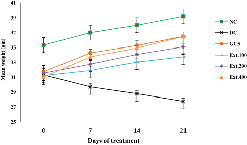 |
Figure 1 Effect of extract on body weight in diabetic mice. Note: Data expressed as means ± SEM (n=5). Abbreviations: Ext.100, 100 mg/kg extract; Ext.200, 200 mg/kg extract; Ext.400, 400 mg/kg extract; GC5, glibenclamide 5 mg/kg; NC, normal control; DC, diabetic control. |
Effects of Extract on Blood-Glucose Levels in Normal Mice
Blood-glucose levels before treatment indicated no significant differences in initial blood glucose among the groups (Figure 2). Mice treated with 100 mg/kg extract showed reduced blood glucose, but did not show statistically significant reduction across any time points compared to normal controls. Similarly, 200 g/kg extract failed to show significant hypoglycemic effects at 1 hours and 2 hours, but did at 3 hours compared to normal controls. The 400 mg/kg dose produced a significant (p<0.05) reduction in blood glucose at 2 and 3 hours compared to normal controls and the 100 mg/kg dose.
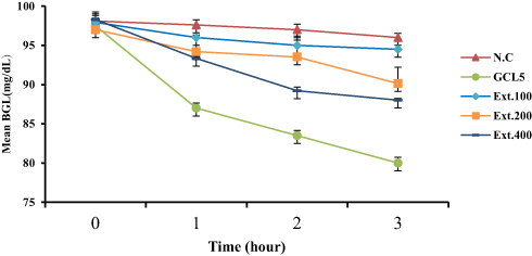 |
Figure 2 Effect of extract on blood-glucose levels in normal mice. Note: Data expressed as means ± SEM (n=5). Abbreviations: Ext.100, 100 mg/kg extract; Ext.200, 200 mg/kg extract; Ext.400, 400 mg/kg extract; GC5, glibenclamide 5 mg/kg; NC, normal control. |
Effects of Extract on Oral Glucose Tolerance in Normal Mice
Administration of glucose (2.5 g/kgorally) produced maximum change in blood glucose of overnight-fasted mice after 30 minutes of glucose challenge in all groups (Figure 3). Compared to normal controls, the two groups taking 200 mg/kg and 400 mg/kg extract showed significant tolerance (p<0.05) of oral glucose load at 1 and 2 hours. The highest dose of the extract (400 mg/kg) produced significant (p<0.05) decreases in blood-glucose levels compared to the lower doses at 1 and 2 hours of glucose loading. It also showed a significant blood glucose–lowering effect (p<0.05) compared to 200 mg/kg after 2 hours of glucose loading.
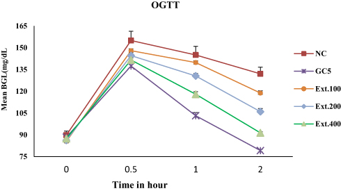 |
Figure 3 Effect of extraction oral glucose tolerance in normal mice. Note: Data expressed as means ± SEM (n=5). Abbreviations: Ext.100, 100 mg/kg extract; Ext.200, 200 mg/kg extract; Ext.400, 400 mg/kg extract; GC5, glibenclamide 5 mg/kg; NC, normal control. |
Effects of Extract on Blood-Glucose Levels in Diabetic Mice
Fasting blood-glucose levels of diabetic control mice were significantly higher than normal control mice throughout the treatment period (Table 1). Those administered 100 mg/kg, 200 mg/kg, and 400 mg/kg extract showed a significant reduction (p<0.05) in glucose levels when compared to diabetic controls at days 7, 14, and 21 of measurement. The 400 mg/kg dose exhibited significant antidiabetic activity compared to 100 mg/kg (p<0.05).
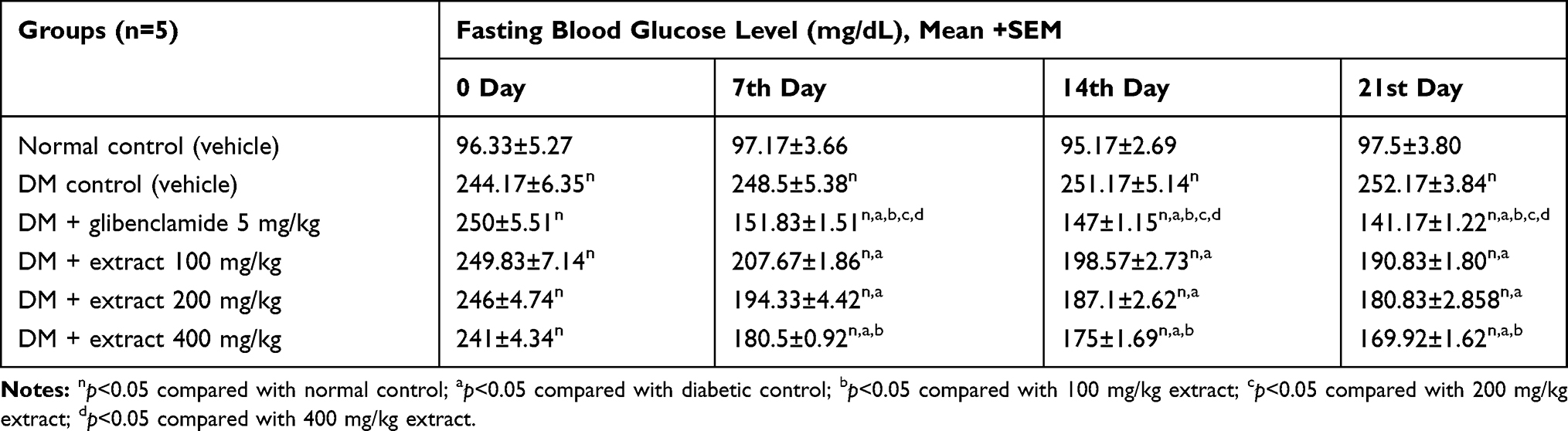 |
Table 1 Effects of Extract on Blood Glucose Level in Diabetic Mice |
Effects of Extract on Lipid Profile
TC, TG, and VLDL cholesterol increased significantly (p<0.05) in diabetic control mice compared to normal control mice (Table 2). Different doses of the extract (100 mg/kg, 200 mg/kg, and 400 mg/kg) significantly decreased (p<0.05) TC, TG, and VLDL compared to diabetic controls. The highest dose of the extract (400 mg/kg) and glibenclamide (5 mg/kg) decreased the three lipid levels significantly (p<0.05) when compared to the lowest dose (100 mg/kg) of the extract (p<0.05). Both 400 mg/kg dose of extract and glibenclamide also reduced TG and VLDL near to values of normal controls.
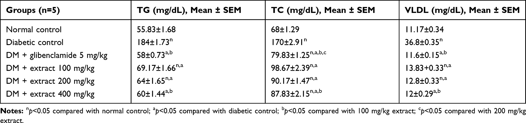 |
Table 2 Effect of Extract on Lipid Profile in Diabetic Mice |
Discussion
In the current study, the effects of germinated L. culinaris seed extract on body weight, lipid profile, and blood-glucose levels were investigated. The findings indicated that administration of L. culinaris aqueous methanolic seed extract produced no observable acute-toxicity signs at doses up to 2 g/kg throughout 2 weeks' follow-up. This indicates that the LD50 of the plant is >2 g/kg.
The 200 mg/kg and 400 mg/kg doses significantly reduced normal blood-glucose levels (hypoglycemic test). Glibenclamide produced a hypoglycemic effect by binding to an ATP-dependent K+ channel. This interaction closes the K+ channel, which inhibits potassium efflux and increase in intracellular calcium levels, causing release of insulin from β cells.26 Though the exact mechanism is not known, the blood glucose–lowering effect of the extract in normal mice may be due to an increase in insulin secretion or improvement in glucose uptake in tissue. The extract at all doses improved glucose tolerance compared to normal controls. Glibenclamide also improved glucose tolerance, with a similar mechanism on hypoglycemic activity.27 The extract may exert its effect due to its inhibitory effect on glucose absorption, increased insulin secretion, or improvement in glucose uptake in tissue, as it contains different bioactive compounds like flavonoids, polyphenols, and saponins that are also present in other plants and produce similar effects.28,29
In this study, intraperitoneal administration of Stz to mice significantly increased blood-glucose levels 3 days after injection, as well as decreased body weight. These results agree with previous observations that have employed this model and that also reported loss of body weight.25 Stz selectively accumulated in pancreatic β cells via the low-affinity GLUT2 glucose transporter. The transfer of the methyl group from Stz nitrosourea moiety to the DNA molecule causes damage, which along a defined chain of events results in the fragmentation of the DNA. Protein glycosylation may be an additional damaging factor for β-cell destruction that ultimately results in induction of diabetes.18 Weight loss is the main sign of diabetes, but its mechanism is not clear. It could be due to many factors, such as loss of appetite, increased muscle waste, and loss of tissue protein.30
Administration of a 5-day germinated L. culinaris seed extract lowered blood-glucose levels and serum lipids in Stz-induced diabetic mice. The lipid-lowering and antidiabetic effect of the extract could be due to different bioactive constituents, such as polyphenols, flavonoids, saponins, which were identified from germinated and raw lentil seed in previous studies. These bioactive constituents increased during germination and reached maximum concentration after 5 days of germination.15,16 Polyphenols and saponins have exhibited antidiabetic, antioxidant, and free radical–scavenging activities and a cholesterol-lowering effect.10,11
L. culinaris seed extract contains small escapable fatty acids, including propionate, that inhibit hepatic cholesterol synthesis, decrease activity of HMG-CoA reductase enzyme, and reduce reabsorbtion of bile acid and cholesterol from the gastrointestinal tract.31,32 It is also rich in antinutritive factors such as phytate, lectins, and tannins,12 which have been shown to reduce blood-glucose level, plasma cholesterol, and TG.33 The extract also increased weight of diabetic mice. The increase in body weight could have been due to improvement in diabetic status or nutritional components of the extract. Similar effects on body-weight increment in study animals has also been seen in other research done on lentil.16
Conclusion
In this study, hydromethanolic extracts of germinated L. culinaris seed extract showed significant reduction in blood-glucose levels in Stz-induced diabetic mice. Similarly, the seed extract caused increases in weight and decreases in TC, TG, and VLDL in Stz induced diabetic mice. It also improved oral glucose tolerance in normal mice. The study thus supports the traditional use of germinated L. culinaris seed for the management of diabetes in Ethiopia.
Availability of Data and Materials
The outcome of this research was generated from the data collected and analyzed based on the specified methods and materials. The original data supporting these findings will be accessible at the time of request.
Acknowledgments
We would like to acknowledge Mekelle University and Bahir Dar Health Science College for sponsoring this study. It is also our pleasure to thank Amhara Regional State Health Bureau for financial and material support. We also appreciate the technical assistance of Mr Mesele Gebresilasie during laboratory work. We are also grateful to Gondar University National Herbarium for authenticating the plant species.
Disclosure
The authors report no conflicts of interest in this work.
References
1. Marin-Penalver JJ, Martin-Timon C, Cristina SC, Del Canizo-Gomez FJ. Update on the treatment of type 2 diabetes mellitus. World J Diabetes. 2016;7:354–395. doi:10.4239/wjd.v7.i17.354
2. Knip M, Simell O. Environmental triggers of Type 1 diabetes. Cold Spring Harb Perspect Med. 2012;29(7):1–15.
3. Alemu F. Diabetes & metabolism prevalence of diabetes mellitus disease and its association with level of education among adult patients attending at dilla referral hospital. Diabetes Metab. 2015;6(4):4–8.
4. Philis-tsimikas A. Type 2 diabetes : limitations of current therapies. Diabetes Care. 2009;31:S150–S154.
5. Demoz MS, Gachoki KP, Mungai KJ, Negusse BG. Evaluation of the anti-diabetic potential of the methanol extracts of aloe camperi, meriandra dianthera and a polyherb. J Diabetes Mellitus. 2015;5:268–276. doi:10.4236/jdm.2015.54033
6. Alamin MA, Yagi AI, Yagi SM. Asian Pacific Journal of Tropical Biomedicine Evaluation of antidiabetic activity of plants used in Western Sudan. Asian Pac J Trop Biomed. 2015;5(5):395–402. doi:10.1016/S2221-1691(15)30375-0
7. Abuajah CI, Ogbonna AC, Osuji CM. Functional components and medicinal properties of food: a Review. J Food Sci Technol. 2015;52:2522–2529. doi:10.1007/s13197-014-1396-5
8. Kang HW. Antioxidant activity of ethanol and water extracts from lentil (Lens Culinaris). Journal of Food and Nutrition Research. 2015;3(10):667–669.
9. Jamirul M, Zehadi A, Masamba K, Li Y, Chen M, Chen X. Identification and purification of antioxidant peptides from lentils (Lens Culinaris) hydrolysates. J Plant Sci. 2015;3(3):123–132.
10. Ganesan K. Polyphenol-rich lentils and their health promoting effects. Int J Mol Sci. 2017;18:2390. doi:10.3390/ijms18112390
11. Shahwar D, Bhat TM, Ansari MYK, Chaudhary S. Health functional compounds of lentil (Lens culinaris Medik): a review. Int J Food Prop. 2017;20(1):1–15. doi:10.1080/10942912.2017.1287192
12. Amita A, Faris MAE Exploring the Nutrition and Health Benefits of Functional Foods. Medical Information Science. Available from: https://www.researchgate.net/publication/307473366.
13. Shams HR. Effects of cooked lentils on glycemic control and blood. ARYA Atheroscler. 2010;4(1):215–218.
14. Giday M, Teklehaymanot T, Animut A, Mekonnen Y. Medicinal plants of the Shinasha, Agew-awi and Amhara peoples in northwest Ethiopia. J Ethnopharmacol. 2007;110:516–525. doi:10.1016/j.jep.2006.10.011
15. Gharachorloo M, Tarzi BG, Baharinia M, Hemaci AH. Antioxidant activity and phenolic content of germinated lentil (Lens culinaris). J Med Plants Res. 2012;6(30):4562–4566.
16. Fouad AA, Rehab FMA. Effect of germination time on proximate analysis, bioactive compounds and antioxidant activity of lentil (lens culinaris medik .) sprouts. Acta Sci Pol Technol Aliment. 2015;14(3):233–246. doi:10.17306/J.AFS
17. Arika WM, Abdirahman YA, Mawia MA, et al. In vivo antidiabetic activity of the aqueous leaf extract of croton macrostachyus in alloxan induced diabetic mice. Pharm Anal Acta. 2015;6:447. doi:10.4172/21532435.1000447
18. Ekeocha PC, Fasola TR, Ekeocha AH. The effect of vernonia amygdalina on alloxan induced diabetic albino rats. Afr J Food Sci Technol. 2012;3:73–77.
19. Miura T, Koike T. Antidiabetic activity of green tea (Thea sinensis L .) in genetically Type 2. J Health Sci. 2005;51(6):708–710. doi:10.1248/jhs.51.708
20. OECD. 2008. Guidelines for the testing of chemicals.
21. Gebeyehu E. Antidiabetic activity of hydroalcoholic extract of the root of Croton macrostachys in Streptozotocin induced diabetic mice. World J Pharm Sci. 2015;3(2):185–191.
22. Tesfaye A, Makonnen E, Gedamu S. Hypoglycemic and antihyperglycemic activity of aqueous extract of Justicia Schimperiana leaves in normal and streptozotocin-induced diabetic mice. Int J Pharma Sci Res. 2016;7(2):107–113.
23. Tafesse TB, Hymete A, Mekonnen Y, Tadesse M. Antidiabetic activity and phytochemical screening of extracts of the leaves of Ajuga remota Benth on alloxan-induced diabetic mice. BMC Complement Altern Med. 2017;17(1):243. doi:10.1186/s12906-017-1757-5
24. Kumar DS, Singh RD, Badoni RS. Antihyperglycemic effect of stephania glabra tubers in alloxan induced diabetic mice. J Med. 2010;11:17–19.
25. Toma A, Makonnen E, Mekonnen Y, Debella A, Adisakwattana S. Antidiabetic activities of aqueous ethanol and n-butanol fraction of Moringa stenopetala leaves in streptozotocin-induced diabetic rats. BMC Complement Altern Med. 2015;15:1–8.
26. Kalra S, Aamir AH, Raza A, et al. Place of sulfonylureas in the management of type 2 diabetes mellitus in South Asia: a consensus statement. Indian J Endocr Metab. 2015;19:577–596. doi:10.4103/2230-8210.163171
27. Lorenzati B, Zucco C, Miglietta S, Lamberti F, Bruno G. Oral hypoglycemic drugs: pathophysiological basis of their mechanism of action. Pharmaceuticals. 2010;3:3005–3020. doi:10.3390/ph3093005
28. Hanamura T, Mayama C, Aok H, Hirayama Y, Shimizu M. Antihyperglycemic effect of polyphenols from Acerola fruit. Biosci Biotechnol Biochem. 2006;70:1813–1820. doi:10.1271/bbb.50592
29. Ekor M. The growing use of herbal medicines : issues relating to adverse reactions and challenges in monitoring safety. Front Pharmacol. 2014;4:1–10.
30. Juarez-Rojop IE, Diaz-Zagoya JC, Ble-Castillo JL, et al. Hypoglycemic effect of Carica papaya leaves in streptozotocin-induced diabetic rats. BMC Complement Altern Med. 2012;12:1–11. doi:10.1186/1472-6882-12-236
31. Al-tibi AMH, Takruri HR, Ahmad MN. Effect of dehulling and cooking of lentils (Lens Culinaris, L.) on serum glucose and lipoprotein levels in streptozotocin-induced diabetic rats. Mal J Nutr. 2010;16(3):409–418.
32. Strowig SM, Raskin P. Combination therapy using metformin or thiazolidinediones and insulin in the treatment of diabetes mellitus. Diabetes Obesity Metab. 2005;79:633–641.
33. Gemede HF, Ratta N. Antinutritional factors in plant foods : potential health benefits and adverse effects. Int J Nutr Food Sci. 2014;3(4):284–289. doi:10.11648/j.ijnfs.20140304.18
 © 2020 The Author(s). This work is published and licensed by Dove Medical Press Limited. The full terms of this license are available at https://www.dovepress.com/terms.php and incorporate the Creative Commons Attribution - Non Commercial (unported, v3.0) License.
By accessing the work you hereby accept the Terms. Non-commercial uses of the work are permitted without any further permission from Dove Medical Press Limited, provided the work is properly attributed. For permission for commercial use of this work, please see paragraphs 4.2 and 5 of our Terms.
© 2020 The Author(s). This work is published and licensed by Dove Medical Press Limited. The full terms of this license are available at https://www.dovepress.com/terms.php and incorporate the Creative Commons Attribution - Non Commercial (unported, v3.0) License.
By accessing the work you hereby accept the Terms. Non-commercial uses of the work are permitted without any further permission from Dove Medical Press Limited, provided the work is properly attributed. For permission for commercial use of this work, please see paragraphs 4.2 and 5 of our Terms.