")
Back to Journals » Clinical, Cosmetic and Investigational Dermatology » Volume 15
Anthocyanin from Lycium ruthenicum Murr. in the Qaidam Basin Alleviates Ultraviolet-Induced Apoptosis of Human Skin Fibroblasts by Regulating the Death Receptor Pathway
Authors Wang L , Wan G, Wang G, Zhang M, Li N, Zhang Q, Yan H
Received 2 September 2022
Accepted for publication 15 November 2022
Published 29 December 2022 Volume 2022:15 Pages 2925—2932
DOI https://doi.org/10.2147/CCID.S388418
Checked for plagiarism Yes
Review by Single anonymous peer review
Peer reviewer comments 4
Editor who approved publication: Dr Jeffrey Weinberg
Liwen Wang,1,* Guangmei Wan,1,* Gang Wang,1 Meihong Zhang,1 Nanxin Li,1 Qinning Zhang,2 Hualing Yan1
1Department of Dermatology, Qinghai University Affiliated Hospital, Xining, People’s Republic of China; 2Shijingshan Teaching Hospital, Capital Medical University, Beijing, People’s Republic of China
*These authors contributed equally to this work
Correspondence: Hualing Yan, Department of Dermatology, Qinghai University Affiliated Hospital, No. 29, Tongren Road, Chengxi District, Xining, Qinghai Province, People’s Republic of China, Email [email protected]
Purpose: The study aimed to investigate the potential protective role of anthocyanin from Lycium ruthenicum Murr. in the Qaidam Basin against ultraviolet B (UVB)-induced apoptosis of human skin fibroblasts (HSFs).
Methods: HSFs cultured in vitro were randomly divided into a control group, UVB group, and anthocyanin groups (0.1, 0.5, and 1.0 mg/mL). HSFs in the UVB and anthocyanin groups were exposed to 30 mJ/cm2 UVB to establish a photoaging model. Then, apoptosis rate, tumor necrosis factor-α (TNF-α), cysteinyl aspartate specific proteinase-3 (caspase-3), cysteinyl aspartate specific proteinase-7 (caspase-7), and survivin expression were evaluated.
Results: UVB irradiation can increase the apoptosis rate of HSFs and expression of TNF-α, caspase-7, and survivin. Anthocyanin pretreatment (0.1, 0.5, and 1.0 mg/mL) decreased UVB-induced apoptosis rate and TNF-α and caspase-7 expression and increased survivin expression. Compared with the control group, the apoptosis rate and expression of TNF-α, caspase-7, and survivin of anthocyanin groups in UVB-irradiated HSFs were high. Among the three doses of anthocyanin (0.1, 0.5, and 1.0 mg/mL) groups, the apoptosis rate and TNF-α expression of anthocyanin at 1.0 mg/mL were the lowest. There was no significant change in caspase-3 expression in each group.
Conclusion: Anthocyanin from Lycium ruthenicum Murr. in the Qaidam Basin could alleviate UVB-induced apoptosis by regulating the death receptor pathway.
Keywords: Lycium ruthenicum Murr., anthocyanin, human skin fibroblasts, ultraviolet B, apoptosis
Introduction
The skin is the body’s first line of defense against the external environment.1 Human skin fibroblasts (HSFs) are the main cell types in the dermis, and they play an important role in the structure and function of the dermis. HSFs are also the main target of skin photoaging.2,3
Skin aging can be classified into intrinsic and extrinsic aging. Intrinsic aging is inevitable, while extrinsic skin aging is mainly attributed to high or low temperature, smoking, ultraviolet (UV) irradiation, etc.4 UVB (280–320 nm) is the most destructive type of UV that can reach the Earth’s surface.5 Long-term ultraviolet radiation causes DNA damage, inflammatory reaction, and apoptosis and induces the expression of apoptosis-related proteins, ultimately leading to skin photoaging and cancer.5,6 Tumor necrosis factor (TNF)-α and other cytokines synthesized and secreted by HSFs play an important role in UV-induced skin photoaging. TNF-α is a cytokine mediating inflammation, immune response, apoptosis, and other biological effects.7,8 TNF-α can initiate a caspase-dependent enzyme cascade through the death receptor pathway to activate apoptosis.9 Caspase-3 and caspase-7 are effector apoptotic proteases.10 Activated caspase-3 and caspase-7 lyse proteins essential for the cell structure and vital functions, leading to apoptosis.11 Survivin, as an inhibitor of apoptosis proteins (IAPs), can bind to caspases or other protein molecules involved in apoptosis and regulate apoptosis by inhibiting the activity of these proteins and promoting their degradation.12,13 The proportion of apoptotic cells in total cells was determined by detecting the apoptosis rate, and the degree of UVB-induced damage to HSFs and the protective effect of anthocyanin pretreatment on HSFs exposed to UVB radiation were inferred.14 Therefore, the apoptosis rate, TNF-α, caspase-3, caspase-7, and survivin were used as indicators to observe UVB-induced apoptosis in this study.
Modern pharmacological studies have proved that anthocyanin of Lycium ruthenicum Murr has various effects, such as anti-aging, vision-improving,15 cardiovascular disease-preventing,16 cognitive ability-improving,17 and anti-tumor effects.18 Lycium barbarum can alleviate UVB-induced apoptosis of human immortalized keratinocytes,19 and studies have shown that anthocyanin can reduce the expression of TNF-α and caspase-3 to alleviate cognitive impairment in rats.20
According to the experiments of Qinning Zhang et al and others,21–24 30 mJ/cm2 UVB was used to irradiate HSFs in this study. According to the research of Li et al,21,25–28 three concentrations of anthocyanin, 0.1, 0.5, and 1.0 mg/mL, were used. This study aimed to investigate whether anthocyanin alleviates UVB-induced apoptosis in HSFs. If yes, we also aimed to clarify whether anthocyanin plays an anti-apoptotic role in HSFs exposed to UVB through the death receptor pathway. In the present study, we provide a basis for anthocyanin as a potential anti-photoaging drug.
Materials and Methods
Study Design
The study was a purely experimental study. The control group, UVB group, and anthocyanin group (0.1, 0.5, and 1.0 mg/mL) were compared with each other. Three doses of anthocyanin were also compared with each other. The experiments were conducted in the central laboratory of the Affiliated Hospital of Qinghai University, China.
Chemicals
Anthocyanin was purchased from Xi’an Tongze Biotech Company (Xi’an, China). The UVB lamp was purchased from Sigma (Shanghai, China).
Subjects
HSFs were obtained from Zhong Qiao Xin Zhou Biotechnology Company (Shanghai, China). According to research on HSFs,23,24,29,30 HSFs were incubated in Dulbecco’s modified Eagle medium (DMEM) supplemented with 10% fetal bovine serum (FBS) and 1% antibodies (streptomycin and penicillin) at 37°C in a 5% CO2 incubator. HSFs were used between passages 3–5 for all experiments. We made the cultured cells into cell suspension with a concentration of 1*105/mL and inoculated them into 6-well plates, with 2.5*105 cells seeded in each well. Then, the cells used were for the experiments when the cell coverage reached 80–90% confluence.
Study Protocol
HSFs were grouped into five groups: control group, UVB group, and anthocyanin groups (0.1, 0.5, and 1.0 mg/mL). Anthocyanin powder was mixed with a complete medium to form different concentrations of anthocyanin solution and added to the anthocyanin group, while only the same volume of complete medium was added to the control and UVB groups. After 24 h of incubation, the cells were exposed to 30 mJ/cm2 UVB. During irradiation, the cells in the control group were covered with tinfoil. After irradiation, a complete medium was added, followed by 24-h culture.
Detection of TNF-α Expression via Enzyme-Linked Immunosorbent Assay (ELISA)
HSFs were disrupted and lysed using ultrasonic equipment and centrifuged. Subsequently, the supernatant was retained and tested following the instructions provided with the human TNF-α ELISA kit. The experiment was repeated three times for each group.
Detection of Caspase-3, Caspase-7, and Survivin Protein Expression via Western Blotting
The cells were lysed on ice with radioimmunoprecipitation assay (RIPA) lysis buffer containing phenylmethanesulfonyl fluoride (PMSF) for 30 min, and the supernatant was collected after centrifugation. A bicinchoninic acid (BCA) protein quantitative kit was used to determine the concentration of protein in each group. The supernatant was diluted with protein loading buffer and boiled in a 95°C water bath for 10 min. The total protein (40 μg) was separated using sodium dodecyl sulfate-polyacrylamide gel electrophoresis (SDS-PAGE). After electrophoresis, the proteins were transferred onto 0.2-μm polyvinylidene fluoride (PVDF) membrane, and the membrane was blocked with 5% skim milk powder at room temperature for 1 h. Then, the membrane was incubated with antibodies against caspase-3 (1:1000), caspase-7 (1:1000), survivin (1:1000), and β-actin (1:1500) at 4°C overnight. The next day, the membrane was washed and incubated with horseradish peroxidase (HRP)-labeled goat anti-rabbit IgG (1:5000) antibody at room temperature for 1 h. After washing again, an enhanced chemiluminescence (ECL) system was employed for protein detection. ImageJ software was used to analyze the grey values of protein bands. The experiment was repeated three times for each group.
Detection of Apoptosis Rate by Flow Cytometry
The cells were diluted using annexin binding buffer (1×) to achieve a cell density of 5×10⁵ cells/mL. Next, 400 μL of annexin binding buffer (1×), 5 μL of annexin V solution, and 5 μL of propidium iodide (PI) solution were added to 100 μL of the cell suspension. The cells were incubated away from light and at room temperature for 15 minutes, after which the apoptosis rate for HSFs in each group was determined by flow cytometry. The experiment was repeated three times for each group.
Statistical Examination
Data represented means ± standard error of the mean (SEM) of at least three separate experiments. SPSS13.0 software was utilized for data processing. One-way analysis of variance (ANOVA) was used for comparison between multiple groups, and an independent sample t-test was used for comparison between two groups. Data with p < 0.05 were considered statistically significant.
Results
Expression of TNF-α
The expression of TNF-α was detected by ELISA. As shown in Table 1 and Figure 1, the expression of TNF-α in UVB-irradiated HSFs was significantly increased compared with the control group (p < 0.01). Moreover, anthocyanin pretreatment significantly attenuated the expression of TNF-α in HSFs (p < 0.01). Among different anthocyanin concentrations (0.1, 0.5, and 1.0 mg/mL), the expression of TNF-α in the 1.0 mg/mL anthocyanin group decreased most obviously, but it was still higher than that in the control group (p < 0.01).
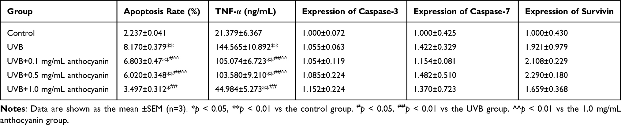 |
Table 1 Effect of Anthocyanin on UVB-Induced Human Skin Fibroblasts (Mean ±SEM, n = 3) |
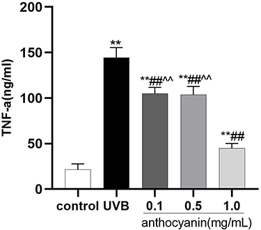 |
Figure 1 Effect of anthocyanin on TNF-α in ultraviolet B (UVB)-induced human skin fibroblasts (HSFs). The expression of TNF-α was detected by ELISA. Data are shown as the mean ±SEM (n = 3). **p < 0.01 vs the control group. ##p < 0.01 vs the UVB group. ^^p < 0.01 vs the 1.0 mg/mL anthocyanin group. |
Protein Expression of Caspase-3, Caspase-7, and Survivin
Western blot was performed to determine the protein expression of caspase-3, caspase-7, and survivin to evaluate the effect of anthocyanin on UVB-induced apoptosis and related proteins in UVB-irradiated HSFs. As shown in Table 1 and Figure 2, there was no significant difference in caspase-3 expression under different treatments (p > 0.05). The expressions of caspase-7 and survivin in UVB-irradiated cells showed an upward trend (p > 0.05). Caspase-7 expression was decreased in UVB + anthocyanin-treated cells, while the expression of survivin was increased (p > 0.05). Among different anthocyanin concentrations (0.1, 0.5, and 1.0 mg/mL), the expression of caspase-7 and survivin was the highest in the 0.5 mg/mL anthocyanin group (p > 0.05).
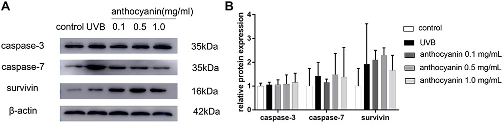 |
Figure 2 Effect of anthocyanin on the expression of caspase-3, caspase-7, and survivin in ultraviolet B (UVB)-induced human skin fibroblasts (HSFs). Western blot was performed to determine the expression of caspase-3, caspase-7, and survivin. (A) Caspase-3, caspase-7, and survivin proteins in HSFs. (B) The expression of caspase-3, caspase-7, and survivin in HSFs. Data are shown as the mean ±SEM (n = 3). |
Apoptosis Rate of HSFs
The apoptosis rate was detected by flow cytometry. As shown in Figure 3 and Table 1, the apoptosis rate in the UVB group was significantly higher than that in the control and anthocyanin groups (p < 0.01). There was no significant difference in apoptosis rate between the 0.1 mg/mL and 0.5 mg/mL anthocyanin groups (p > 0.05), while the apoptosis rate in the 1.0 mg/mL anthocyanin group was lower than that in the 0.1 mg/mL and 0.5 mg/mL anthocyanin groups (p < 0.05). At the same time, we noticed that the apoptosis rate in the anthocyanin (0.1, 0.5, and 1.0 mg/mL) group was higher than that in the control group (p < 0.05).
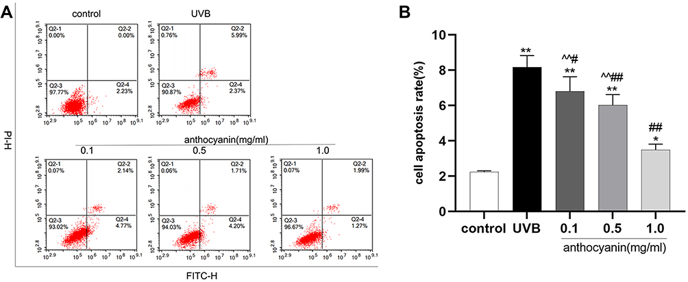 |
Figure 3 Effect of anthocyanin on apoptosis rate in human skin fibroblasts (HSFs). The apoptosis rate was detected by flow cytometry. (A) Apoptosis rate in HSFs. (B) Comparison of apoptosis rate in HSFs. Data are shown as the mean ±SEM (n = 3). *p < 0.05,**p < 0.01 vs the control group. #p < 0.05, ##p < 0.01 vs the UVB group. ^^p < 0.01 vs the 1.0 mg/mL anthocyanin group. |
Discussion
In this study, we investigated the protective effect of anthocyanin on UVB-exposed HSFs. According to N. Xie ‘s research, anthocyanins can reduce arsanilic acid-induced apoptosis.31 UVB irradiation increases the apoptosis rate of HSFs and TNF-α production, suggesting that UVB irradiation induces apoptosis in HSFs cells;30 thus, TNF-α-mediated apoptosis could mimic the risk of photoaging in HSFs. TNF-α is an important mediator of UVB-induced apoptosis in HSFs. Nizamutdinova et al found that anthocyanin could reduce inflammation by inhibiting TNF-α production in fibroblasts.32 The apoptosis rate in the anthocyanin (0.1, 0.5, and 1.0 mg/mL) group was lower than that in the UVB group and higher compared to the control group. These results indicated that anthocyanin pretreatment could reduce UVB-induced apoptotic cells but could not achieve complete remission. At the same time, we noticed that the apoptosis rate in the 1.0 mg/mL anthocyanin group was lower than that in the 0.1 and 0.5 mg/mL anthocyanin groups, and there was no significant difference in the apoptosis rate in the 0.1 and 0.5 mg/mL anthocyanin groups. Therefore, 1.0 mg/mL anthocyanin had the best protective effect. In this study, anthocyanin was used as a drug with inhibitory effects on UVB-induced apoptosis. The anti-apoptotic effect of anthocyanin may be related to the regulation of external apoptotic pathways mediated by death receptors.
Apoptosis, also known as programmed cell death, is a programmed autonomous gene-regulated ablation process, which is normal and advantageous in the cellular life cycle and survival.33 Apoptosis is strictly regulated by intracellular signal transduction factors; hence, apoptosis can respond to physiological and pathological stimuli in a regulated manner.34 TNF-α, as an inflammation-related cytokine, can not only induce inflammation but also induce apoptosis through the death receptor pathway.35 When TNF-α is recognized by TNF receptor 1 (TNFR1) on the surface of the cell membrane, TNF receptor-associated death domain (TRADD) and Fas-associated death domain protein (FADD) are recruited to transmit death signals and activate the upstream promoter caspase-8.36,37 Activated caspase-8 directly activates caspase-3, 6, and 7 through a cascade amplification reaction. Activated caspase-3 and 7 can cleave DNA molecules, apoptosis inhibitory effector proteins, extracellular matrix proteins, and cytoskeleton proteins and then promote apoptosis.34,38 TNF-α is an important factor mediating apoptosis, and caspase-3 and caspase-7 are effector apoptotic proteases. They all play an important role in the external apoptotic pathway mediated by death receptors and can be regarded as relevant factors promoting apoptosis.
We found that the apoptosis rate and TNF-α and caspase-7 expression of UVB-irradiated HSFs increased, indicating that UVB irradiation could activate the caspase protein family through the death receptor pathway and induce apoptosis. Compared with the UVB group, the apoptosis rate and TNF-α and caspase-7 expression in HSFs exposed to anthocyanin were decreased, suggesting that anthocyanin reduced UVB-induced apoptosis.39 However, compared with the control group, the apoptosis rate and TNF-α and caspase-7 expression in the anthocyanin group were still higher. This suggests that anthocyanin can only alleviate UVB-mediated damage to a certain extent but not completely repair it. Among the three doses of anthocyanin (0.1 mg/mL, 0.5 mg/mL, and 1.0 mg/mL), 1.0 mg/mL anthocyanin led to the lowest apoptosis rate and TNF-α expression and the best photoprotective effect.
Survivin is a bifunctional member of the inhibitor family of apoptosis proteins,40,41 which can not only inhibit apoptosis but also regulate the cell cycle. Survivin can inhibit apoptosis by inhibiting caspase-3 and caspase-7.42,43 Overexpression of survivin protects keratinocytes from UVB-induced apoptosis.44 The expression of survivin in the control group was the lowest among all the study groups, which was related to the low expression of survivin in normal cells.45 UVB irradiation-induced apoptosis, and the apoptosis rate and survivin protein expression increased in the UVB group. The expression of survivin in HSFs pretreated with anthocyanin was higher than that in the UVB group, suggesting that anthocyanin could promote survivin of HSFs to inhibit UVB-induced apoptosis and reduce UV-induced cell damage. The result was consistent with H. Cai’s study, in which they found that anthocyanin increased survivin expression to enhance the viability of mouse islets.46 Among the three doses of anthocyanin, the expression of survivin with 0.5 mg/mL anthocyanin was the highest, and the apoptosis rate was also low. This suggests that this dose of anthocyanin provided good protection against UVB-induced apoptosis.
In this study, there was no significant difference in the protein expression of caspase-3 in each group. This might be because caspase-8 does not directly activate caspase-3 but activates BH3-only protein, which promotes the subsequent cascade reaction mediated by Bax and Bak, leading to apoptosis.47 At the same time, it was observed that caspase-3 expression in the 1.0 mg/mL anthocyanin group was the highest among the three anthocyanin groups, but the survivin expression in this group was the lowest among the three doses. Dallaglio et al have shown that silenced survivin could mediate caspase-3 activation.44 This might be the reason for the highest expression of caspase-3 in the 1.0 mg/mL anthocyanin group. Additionally, although the expression levels of caspase-7 and survivin in each group increased or decreased, the differences were not statistically significant, requiring further exploration.
In conclusion, we reported that anthocyanin of Lycium ruthenicum Murr. had photoprotective effects on UVB-irradiated HSFs, such as reducing the apoptosis rate and TNF-α and caspase-7 expression through the death receptor pathway and increasing survivin expression. These results indicated the potential application of anthocyanin from Lycium ruthenicum Murr. in the Qaidam Basin as a drug to combat skin photodamage and protect photoaging cells. However, this was an in vitro study, and the protective effect of anthocyanin on photodamaged cells needs to be confirmed by further studies in vivo, with further exploration in the future.
Abbreviations
UV, ultraviolet; HSF, human skin fibroblast; TNF, tumor necrosis factor; SEM, standard error of the mean; ANOVA, analysis of variance; IAP, inhibitor of apoptosis protein; DMEM, Dulbecco’s modified Eagle medium; FBS, fetal bovine serum; ELISA, enzyme-linked immunosorbent assay; PMSF, phenylmethanesulfonyl fluoride; BCA, bicinchoninic acid; SDS-PAGE, sodium dodecyl sulfate-polyacrylamide gel electrophoresis; PVDF, polyvinylidene fluoride; HRP, horseradish peroxidase; ECL, enhanced chemiluminescence; PI, propidium iodide; TNFR1, TNF receptor 1; TRADD, TNF receptor-associated death domain; FADD, Fas-associated death domain protein.
Ethics Approval
The cells we used were provided by the biological company, and the study did not require review and approval of the Ethics Committee of Qinghai University Affiliated Hospital based on 2019 Qinghai Province research guidelines.
Data Sharing Statement
All data used during the current study are available from the corresponding author upon reasonable request.
Acknowledgment
The authors would like to thank all the laboratory staff and assistants for their technical assistance.
Author Contributions
All authors made significant contributions to the work reported, whether that is in the conception, study design, execution, acquisition of data, analysis, or interpretation. All authors took part in drafting, revising, or critically reviewing the article; gave final approval of the version to be published; agreed on the journal to which the article has been submitted; and agreed to be accountable for all aspects of the work.
Funding
This work was supported by the Applied Basic Research Program of Department of Science and Technology of Qinghai Province (2019-ZJ-7058).
Disclosure
All authors declare that they have no conflicts of interest in this work.
References
1. Swaney MH, Kalan LR, Richardson AR. Living in your skin: microbes, molecules, and mechanisms. Infect Immun. 2021;89(4):e00695–20. doi:10.1128/IAI.00695-20
2. Li Q, Chen Y, Ma K, Zhao A, Zhang C, Fu X. Regenerative and reparative effects of human chorion-derived stem cell conditioned medium on photo-aged epidermal cells. Cell Cycle. 2016;15(8):1144–1155. doi:10.1080/15384101.2016.1158376
3. Shin J-W, Kwon S-H, Choi J-Y, et al. Molecular mechanisms of dermal aging and antiaging approaches. Int J Mol Sci. 2019;20(9):2126. doi:10.3390/ijms20092126
4. Michalak M, Pierzak M, Kręcisz B, Suliga E. Bioactive compounds for skin health: a review. Nutrients. 2021;13(1):203. doi:10.3390/nu13010203
5. Widel M, Krzywon A, Gajda K, Skonieczna M, Rzeszowska-Wolny J. Induction of bystander effects by UVA, UVB, and UVC radiation in human fibroblasts and the implication of reactive oxygen species. Free Radic Biol Med. 2014;68:278–287. doi:10.1016/j.freeradbiomed.2013.12.021
6. Mu J, Ma H, Chen H, Zhang X, Ye M. Luteolin prevents UVB-induced skin photoaging damage by modulating SIRT3/ROS/MAPK signaling: an in vitro and in vivo studies. Front Pharmacol. 2021;12:2236. doi:10.3389/fphar.2021.728261
7. Balzano T, Arenas YM, Dadsetan S, et al. Sustained hyperammonemia induces TNF-a IN Purkinje neurons by activating the TNFR1-NF-κB pathway. J Neuroinflammation. 2020;17(1):1–22. doi:10.1186/s12974-020-01746-z
8. Hong JJ, Hadeler EK, Mosca ML, Brownstone ND, Bhutani T, Liao WJ. Off-label uses of TNF-a inhibitors and IL-12/23 inhibitors in dermatology. Dermatol Online J. 2021;27:11.
9. Moriwaki K, Chan FK, Miyoshi E. Sweet modification and regulation of death receptor signalling pathway. J Biochem. 2021;169(6):643–652. doi:10.1093/jb/mvab034
10. Asadi M, Taghizadeh S, Kaviani E, et al. Caspase‐3: structure, function, and biotechnological aspects. Biotechnol Appl Biochem. 2021;9(4):1633–1645.
11. Yadav P, Yadav R, Jain S, Vaidya A. Caspase‐3: a primary target for natural and synthetic compounds for cancer therapy. Chem Biol Drug Des. 2021;98(1):144–165. doi:10.1111/cbdd.13860
12. Gąsowska-Bajger B, Gąsowska-Bodnar A, Knapp P, Bodnar L. Prognostic Significance of survivin expression in patients with ovarian carcinoma: a meta-analysis. J Clin Med. 2021;10(4):879. doi:10.3390/jcm10040879
13. Shojaei F, Yazdani-Nafchi F, Banitalebi-Dehkordi M, Chehelgerdi M, Khorramian-Ghahfarokhi M. Trace of survivin in cancer. Eur J Cancer Prev. 2019;28(4):365–372. doi:10.1097/CEJ.0000000000000453
14. Manohar SM, Shah P, Nair A. Flow cytometry: principles, applications and recent advances. Bioanalysis. 2021;13(3):181–198. doi:10.4155/bio-2020-0267
15. Nomi Y, Iwasaki-Kurashige K, Matsumoto H. Therapeutic effects of anthocyanins for vision and eye health. Molecules. 2019;24(18):3311. doi:10.3390/molecules24183311
16. Dong Y, Wu X, Han L, et al. The potential roles of dietary anthocyanins in inhibiting vascular endothelial cell senescence and preventing cardiovascular diseases. Nutrients. 2022;14(14):2836. doi:10.3390/nu14142836
17. Mattioli R, Francioso A, Mosca L, Silva P. Anthocyanins: a comprehensive review of their chemical properties and health effects on cardiovascular and neurodegenerative diseases. Molecules. 2020;25(17):3809. doi:10.3390/molecules25173809
18. Diaconeasa Z, Știrbu I, Xiao J, et al. Anthocyanins, vibrant color pigments, and their role in skin cancer prevention. Biomedicines. 2020;8(9):336. doi:10.3390/biomedicines8090336
19. Li H, Li Z, Peng L, et al. Lycium barbarum polysaccharide protects human keratinocytes against UVB-induced photo-damage. Free Radic Res. 2017;51(2):200–210. doi:10.1080/10715762.2017.1294755
20. Chen S, Zhou H, Zhang G, et al. Anthocyanins from Lycium ruthenicum Murr. ameliorated d-galactose-induced memory impairment, oxidative stress, and neuroinflammation in adult rats. J Agric Food Chem. 2019;67(11):3140–3149. doi:10.1021/acs.jafc.8b06402
21. Zhang Q, Yan H, Ren L, Jia Y, Wang R. Inhibition effect of Lycium ruthenicum murr aqueous extract on the apotosis and expression of p16 and p53 in HaCaT cell induced by UVB irradiation. China J Lepr Skin Dis. 2018;34(11):645–648.
22. Oh M, Lee J, Kim YJ, Rhee WJ, Park JH. Exosomes derived from human induced pluripotent stem cells ameliorate the aging of skin fibroblasts. Int J Mol Sci. 2018;19(6):1715. doi:10.3390/ijms19061715
23. Kang W, Choi D, Park T. Decanal protects against UVB-induced photoaging in human dermal fibroblasts via the cAMP pathway. Nutrients. 2020;12(5):1214. doi:10.3390/nu12051214
24. Kim H-R, Jeong D-H, Kim S, et al. Fermentation of blackberry with L. plantarum JBMI F5 enhance the protection effect on UVB-mediated photoaging in human foreskin fibroblast and hairless mice through regulation of MAPK/NF-κB signaling. Nutrients. 2019;11(10):2429. doi:10.3390/nu11102429
25. Li W, Ji G, Zhang X, Yu H. The influence and mechanisms of purple sweet potato anthocyanins on the growth of bladder cancer BIU87 cell. Zhonghua Yi Xue Za Zhi. 2018;98(6):457–459. doi:10.3760/cma.j.iss.0376-2491.2018.06.013
26. Zhang L, Xing Q, Shen T, et al. Black Wolfberry anthocyanins inhibits apoptosis of mouse macrophage RAW264.7 through JNK pathway. J Yunnan Univ Nat Sci Ed. 2022;37(01):111–119.
27. Giampieri F, Alvarez-Suarez JM, Mazzoni L, et al. An anthocyanin-rich strawberry extract protects against oxidative stress damage and improves mitochondrial functionality in human dermal fibroblasts exposed to an oxidizing agent. Food Funct. 2014;5(8):1939–1948. doi:10.1039/C4FO00048J
28. Yue E, Yu Y, Wang X, Liu B, Bai Y, Yang B. Anthocyanin protects cardiac function and cardiac fibroblasts from high-glucose induced inflammation and myocardial fibrosis by inhibiting IL-17. Front Pharmacol. 2021;11:593633. doi:10.3389/fphar.2020.593633
29. Kamiya Y, Odama M, Mizuguti A, Murakami S, Ito T, Pintus G. Puerarin blocks the aging phenotype in human dermal fibroblasts. PLoS One. 2021;16(4):e0249367. doi:10.1371/journal.pone.0249367
30. Mavrogonatou E, Angelopoulou M, Rizou SV, Pratsinis H, Gorgoulis VG, Kletsas D. Activation of the JNKs/ATM-p53 axis is indispensable for the cytoprotection of dermal fibroblasts exposed to UVB radiation. Cell Death Dis. 2022;13(7):1–14. doi:10.1038/s41419-022-05106-y
31. Xie N, Geng N, Zhou D, et al. Protective effects of anthocyanin against apoptosis and oxidative stress induced by arsanilic acid in DF-1 cells. Mol Biol Rep. 2019;46(1):301–308. doi:10.1007/s11033-018-4472-5
32. Nizamutdinova IT, Kim YM, Chung JI, et al. Anthocyanins from black soybean seed coats stimulate wound healing in fibroblasts and keratinocytes and prevent inflammation in endothelial cells. Food Chem Toxicol. 2009;47(11):2806–2812. doi:10.1016/j.fct.2009.08.016
33. Xu X, Lai Y, Hua Z-C. Apoptosis and apoptotic body: disease message and therapeutic target potentials. Biosci Rep. 2019;39:1. doi:10.1042/BSR20180992
34. Obeng E. Apoptosis (programmed cell death) and its signals-A review. Braz J Biol. 2020;81:1133–1143. doi:10.1590/1519-6984.228437
35. Yuan X, Gajan A, Chu Q, Xiong H, Wu K, Wu GS. Developing TRAIL/TRAIL death receptor-based cancer therapies. Cancer Metastasis Rev. 2018;37(4):733–748. doi:10.1007/s10555-018-9728-y
36. Fu Y-C, Chi C-S, Yin S-C, Hwang B, Chiu Y-T, Hsu S-L. Norepinephrine induces apoptosis in neonatal rat cardiomyocytes through a reactive oxygen species–TNFα–caspase signaling pathway. Cardiovasc Res. 2004;62(3):558–567. doi:10.1016/j.cardiores.2004.01.039
37. Zhang C, Ma K, Yang Y, Wang F, Li W. Glaucocalyxin A suppresses inflammatory responses and induces apoptosis in TNF-a-induced human rheumatoid arthritis via modulation of the STAT3 pathway. Chem Biol Interact. 2021;341:109451. doi:10.1016/j.cbi.2021.109451
38. Zhao X, Liu L, Li R, et al. Hypoxia-inducible factor 1-α (HIF-1α) induces apoptosis of human uterosacral ligament fibroblasts through the death receptor and mitochondrial pathways. Med Sci Monit. 2018;24:8722. doi:10.12659/MSM.913384
39. Bae JY, Lim SS, Kim SJ, et al. Bog blueberry anthocyanins alleviate photoaging in ultraviolet‐B irradiation‐induced human dermal fibroblasts. Mol Nutr Food Res. 2009;53(6):726–738. doi:10.1002/mnfr.200800245
40. Quispe PA, Lavecchia MJ, León IE. On the discovery of a potential survivin inhibitor combining computational tools and cytotoxicity studies. Heliyon. 2019;5(8):e02238. doi:10.1016/j.heliyon.2019.e02238
41. Lin T-Y, Chan -H-H, Chen S-H, et al. BIRC5/Survivin is a novel ATG12–ATG5 conjugate interactor and an autophagy-induced DNA damage suppressor in human cancer and mouse embryonic fibroblast cells. Autophagy. 2020;16(7):1296–1313. doi:10.1080/15548627.2019.1671643
42. Miliaraki M, Briassoulis P, Ilia S, et al. Survivin and caspases serum protein levels and survivin variants mRNA expression in sepsis. Sci Rep. 2021;11(1):1–10. doi:10.1038/s41598-020-78208-2
43. Wangyan T-C. Inhibitory effect of exopolysaccharide from Rhizopus nigricans combined with oxaliplatin on dimethylhydrazine-induced colon cancer in rats and its effect on Survivin/caspase-3/caspase-7. Chin Pharmacol Bull. 2019;12:690–694.
44. Dallaglio K, Palazzo E, Marconi A, et al. Endogenous survivin modulates survival and proliferation in UVB‐treated human keratinocytes. Exp Dermatol. 2009;18(5):464–471. doi:10.1111/j.1600-0625.2008.00819.x
45. Wheatley SP, Altieri DC. Survivin at a glance. J Cell Sci. 2019;132(7):jcs223826. doi:10.1242/jcs.223826
46. Cai H, Yang B, Xu Z, et al. Cyanidin-3-O-glucoside enhanced the function of syngeneic mouse islets transplanted under the kidney capsule or into the portal vein. Transplantation. 2015;99(3):508–514. doi:10.1097/TP.0000000000000628
47. Muñoz-Pinedo C. Signaling pathways that regulate life and cell death: evolution of apoptosis in the context of self-defense. Self Nonself. 2012;738:124–143.
 © 2022 The Author(s). This work is published and licensed by Dove Medical Press Limited. The full terms of this license are available at https://www.dovepress.com/terms.php and incorporate the Creative Commons Attribution - Non Commercial (unported, v3.0) License.
By accessing the work you hereby accept the Terms. Non-commercial uses of the work are permitted without any further permission from Dove Medical Press Limited, provided the work is properly attributed. For permission for commercial use of this work, please see paragraphs 4.2 and 5 of our Terms.
© 2022 The Author(s). This work is published and licensed by Dove Medical Press Limited. The full terms of this license are available at https://www.dovepress.com/terms.php and incorporate the Creative Commons Attribution - Non Commercial (unported, v3.0) License.
By accessing the work you hereby accept the Terms. Non-commercial uses of the work are permitted without any further permission from Dove Medical Press Limited, provided the work is properly attributed. For permission for commercial use of this work, please see paragraphs 4.2 and 5 of our Terms.