")
Back to Journals » International Journal of Nanomedicine » Volume 17
A Comprehensive Review of the Application of Nanoparticles in Diabetic Wound Healing: Therapeutic Potential and Future Perspectives
Authors Qin W, Wu Y , Liu J, Yuan X, Gao J
Received 25 August 2022
Accepted for publication 23 November 2022
Published 5 December 2022 Volume 2022:17 Pages 6007—6029
DOI https://doi.org/10.2147/IJN.S386585
Checked for plagiarism Yes
Review by Single anonymous peer review
Peer reviewer comments 2
Editor who approved publication: Dr Phong A Tran
Wenqi Qin,1 Yan Wu,1 Jieting Liu,1 Xiaohuan Yuan,1 Jie Gao2
1College of Life Science, Mudanjiang Medical University, Mudanjiang, People’s Republic of China; 2Changhai Clinical Research Unit, Shanghai Changhai Hospital, Naval Medical University, Shanghai, People’s Republic of China
Correspondence: Xiaohuan Yuan, College of Life Science, Mudanjiang Medical University, Mudanjiang, Heilongjiang, 157001, People’s Republic of China, Tel/Fax +86 453 6984647, Email [email protected] Jie Gao, Changhai Clinical Research Unit, Shanghai Changhai Hospital, Naval Medical University, Shanghai, 200433, People’s Republic of China, Tel/Fax +86 021 31166666, Email [email protected]
Abstract: Diabetic wounds are one of the most challenging public health issues of the 21st century due to their inadequate vascular supply, bacterial infections, high levels of oxidative stress, and abnormalities in antioxidant defenses, whereas there is no effective treatment for diabetic wounds. Due to the distinct properties of nanoparticles, such as their small particle size, elevated cellular uptake, low cytotoxicity, antibacterial activity, good biocompatibility, and biodegradability. The application of nanoparticles has been widely used in the treatment of diabetic wound healing due to their superior anti-inflammatory, antibacterial, and antioxidant activities. These nanoparticles can also be loaded with various agents, such as organic molecules (eg, exosomes, small molecule compounds, etc.), inorganic molecules (metals, nonmetals, etc.), or complexed with various biomaterials, such as smart hydrogels (HG), chitosan (CS), and hyaluronic acid (HA), to augment their therapeutic potential in diabetic wounds. This paper reviews the therapeutic potential and future perspective of nanoparticles in the treatment of diabetic wounds. Together, nanoparticles represent a promising strategy in the treatment of diabetic wound healing. The future direction may be to develop novel nanoparticles with multiple effects that not only act in wound healing at all stages of diabetes but also provide a stable physiological environment throughout the wound-healing process.
Keywords: nanoparticles, biomaterials, wound healing, diabetic wound, diabetic complications
Introduction
Diabetes mellitus is a metabolic disorder characterized by abnormal glucose metabolism and consists of two types, including type I diabetes mellitus, which is due to the destruction of the beta cells in the pancreas by the autoimmune system, and type II diabetes mellitus, which involves insulin resistance and subsequent pancreatic beta cell failure.1 Elevated blood glucose levels, characterized by insulin resistance or reduced insulin secretion, cause a variety of complications, including diabetic nephropathy, neuropathy, cardiomyopathy, hearing loss, and skin ulcers. Currently, with the development of society and changes in people’s lifestyles, as time passes, the prevalence of diabetes is increasing worldwide, affecting approximately hundreds of million of people.2,3 One of the most troublesome complications for diabetic patients is diabetic wounds, which have emerged as one of the most prominent threats to human health worldwide. Diabetic wounds can develop into chronic intractable ulcers due to persistent infection caused by cellular dysfunction, microcirculatory disorders, high levels of oxidative stress and hypoxia, which are the main causes of amputation and disability.4 The traditional clinical treatments include controlling blood glucose, surgical debridement, skin transplantation, wound dressing, and hyperbaric oxygen therapy.5–7 Although these clinical treatments can better achieve symptom better control, they have a limited therapeutic effect on diabetic wound healing. Additionally, given the long duration of diabetic treatments, it is prone to secondary damage, which has a tremendous psychological and physical impact on the patients. Thus, there is an urgent need for modern treatments that are potentially effective, painless, and scar-free for diabetic wounds. The literature reports that nanoparticles have long been used as excellent biologically active delivery systems for wound healing.8 Nanotechnology-based nanoparticles (NPs) have gained attention for their breakthrough potential in deciphering the biological environment and providing personalized therapeutic approaches for wound healing.9 They are not only small, steady in nature, and easily absorbed by cells but also have good biocompatibility or targeting properties and effectively control drug delivery and release.10 These properties of NPs are the main driving force for the development of novel nanotechnology-based platforms that have a crucial impact on angiogenesis, collagen production and extracellular matrix (ECM) production, all of which are essential properties to promote wound healing. NPs and different NPs-based platforms can interact with the word healing process, affecting many cellular and molecular processes and aiding diabetic wound healing due to their intrinsic nanoscale characteristics.9 Therefore, they represent a promising strategy in the treatment of diabetic wound healing due to their potential to improve the stability and bioavailability of various bioactive substances.11
Over the past decade, nanoparticles have attracted considerable attention in diabetic wound therapy due to their unique advantages, and several research groups have reported encouraging preclinical results. Many reviews have discussed the potential and significance of a single or several kinds of nanoparticles (eg, polymeric nanoparticles, silver nanoparticles),12–16 and future prospects for gene therapy and nanotechnology to extend the bioavailability of drugs for diabetic wound healing were also studied.7 However, to the best of our knowledge, none of them has given us a comprehensive summary of nanoparticles. Based on the literature, we summarized the recent research progress of nanoparticles in diabetic wound treatment (Tables 1 and 2).
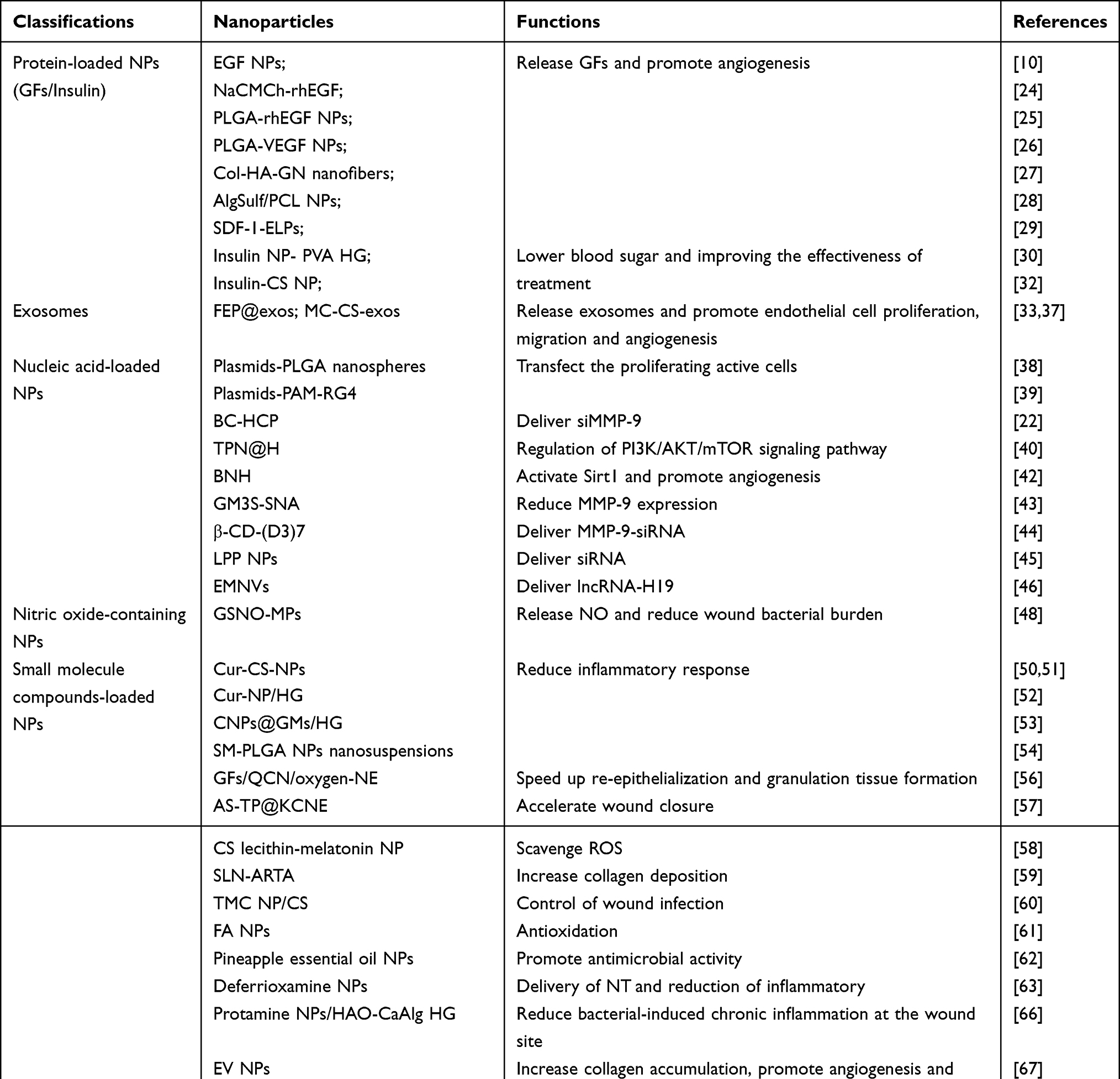 |
Table 1 To Summarize the Classifications of Organic Nanoparticles in Diabetic Wound Healing |
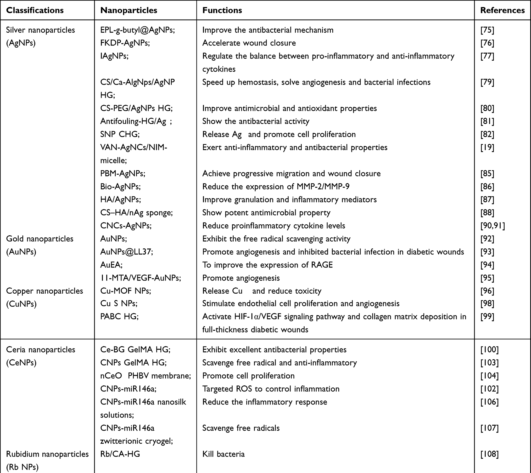 |
Table 2 To Summarize the Classifications of Inorganic Nanoparticles in Diabetic Wound Healing |
Wound Healing Process
The Normal Wound-Healing Process
During wound healing, the recovery of the physiological process is highly complicated and dynamic, consisting of four stages: the hemostasis phase, the inflammatory phase, the proliferative phase and the remodeling phase. The hemostasis phase (0 hours after injury) occurs when vascular constriction and thrombin are activated to promote platelet aggregation, which leads to the formation of thrombi and the release of some proinflammatory mediators, such as cytokines, proceeding to the next phase. The inflammatory phase (1–3 days) occurs when mast cells release 5-hydroxytryptamine and histamine, increasing vascular permeability at the wound site, which promotes the migration of neutrophils, monocytes and chemokines to the injury area and produces an inflammatory response.17 The proliferative phase (4–21 days) involves the migration of lymphocytes and macrophages to the wound to facilitate infection control and the degradation of cellular fragments, which release certain cytokines and GFs that promote cell proliferation.18 The remodeling period (21 days or more) occurs when new epithelial tissue forms at the injured location. New blood vessels are also generated, fibroblasts are converted into muscle fibroblasts in great numbers, and the wound contracts. This remodeling leads to the formation of a scar.16,19,20
The Diabetic Wound-Healing Process
Under normal physiological circumstances, the injured organism is expected to comply with the normal wound processing. Nevertheless, when recovery is constantly stimulated or affected by abnormal factors, a chronic wound (eg, diabetic wound) develops. There are several reasons for which diabetic wounds develop into chronic wounds that are difficult to heal.
First, hypoxia is the main cause of diabetic wound damage. It is becoming clearer and clearer how important oxygen is to wound healing, as it promotes fibroblast proliferation, enhances immune function, and stimulates angiogenesis.16 However, diabetic patients not only have an insufficient oxygen supply but also consume more oxygen in the wound areas.21 Hypoxia enhances the inflammatory response, thereby prolonging effective wound healing by increasing oxygen-free radical levels.
Second, diabetic wounds are characterized by decreased local angiogenesis and a diminished blood supply due to disturbed pathological mechanisms, persistent infections, and imbalanced angiogenic factors (eg, transforming growth factor-α (TGF-α), TGF-β, fibroblast growth factor-2 (FGF-2), vascular endothelial growth factor (VEGF), epidermal growth factor (EGF), hypoxia-inducible factor (HIF-1α) and vascular inhibitory factors (eg, platelet-activating protein, endothelial inhibitor, vasopressor). As a result, there may be a link between prolonged wound inflammation and delayed wound healing.16,20 In addition, diabetic patients develop macroangiopathy, which leads to reduced blood flow. At the microvascular level, because of abnormal capillary regulation and reduced levels of nitric oxide synthase (NOS), the production of NO decreases, and the diastolic function of the vasculature is impaired, which leads to microvascular malfunction.2
Third, in contrast to the normal healing process, diabetic wounds go directly to a chronic inflammatory phase. If inflammation is not controlled and inflammatory cells persist in the wound site, it may lead to chronic inflammation, which is characterized by the excessive accumulation of M1 macrophages, significantly reduced differentiation of fibroblasts into myofibroblasts, inhibition of the expression of TGF-β type II receptor, and decreased collagen synthesis, thus hindering the reshaping phase of the skin.20
Fourth, matrix metalloproteinases (MMPs) are responsible for degrading collagen and regulating the migration of keratinocytes. However, excessive glucose levels lead to the excessive expression of MMPs, slowing down the healing of diabetic wounds by the excessive degradation of extracellular matrix, growth factors, growth factor receptors, integrins, and their receptors and increasing the local inflammatory response to the wound.22
In summary, diabetic wounds are more challenging to heal than normal wounds. Unlike normal wounds, diabetic wounds are characterized by hypoxia, impaired angiogenesis, chronic inflammation, and excessive expression of matrix metalloproteinases (MMPs), leading to delayed diabetic wounds healing (Figure 1). In clinical practice, we generally use symptomatic treatments, such as infection control, anti-inflammation, surgical debridement, and the use of GFs, but the effectiveness of these treatments is limited. Thus, the utilization of nanoparticles to address this difficult health problem is important.
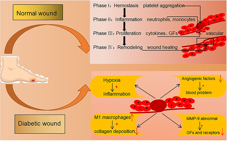 |
Figure 1 The healing process of diabetic wounds and normal wounds. Unlike normal wounds, diabetic wounds are characterized by hypoxia, impaired angiogenesis, chronic inflammation, and excessive expression of matrix metalloproteinases (MMPs), leading to the delayed healing of diabetic wounds. |
Application of Nanoparticles to Treat Diabetic Wounds
Considering that organic nanoparticles have abundant functional group structures that can better bind to drugs and GFs and the ions brought by metal nanoparticles have good antibacterial properties, they show great potential in the treatment of diabetic wounds.23 With the advancement of society and technology, researchers have discovered nanoparticles with numerous biological properties that can be applied to diabetic wound therapy to remedy some of the drawbacks of clinical treatment. This review summarizes the recent applications of organic nanoparticles (eg, polymeric nanoparticles, exosomes, etc.) and inorganic nanoparticles (metal, non metal nanoparticles, etc.) in the treatment of diabetic wound healing, as shown in Figure 2.
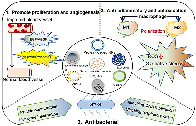 |
Figure 2 Schematic illustration of the categories and therapeutic mechanism of biomaterials used on diabetic wounds. Biomaterials are loaded with organic molecules (including protein-loaded, exosomes, and small molecule compounds, etc.) and inorganic molecules (including AgNPs, CeNPs, SiO2NPs, etc.) to promote diabetic wound healing. |
Organic Nanoparticles in Diabetic Wound Healing
Protein-Loaded Nanoparticles
Protein-loaded nanoparticles can release various growth factors and also act as carriers to improve their stability in the wound area and control their release.8 Diabetic wounds lack EGF and its receptor, resulting in less matrix deposition, poor tissue repair function, and difficult wound healing. Although EGF has been widely used in the treatment of diabetic wounds, it has a short half-life.10 Qi et al successfully prepared a stable EGF nanoparticle via a polylactic acid/glycolic acid copolymer using composite emulsion that had no toxic effect on cells, and it not only preserved the biological activity of EGF but also promoted fibroblast proliferation.10 Hajimiri et al developed a sodium carboxymethyl chitosan recombinant human epidermal growth factor coupling (NaCMCh-rhEGF) as a delivery system for EGFs that protected rhEGF from hydrolysis.24 Likewise, to protect EGF, Chu et al prepared rhEGF nanoparticles using poly (lactic-co-glycolic acid) (PLGA) as a carrier by a modified compound emulsion method. The release of rhEGF lasted for 24 hours, and its biological activity did not change.25
In addition to EGF delivery, nanoparticles are also effective in delivering VEGF. There are serious limitations in the delivery of VEGF by local injection. Therefore, the development of new effective and biologically active drug delivery systems is inevitable Capitalizing on the fact that PLGA provides lactic acid which promotes the function of promoting angiogenesis, activating precollagen factors and improving the recruitment of endothelial cells to wounds, Chereddy et al encapsulated VEGF in PLGA nanoparticles (PLGA-VEGF NPs). Compared to the controls, placing the PLGA-VEGF NPs on the active healing of nondiabetic and diabetic wounds not only enhanced the proliferation and migration of keratin-forming cells but also upregulated VEGFR2 expression at the mRNA level.26
Nanoparticles can synergistically deliver multiple growth factors in addition to the pure delivery of EGF and VEGF. Given this, research has focused on strategies for the effective delivery of multiple vascular factors based on nanofibers/polymers to achieve regeneration of the whole skin layer by promoting angiogenesis. For example, to design collagen (Col), hyaluronic acid (HA), and gelatin nanoparticle (GN) (Col-HA-GN) nanofiber films, Lai et al developed Col and HA nanofibers stacked on top of each other with multiple angiogenic growth factors (VEGF, bFGF, and EGF) encapsulated in the nanofibers and embedded them in gelatin nanoparticles (GNs) using electrostatic spinning technology. Col-HA-GN nanofiber films are mechanically comparable to those in naturally occurring human skin and allow for the release of growth factors (GFs) for up to 1 month.27 To improve the utilization of growth factors, Maatouk et al synthesized a bisemulsion of sulfated alginate/polycaprolactone (AlgSulf/PCL) NPs that are capable of delivering heparin (HN) and demonstrated that PCL and AlgSulf effectively deliver CTGF and IGF-I, a family of GFs, and prevent them from being degraded. Both proteins are HN conjugating proteins and show a high affinity for sulfated glycosaminoglycans.28 As a consequence, the use of GF therapy is essential to overcome the difficulties of vascular damage in diabetic wounds.
There is also local stromal cell-derived growth factor-1 (SDF-1), which contributes to faster wound revascularization and promotes re-epithelialization in diabetic mice, but its susceptibility to degradation limits its clinical applications. Elastin-like peptides (ELPs) are nonimmunogenic, nonpyrogenic, and biocompatible proprotein derivatives. Yeboah et al formed a recombinant fusion protein composed of SDF-1 and ELPs that conferred the ability to self-assemble into nanoparticles and showed a good ability to improve wound healing.29 Obviously, GFs have the advantages of promoting angiogenesis and cell proliferation, and the therapeutic regimen of promoting diabetic wound healing with GFs therapy is feasible. However, due to the local defects of GFs themselves, such as short half-life and degradability, their efficacy is limited. Thus, we developed a variety of nanoparticles to deliver various GFs, not only protecting GFs from degradation but also maximizing the benefits of GFs.
In addition to GF-like protein nanoparticles, there is also an insulin-like protein nanoparticle. Insulin has a good hypoglycemic effect, and its mode of delivery in the wound bed has not been well investigated. Since topically applied insulin is prone to degradation, Abdelkader et al encapsulated insulin in a nanoparticle carrier using a compound emulsion method, which was then loaded into a novel poly (vinyl alcohol) (PVA)-borate hydrogel. Group members evaluated the therapeutic efficacy of the drug delivery system.30 Insulin promotes tissue repair through the stimulation of keratin-forming cell migration via the PI3-K/AKT-Rac1 (phosphatidylinositol-3 kinase and protein kinase B-Ras-related C3 botulinum toxin substrate 1) signaling pathway.31 A local injection is mostly used for clinical administration, but its effect is always limited. Ribeiro et al prepared an insulin-CS nanoparticle that promoted increased expression levels of signaling molecules related to wound healing, such as VEGF and protein kinase B (called Akt), which stimulate angiogenesis and cell differentiation.32 Regardless of the type of protein-loaded nanoparticles, these nanoparticles can act as suitable carriers and take advantage of their controlled and slow release to prolong the action of protein-based substances.
Exosomes
Mesenchymal stem cells (MSCs) from bone marrow have been shown to produce higher levels of collagen, FGF, and VEGF than fibroblasts and induce angiogenesis at a faster rate than fibroblasts.7 MSCs-derived exosomes, as important bioactive molecules, offer significant advantages.34 For wound healing, since serum half-lives in vivo are relatively short, it is important to build a biocompatible scaffold that can sustain exosome functionality and sustained release. Wang et al prepared a polysaccharide-based fluorinated ethylene propylene (FEP) scaffold dressing with thermosensitive, injectable, self-healing, adhesive, antimicrobial, hemostatic, and UV-shielded properties using the good biocompatibility conferred by the amino group of polyethyleneimine (PEI) and the aldehyde group of aldehyde pullulan (APu) grafted to Pluronic F127 (F127) via a reversible Schiff base reaction, and this FEP scaffold exhibited good tissue adhesion through hydrogen bonding and the Schiff base reaction with tissue. The reversible Schiff base bonds (PEI and APu) provided self-healing and injectability of the scaffold. The electrostatic interaction between adipose mesenchymal stem cell (ADSC)-derived exosomes and F127-PEI contributes to the loading of exosomes into the FEP scaffold dressing (FEP@exos) and their release with a pH response. FEP@exos continuously release exosomes and maintains the biological activity of exosomes, which significantly accelerate the proliferation, migration, and tube formation abilities of endothelial cells in vitro and promote angiogenesis and diabetic wound healing.33 (Figure 3).
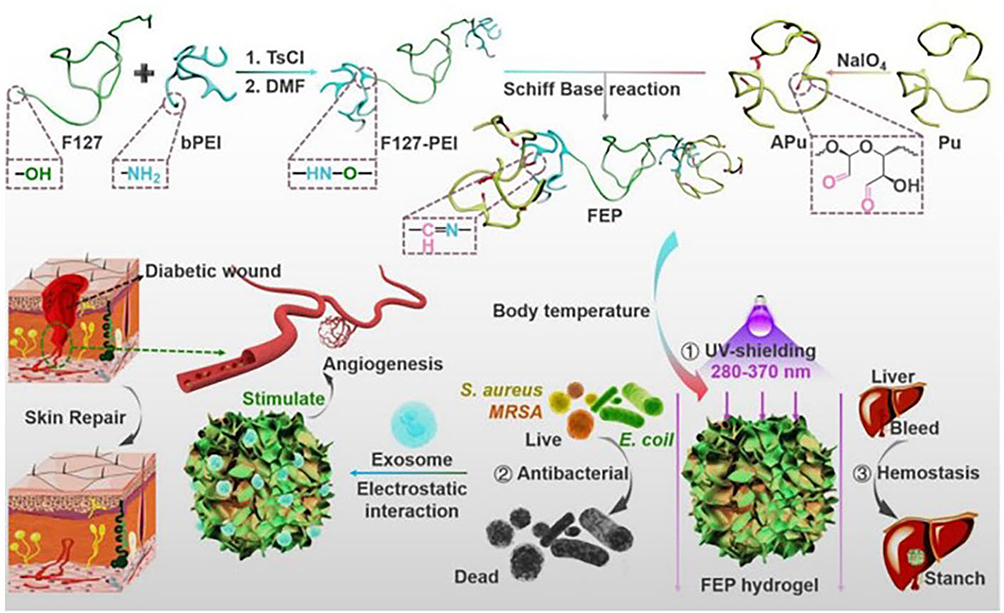 |
Figure 3 Schematic illustration showing the synthesis of F127-PEI, APu, multifunctional FEP scaffold dressing, nanoscale exosome-loaded FEP scaffold dressing and the potential application in diabetic wound healing and skin reconstruction.34 Notes: Reproduced from Wang M, Wang C, Chen M, et al. Efficient angiogenesis-based diabetic wound healing/skin reconstruction through bioactive antibacterial adhesive ultraviolet shielding nanodressing with exosome release. ACS Nano. 2019;13(9):10279–10293. Copyright © 2019, American Chemical Society.34 |
Compared to cell-based therapies, exosomes exhibit the advantages of easier endocytosis and a higher loading efficiency as a result of their unique structure. In the healing process, exosomes not only transfer their loading content to recipient cells and promote fibroblast proliferation and migration but also regulate the expression of type I and III collagen and fibronectin.35,36 Materials such as methylcellulose (MC) and chitosan (CS) and their complimentary derivatives of natural polysaccharides are considered optimal for use as a dressing. Wang et al engineered a methylcellulose-chitosan hydrogel loaded with exosomes (MC-CS-exos) as an efficient injectable self-healing candidate by linking the dynamic covalent Schiff bases between the aldehyde of MC and grafted poly (ethylene glycol) of CS. As a result of the synergistic actions of MC-CS-exos, intact structural and functional recovery was observed in mice with diabetes.37 We note that exosomes as delivery carriers are not only cell-derived and high yielding but also have excellent bioactivity and biocompatibility. Thus, they should have great potential in obtaining successful healing of diabetic and other vascular compromised wounds.
Nucleic Acid-Loaded Nanoparticles
Cytokines and GFs are critical for chronic and nonhealing diabetic wounds, and several plasmids expressing related genes also have a beneficial effect on wound healing. A plasmid expressing both VEGF-A and platelet-derived growth factor-B (PDGF-B) was compounded with PLGA nanospheres. Transfected VEGF-A and PDGF-B upregulated gene expression in DFU wounds and promoted wound healing.38 In addition, Kwon et al combined microcyclic plasmid DNA encoding VEGF with the arginine-tagged PAM-RG4 that is designed to improve nonviral vector delivery and toxicity by improving its surface cationic properties and injected the resulting complex under skin wounds in diabetic mice, which effectively transfected the proliferating active cells in the wound tissue and synergistically regulated the duration and therapeutic range of VEGF. Moreover, there was a strong promotion of angiogenesis and diabetic wound healing.39
To suppress the abnormal expression of MMP-9 at the wound site, BC-HCP/siMMP-9 formed a wound dressing that was synthesized by encapsulating siMMP with a bacterial cellulose hyperbranched cationic polysaccharide (BC-HCP), which released siMMP-9 to effectively reduce the gene and protein expression of MMP-9 in diabetic rat wounds. Bacterial cellulose (BC) is the primary material used to cover HCP/siMMP9 because it is an ideal wound dressing providing a barrier to control infection, controlling fluid loss, and reducing pain in the body, in addition to maintaining a moist environment at the wound site and absorbing inflammatory exudate.22
Some nanoparticles regulate wound healing by modulating signaling pathways and related genes. The phosphatidylinositol 3-hydroxyl kinase/protein kinase B/rapamycin mammalian target protein (PI3K/AKT/mTOR) pathway is a critically valuable intracellular signaling pathway associated with cell growth and proliferation. Chen et al synthesized novel tea polyphenol nanomicrospheres (TPN) and encapsulated them in PV a/alginate hydrogel (TPN@H), which modified the above signaling pathway to promote wound healing in rats with diabetes.40 Silent information regulator 1 (Sirt1), which is associated with cellular senescence, resistance to oxidative stress, and inflammation inhibition, is a nicotinamide adenine dinucleotide (NAD+)-dependent type III histone deacetylase.41 By enhancing wound healing in diabetic rats, Zhang et al prepared a novel flavopiridol nanocolloidal hydrogel (BNH) that, through the activation of Sirt1, increased the expression of F-VEGF, CD31, and SMA while inhibiting the expression of NF-κB, TNF-α and IL-6.42 Ganglioside monosialic acid 3 synthase (GM3S) is a key mediator of insulin resistance, as it is an overexpressed target in diabetic mice that may impede wound healing. Spherical nucleic acids (SNAs) are an up-and-coming group of gene-regulating bodies, and Randeria et al developed an SNA-based GM3S (GM3S-SNA), by which granulation tissue formation, vascular proliferation, and phosphorylation of IGF1 and EGF receptors were observed in treated sites.43 Some other nanoparticles can also act as carriers that deliver genes. For example, β-CD-(D3)7 is a novel cationic stellate polymer with good biocompatibility and a strong complexation ability that can be used as an effective carrier of MMP-9-siRNA to reduce the expression of MMP-9 in diabetic rat skin fibroblasts and promote wound healing.44 Moreover, Rabbani et al optimized an LPP nanoparticle composed of DNC cationic liposomes and CSP proteins as a suitable system for the effective delivery of siRNA into cells, successfully delivering siRNA targeting Keap1 (a central regulator in the redox response).45 Moreover, Tao et al studied extracellular mimic nanovesicles (EMNVs) as an efficient lncRNA nanodelivery system to promote wound healing. Among patients with diabetes, lncRNA-H19 expression is significantly decreased, inhibiting the insulin-phosphatidylinositol 3-kinase (PI3K)-Akt pathway, which leads to angiogenesis obstruction.46
Nitric Oxide-Containing Nanoparticles
NO has important biological functions as a potent biofilm-resistant, antibacterial, and wound-healing agent. It dilates blood vessels, acts as a signaling molecule, regulates the intracellular secondary messenger cycle GMP, and kills MRSA, the active component of biofilms, by attacking bacterial DNA, interfering with protein synthesis, and disrupting bacterial cell walls.47 Due to the short half-life and limited spreading distance of NO, it is necessary to supply NO to the wound site. Therefore, Hasan et al designed S-nitrosoglutathione (GSNO, an endogenous NO donor) loaded with PLGA particles (GSNO-MPs) by solvent evaporation of a water-in-oil emulsion and released NO for a longer period of time. In addition, GSNO-MPs significantly reduced the viability of MRSA and effectively enhanced wound healing.48
Small Molecule Compound-Loaded Nanoparticles
Studies reveal that curcumin (Cur) is a natural hydrophobic polyphenol with important anti-inflammatory, antioxidant, antibacterial, and antitumor effects.49 However, its application is hindered by its low bioavailability and poor stability.50 To suppress macrophage-mediated inflammation, Li et al and Karri et al both prepared CS nanoparticles equipped with Cur (Cur-CS-NPs), which improved the stability and solubility of Cur.50,51 The results showed that the physical properties of Cur-CS-NPs as a treatment in a diabetic model were superior to those of the control group to the extent that the model group ensured the release of sustained Cur and its uptake into cells. Finally, Cur-CS-NPs were effective not only in reducing macrophage-mediated inflammation in vivo and in vitro but also in promoting angiogenesis.51 To better exploit the efficacy of Cur, Kamar et al loaded Cur onto hydrogels to obtain a Cur nanoparticle/hydrogel (Cur-NP/HG). The Cur-NP/HG composite improved the diabetic skin wound healing rate, granulation tissue formation, collagen deposition, VEGF production, and AQP3 expression compared to conventional Cur/HG.52 Furthermore, Liu et al first self-assembled Cur into nanoparticles (CNPs) by using a precipitation method that improved the solubility and stability of curcumin. Afterward, CNPs were wrapped in gelatin microspheres (GMs) (CNPs@GMs), which stimulate the production of MMP-9 overexpressed in diabetic trauma as a carrier and release the therapeutic drug Cur into the wound bed. Finally, CNPs@GMs mixed with a thermoresponsive hydrogel (CNPs@GMs/HG) applied to the skin injury of diabetic patients are expected to be a wound dressing with antioxidant and cell migration-promoting functions. This thermoresponsive hydrogel is capable of transitioning from a solution to a gel state at different temperatures, ultimately inducing drug release at the wound surface.53 (Figure 4). Sesamol (SM) is a phenolic compound that possesses powerful antioxidant, anti-inflammatory, and anti-hyperglycemic properties, and it accelerates wound healing by increasing ECM deposition and downregulating the expression of inflammatory mediators. To address the issue of SM bioavailability, Gourishetti et al used PLGA nanoparticles to encapsulate SM and prepared SM-PLGA NPs.54 In summary, both Cur and SM, as natural organic phenolic compounds, have been maximized by processing and assembling them into nanoparticles or selecting suitable carriers for delivery due to their poor therapeutic efficacy when applied alone. This is significantly observed in the treatment of diabetic wounds.
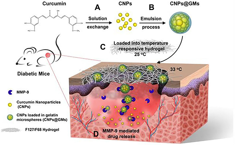 |
Figure 4 Schematic representations of the CNPs@GMs/hydrogel preparation and the process of drug release at the wound bed in diabetic mice. (A) Preparation of pure CNPs via a solution exchange method. (B) CNPs loaded into GMs by an emulsion process to obtain CNPs@GMs. (C) The CNPs@GMs mixed with thermosensitive hydrogel and covered the wound in diabetic mice. (D) Under the microenvironment of a nonhealing wound, GMs were degraded by MMPs and specifically released the drug.53 Notes: Reproduced from Liu J, Chen Z, Wang J, et al. Encapsulation of curcumin nanoparticles with MMP9-responsive and thermos-sensitive hydrogel improves diabetic wound healing. ACS Appl Mater Interfaces. 2018;10(19):16315–16326. Copyright © 2018, American Chemical Society.53 |
In addition to natural phenolic compounds, there are also some natural compound products that are obtained by hydrolysis or other chemical processes. The hypoxic and inflammatory environment in persistent trauma induces increased ROS production and the destruction of ECM proteins, which in turn leads to cellular damage and triggers proteases and inflammatory cytokines.55 Exploiting the ability of quercetin (QCN) to scavenge oxygen free radicals and inhibit lipid peroxidation, which is poorly permeable and does not penetrate easily into the skin when applied alone, Jee et al investigated a new complex of highly permeable GFs, including epidermal growth factor (EGF), insulin-like growth factor-I (IGF-I), platelet-derived growth factor-A (PDGF-A), and basic fibroblast growth factor (bFGF), forming low-molecular-weight fisetin (LMWP) (LMWP-GFs) and prepared a quercetin (QCN) and oxygen-carrying 1-bromofluorooctane (PFOB) nanoemulsion (QCN-NE and oxygen-carrying PFOBE) to improve the local transport of QCN and oxygen.56 (Figure 5). Additionally, Shanmugapriya et al combined astaxanthin and α-tocopherol (TP), which improved the chemical stability with a κ-carrageenan NE, to prepare nanomicrospheres (AS-TP@KCNE) with high- and low-energy emulsification methods. These resulted in ROS reduction and an improved immune response.57 Melatonin is an indoleamine with a reactive oxygen species (ROS)-scavenging effect. However, due to its nature, it is easily oxidized and poorly water-soluble. Correa et al designed a CS-lecithin nanoparticle to encapsulate melatonin and prevent its degradation.58 All-trans retinoic acid (ATRA) is reported to stimulate fibroblast proliferation and reduce MMP expression, which has generated interest in its use to promote skin wound healing. However, it has limitations due to its poor water solubility when used alone in skin wounds. Therefore, Arantes et al designed a solid lipid nanoparticle (SLN) wrapped in a CS membrane loaded with ATRA (SLN-ARTA) to modulate the release of ATRA. SLN has general biomaterial activity, and CS membrane SLN-ATRA increases collagen deposition in diabetic wounds.59 To control chronic wound infection, Xia et al successfully prepared a novel quaternary ammonium chitosan nanoparticle (TMC NP/CS) composite sponge, which possessed an asymmetric surface with a hydrophobic outer surface and a hydrophilic inner surface. The former repelled water as well as some contaminants, and the latter provided a good water absorption ability to maintain a comfortable, humid environment.60
 |
Figure 5 Schematic illustration of LMWP-GFs, QCN-NE, OXY-PFOB-NE, and a hydrogel comprising LMWP-GFs, QCN-NE, and OXY-PFOB-NE. Dove Medical Press Ltd.56 Notes:Reproduced from Jee JP, Pangeni R, Jha SK, Byun Y, Park JW. Preparation and in vivo evaluation of a topical hydrogel system incorporating highly skin-permeable growth factors, quercetin, and oxygen carriers for enhanced diabetic wound-healing therapy. Int J Nanomedicine. 2019;14:5449–5475. Copyright © 2019 Dove Medical Press Ltd.56 |
Moreover, some are extracted from plants. For example, ferulic acid (FA) has been previously reported to have dual properties, ie, antidiabetic and antioxidant properties. FA NPs were prepared by the nanoprecipitation method, and their hypoglycemic and wound-healing effects were assessed.61 Moreover, homalomena pineapple essential oil demonstrates good bactericidal activity against traumatic diabetic pathogens. However, it shows an unsustainable antibacterial effect. Thus, Rozman et al used CS as an encapsulating material to maintain its antimicrobial activity.62
In addition to these nanoparticles, synthetic compound nanoparticles are also used. Under hypoxia-inducible factor-1α (HIF-1α) induction, VEGF, SDF-1, and heme oxygenase-1 (HO-1) are synthesized more rapidly. Diabetic wounds have reduced expression of VEGF and SDF-1α due to significantly lower levels of HIF-1α, and in addition, the hyperglycemic environment at the wound site produces more ROS, modifying the HIF-1α coactivator p300, resulting in reduced HIF-1α-mediated trans-activation of target genes, both of which contribute to the difficulty of wound healing in diabetic patients. By competitively contradicting proline hydroxylase (PHD) and iron chelation from the wound area, desferrioxamine prevents the hydroxylation of HIF-1α and its subsequent proteasomal degradation, which subsequently increases angiogenesis, reduces inflammation, and promotes wound maturation. (Figure 6). Qayoom et al prepared and characterized lecithin-based desferrioxamine NPs and found that desferrioxamine NPs, when applied topically to diabetic wounds, resulted in faster wound healing than desferrioxamine alone.63
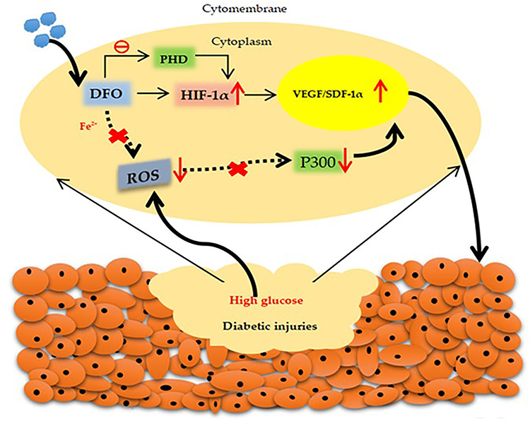 |
Figure 6 Development of a transdermal drug delivery system for DFO and its regulation in the HIF-1a signaling pathway. |
Diabetic ulcer wounds are associated with an alkaline pH, accelerating bacterial infection and causing varying degrees of cell degeneration and necrosis.64,65 The combined addition of hyaluronic acid oligosaccharide (HAO) and VEGF was shown to be effective, and the Ca-alginate (CaAlg) hydrogel as a pH-sensitive dressing disrupted the basic injured solution in diabetic ulcers, thereby triggering drug release. A pH-responsive CaAlg hydrogel containing protamine NPs and HAO (protamine NPs/HAO CaAlg hydrogel) exhibits a significant strong bactericidal effect against both Gram-positive and Gram-negative bacteria.66 To promote collagen accumulation, angiogenesis, and complete re-epithelialization, many researchers have made substantial contributions. For instance, Sun et al prepared epigallocatechin gallate, ascorbic acid, gelatin and chitosan nanoparticles (EV NPs) by an ionic cross-linking method to accelerate wound healing.67
Moreover, there are some more small molecule nanoparticles with pharmacological effects. For example, the drug edaravone possesses strong wound-healing properties by scavenging free radicals, but its partial application is limited because of its poor stability and low solubility. To scavenge excessive ROS from the wound site, Fan et al investigated the incorporation of edaravone into a nanocomposite hydrogel (HG) based on alginate and positively charged Eudragit nanoparticles.68 In addition, flavonoids, such as diosmin, are used in the clinic to enhance venous tone and protect blood vessels. In addition to their potential metal chelating and free radical scavenging effects, they have strong anti-inflammatory, antioxidant, anti-ulcer, anti-cancer, anti-atherosclerotic, hepatoprotective, and neuroprotective effects. To date, no topical use of diosmin nanocrystals in diabetic wounds or their placement in sodium alginate (SA) wound dressings have been reported. For this reason, Atia et al made an innovative attempt to load diosmin nanocrystals into SA wafers with gelatin.69 Astragaloside (ASI) is involved in various signaling pathways and has pharmacological effects, such as anti-inflammatory, immunomodulatory, and antifibrotic effects. To tackle the series of side effects caused by abnormal MMP expression, Zhang et al linked the dendrimer polyamidoamine (PAMAM) to the polysaccharide HA via the substrate peptide of MMP-2 to obtain the nanocarrier HA-pep-PAMAM in response to MMP-2, in which insoluble ASI was encapsulated. The biodegradable polysaccharide HA has a moisturizing and lubricating effect on wounds and protects against the release of drugs.70 Several reports show that solid tranilast (TS, a kind of o-aminobenzoic acid) prevents or improves proliferative scarring by carrageenan-induced granulation tissue in rats. Nagai et al designed a combination ointment containing TS nanoparticles and dissolved silk sericin (TS-combination ointment) as a wound healing agent. The combination of tranilast and silk sericin not only improved the skin healing rate but also reduced redness and swelling.71 Some of these small-molecule nanoparticles are used in clinical applications where their pharmacological effects are confirmed and can also use their pharmacological advantages to fulfill their therapeutic potential in diabetic wounds.
Finally, there are small molecule NPs that increase the expression of GFs and cytokines that promote healing. For example, Kaymakcalan et al investigated α-gal nanoparticles (AGNs) that led to keratinocyte migration, granulation tissue deposition, and increased endothelial cell density and ultimately promoted diabetic wound healing.72 Recently, neurohypocretin (NT), an inflammatory regulator in wound healing, was shown to promote diabetic wound healing by downregulating proinflammatory factors and increasing EGF expression. PLGA/cellulose nanocrystal (CNC) (PLGA/CNC) nanofiber membranes are novel materials that have not been previously used as an NT carrier for diabetic wounds. Zheng et al prepared NT-loaded nanofiber membranes by combining PLGA/CNC nanofibers with NT. Compared to other controls, the NT-loaded PLGA/CNC composite nanofibrous membranes healed faster due to their ability to reduce the expression of the inflammatory cytokines IL-1b and IL-6, and they also effectively boosted collagen deposition and re-epithelialization in diabetic wounds.73
In summary, regardless of the class of organic nanoparticles, research has been conducted to target the mechanisms of diabetic trauma refractory to healing, with single and combined actions to address a particular type of problem, such as impaired angiogenesis, hypoxia, and infection. Moreover, suitable delivery vehicles have been investigated to deliver some potent but limited substances to achieve better release rates. The advantages of inorganic nanoparticles for diabetic wound healing are also analyzed in the following sections. (Table 1).
Inorganic Nanoparticles in Diabetic Wound Healing
Silver Nanoparticles
It is well known that silver nanoparticles (AgNPs), as metallic nanoparticles, have powerful antibacterial effects due to their physical and biological characteristics. Li et al explored the antibacterial mechanism of AgNPs and found that AgNPs disrupt the intracellular membrane of bacterial cells by anchoring, leading to the flow of substances inside and outside the membrane. Furthermore, AgNPs interfere with important intracellular life activities, such as blocking the respiratory chain, attacking DNA replication, interfering with cell division, and expediting protein denaturation and enzyme inactivation.74 Diabetic patients struggle with wound healing due to the prolonged inflammatory period and persistent wound infection. Researchers have used the anti-infective properties of silver to develop several silver-containing protein-based NPs. For instance, ε-Polylysine is a natural peptide with superior insolubility in water, outstanding biocompatibility, and infection-fighting behavior. Dai et al designed a simple and novel ε-polylysine/AgNPs nanocomposite (EPL-g-butyl@AgNPs) in which alkylated ε-polylysine acted as a bacterial affinity ligand to modify the AgNPs, and its bactericidal effects were achieved by attaching it to the bacterial structure, irreversibly breaking the bacterial membrane of the surface and inhibiting protein activity.75 Moreover, Konop et al used an insoluble fur keratin-derived powder containing AgNPs (FKDP-AgNPs) to accelerate diabetic wound healing.76 Insulin is most commonly used clinically to lower blood glucose and is one of the most common treatments. Kaur et al combined insulin and AgNPs (IAgNPs), which exhibited significant healing activity in vitro and in vivo compared to the experimental control group. The mechanism involved promoting wound remodeling by regulating the relationship between positive inflammatory factors (IL-6, TNFα) and negative inflammatory factors (IL-10).77 Thus, these protein-based silver-containing nanoparticles not only have antibacterial effects but also exert anti-inflammatory properties.
Not only are there protein-loaded silver nanoparticles, but researchers have also developed several silver-containing hydrogel dressings. The ideal dressings, in addition to their biocompatibility and antimicrobial properties, also provide a moist wound environment to help remove wound exudate and promote tissue regeneration.78 HGs are one of the most commonly used scaffolds because they offer many unique advantages at the wound site, such as zero irritation, the ability to adhere to cells, hemostatic properties, and moisturizing effects. One of the most attractive irritation-responsive HGs, which has different properties and states depending on the external environment (eg, temperature, enzymes, oxygen), can very easily achieve drug-only release at the wound site.19 Considering the good biodegradability and biocompatibility of CS, Choudhary et al prepared a CS/calcium alginate nanoparticle (Ca-AlgNP)/AgNP HG to release calcium ions to activate platelets, accelerate hemostasis, and solve the problem of the poor vascular supply and bacterial infection in diabetic wounds.79 To achieve the controlled release of silver at the wound site, Masood et al prepared a CS-polyethylene glycol (PEG)-silver nitrate-based HG. The results showed that the AgNP-loaded CS-PEG/HG not only had a higher porosity but the swellability and water vapor transition rate (WVTR) were better than those of bare CS-PEG and showed enhanced antibacterial and antioxidant properties, which contributed to wound healing.80 In addition, Shi et al synthesized an antifouling hybrid HG from maleic acid-grafted dextrose and thiosemicarbazone, which not only prevented wound adhesion and provided a slow and sustained Ag+ release to exert antibacterial properties but also modulated the immune response, inhibited inflammation, and accelerated wound healing.81 Furthermore, in wound healing studies, researchers have combined HGs with various GFs and cytokines to identify breakthroughs.82 Lee et al synthesized an innovative CS-based composite HG (SNPECHG) that not only had a continuous supply of silver ions (Ag+) but also a sustainable release of nanoparticle-encapsulated EGF, ensuring stable antibacterial and cell growth-promoting performance.82 In addition, there is a superimposed multifunctional nano silver hydrogel. For instance, nimesulide (NIM), an anti-inflammatory drug, was programmed to be released in an inflammatory acidic environment. With these properties, Wang et al cross-linked 3-carboxy-phenylboronic acid (BA), a boronic acid-containing molecule with the ability to alter pH and ROS sensitivity, to the gelatin molecular backbone by attaching it to PVA (polyvinyl alcohol) and successfully synthesized inflammation-responsive drug-loaded HGs containing vancomycin silver nanoclusters (VAN-AgNCs) and pH-sensitive micelles carrying nimesulide (MIC@NIM). Among these, the combination of AgNPs and the multidrug-resistant drug vancomycin formed a hybrid antibacterial system with significant anti-inflammatory and antibacterial effects19 (Figure 7). In conclusion, HG dressings are a suitable platform for loading antioxidants, antimicrobial agents, growth factors, and biomolecules.83
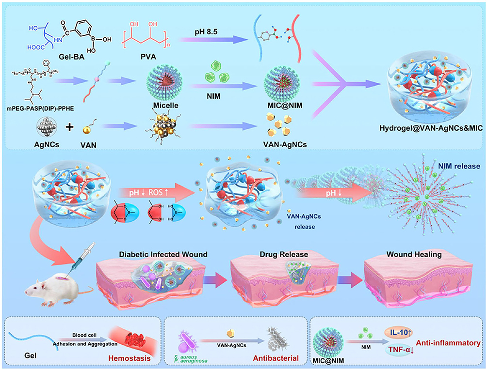 |
Figure 7 Diagrams displaying the formation of NIM-loaded micelles, VAN-AgNCs, hydrogels, and mechanisms of accelerating wound healing.19 Notes: Reproduced from Wang Y, Wu Y, Long L, et al. Inflammation-responsive drug-loaded hydrogels with sequential hemostasis, antibacterial, and anti-inflammatory behavior for chronically infected diabetic wound treatment. ACS Appl Mater Interfaces. 2021;13(28):33584–33599. Copyright © 2021, American Chemical Society.19 |
However, since the currently used silver-containing wound dressings cannot control the dosage of silver, they tend to release too much silver, which may be toxic to cells. Therefore, Guthrie et al used a polyelectrolyte multilayer (PEM) attachment technique to load silver onto a proton exchange membrane to release a certain amount of silver to the wound site, which not only avoided cytotoxicity but also had a bactericidal effect.84 Photobiomodulation (PBM) treats normal and diabetic wounds by stimulating cellular processes. To take full advantage of this, Kumar et al studied whether silver nanoparticles plus PBM could better promote wound healing.85 Moreover, to reduce the abnormal expression of MMP-2 and MMP-9 at the mRNA and protein level in the granulation tissue of diabetic patients, Krishnan et al synthesized bio-AgNPs from the culture supernatant of B. brevis KN8(2).86 Thus, it is possible to control the appropriate dose of silver using certain techniques or means.
Many biomaterials, including cellulose, gelatin, CS, HA, and alginate, have also been widely used in biomedicine because of their inherent biocompatibility, biodegradability, mechanical durability, etc. HA, a linear glycosaminoglycan polymer, is a major component of the ECM of the skin. It has good hydrophilicity and unique viscoelasticity, so it provides a moist environment for the injured tissue and protects the skin surface from injury. Based on this advantage, Fouda et al attempted to apply nanocomposites combining HA and AgNPs (HA/AgNPs) to diabetic wounds.87 Based on previous experience, Anisha developed a flexible and porous antibacterial sponge composed of CS, HA, and nano silver (nAg) (CS–HA/nAg), which provided an optimal environment for wound healing when applied to the affected area.88 Another of the major obstacles to diabetic wound healing is the impairment of angiogenesis and the high levels of proinflammatory cytokines, such as TNF-α and IL-6.89 Singla et al isolated cellulose nanocrystals (CNCs) from cucumber leaves using an environmentally friendly method and then immersed AgNPs in a matrix of CNCs.90,91 The experimental data demonstrated that the level of inflammatory molecules was significantly lower after the CNC-AgNP treatment than in the control group, which not only reduced inflammation at the wound site but also increased the expression of collagen and growth factors (FGF, PDGF, and VEGF).90 In summary, combining biomaterials and AgNPs are another innovation in treating diabetic wounds.
Gold Nanoparticles
Gold nanoparticles (AuNPs) are distinguished by two major properties: free radical scavenging and antibacterial action. Ponnanikajamideen et al prepared green synthetic AuNPs from plant extracts and found them to have free radical scavenging activity.92 Moreover, the antimicrobial peptide LL37, which is a natural antibiotic that can be inserted into the surface of bacterial cells and subsequently decomposed was demonstrated. To better investigate the bactericidal activity of AuNPs, Wang et al pioneered a new genetic delivery system for AuNPs bearing LL37 (AuNPs@LL37).93 In addition to these simple AuNPs, there is a more complex type of AuNPs. End products of advanced glycosylation, AGEs, and the receptor for AGEs, RAGE, contribute to oxidative stimulation and give rise to abnormal angiogenesis. α-Lipoic acid (ALA) is functionally powerful as a well-known purifier of ROS and is a renewable agent for some antioxidants (eg, vitamin C). Chen et al mixed AuNPs, epigallocatechin gallate (EGCG), and α-lipoic acid (ALA) (AuEA) and investigated their effect on the expression of RAGE, revealing a restorative effect for diabetic wounds.94 VEGF-A165 is a common proangiogenic growth factor that stimulates multiple components during wound healing. Wei et al combined AuNPs with VEGF-A165 and (11mercapto-undecyl)-N,N,N-trimethylammonium (11-MTA) cations (where 11-MTA has antimicrobial activity and binds to AuNPs via Au-S bonds) to form bifunctional AuNPs (11-MTA/VEGF-AuNPs) that promoted antibacterial and angiogenic activities for the treatment of chronic wound infections.95
Copper Nanoparticles
The literature reveals that copper ions (Cu2+) spur angiogenesis and collagen deposition. Metal organic backbones (MOFs, also known as HKUST-1) are a class of polymers formed by the interaction of inorganic metal ions and organic ligands. Due to their structural characteristics, MOFs are good carriers that store and release Cu2+. Xiao et al added folic acid (a vitamin with good biocompatibility) to HKUST-1, slowly releasing Cu2+, which reduced cytotoxicity and boosted cell migration in vitro.96 Electrospun membranes have an ECM-like structure, high surface area ratio, and biocompatible components that provide mechanical support and biostimulation for cell adhesion, migration, and proliferation.97 Wang et al successfully prepared a novel nanocomposite membrane by incorporating Cu2S nanoflowers into biopolymer fibers using an improved electrostatic spinning method. This membrane releases Cu2+, thus stimulating endothelial cell proliferation and angiogenesis.98 Moreover, since Cu is an essential micronutrient, it stabilizes the expression of extracellular skin proteins, and in this way, Cu2+ is recognized to have excellent antibacterial properties, which greatly minimize wound infection and aid in wound healing. To ensure the stable release of Cu2+, BGNC was formed by wrapping Cu with bioactive glass nanoparticles (BGNs). Li et al developed a self-healing injectable composite (PABC) HG composed of polyethylene glycol diacrylate (PEGDA) to which BGNC and sodium alginate (ALG) were added to assist in the formation of a dynamic reticular structure. This innovative PABC HG not only promoted HIF-1α/VEGF expression and collagen matrix deposition on target wounds but also effectively regenerated crucial blood vessels and skin tissue.99
Ceria Nanoparticles
It is well known that cerium (Ce), as a rare earth element, possesses antibacterial ability. To overcome the troubles of diabetic wound blood flow reconstruction and infection control, Chen et al infused cerium-containing bioactive glass (Ce-BG) into a gelatin methacryloyl (GelMA) HG and synthesized a composite HG with good biocompatibility that also released Ce4+ to show high antimicrobial activity. GelMA HG loaded with Ce-BG has good moisture retention to avoid tearing in the wound.100 Cerium oxide is a material that occurs regularly in nature. The specificity of its nanocrystal structure and the flexible shift in valence potential of cerium atoms are properties that give cerium oxide nanoparticles (CNPs) the ability to automatically regenerate and scavenge free radicals. CNPs also have anti-inflammatory, anticancer, and proangiogenic properties.101 In the presence of ROS, cerium atoms switch reversibly between 3+ and 4+, thereby coordinating the balance between oxidative and complimentary enzymes in diabetic trauma.102 Augustine et al introduced CNPs into a biodegradable GelMA HG patches to generate a novel patch loaded with CNPs103 and synthesized a new electrospun poly (3-hydroxybutyrate-3-hydroxypentanoate) (PHBV) film containing nCeO2 using electrostatic spinning technology,104 which effectively scavenged free radicals, reduced oxidative stress, and promoted cell proliferation associated with skin healing. Previous studies have shown that decreased microRNA miR-146a expression increases the inflammatory response and delays healing in diabetic wounds.102 Currently, CNPs are thought to have peroxidase-like and superoxide-like dismutase activities. The penetration of CNPs into wound tissue prevents secondary infection, may reduce oxidative damage, and protects regenerating tissues.105 Considering the abovementioned unique functions of CNPs, Zgheib and team first coupled miR-146a to CNPs (CNPs-miR146a) and designed the optimal material concentration to improve the biomechanical properties of healing skin.102 Based on previous experiments, many researchers are investigating suitable vectors hoping to efficiently deliver CNPs-miR146a. For instance, Niemiec et al utilized nanosilk solutions as a novel delivery system for topical administration, which effectively delivered therapeutic CNPs-miR146a to reduce the inflammatory response and excessive oxidative stress at the injury location. This nanoserum solution was chosen because it contained sericin, a protein that not only promoted the aggregation of relevant cells and improved the diffusion of type I collagen in the relevant cells but also protected the wound edges from unnecessary damage and sped up healing.106 Sener et al used a zwitterionic cryogel (a gel formed below the freezing point) biomaterial system loaded with CNPs-miR146a to scavenge free radicals from the wound surface and eliminate oxidative stress.107
Rubidium Nanoparticles
Rubidium (Rb) is one of the most abundant trace elements in the human system, and it has been previously reported in the literature that Rb+ inhibits/kills Staphylococcus aureus and Pseudomonas aeruginosa through its effect on membrane potential. The successful synthesis of Rb calcium alginate hydrogel (Rb/CA-HG) with good homogeneity and integrity was achieved. Rb/CA-HG, as a novel therapeutic approach to promote diabetic wound healing, not only facilitates the growth and migration of human umbilical vein endothelial cells (HUVECs), fibroblasts and keratin-forming cells and promotes wound angiogenesis, re-epithelialization and collagen deposition compared to controls but also regulates nuclear factor (erythroid-derived 2)-like 2 (NRF2)/heme-oxygenase-1 (HO-1) secretion and inhibits oxidative stress in traumatic surfaces.108
Silicon Dioxide Nanoparticles
Silicon dioxide (SiO2) is a biomaterial with several unique properties. Porous silica is widely attractive for both in vivo and ex vivo applications and has an excellent specific surface area and large pore capacity; thus, it is widely used as a controlled drug carrier.109,110 M1-type macrophages induce an inflammatory response, whereas M2-type macrophages suppress the inflammatory response and promote wound repair. Gan et al modified SiO2 NPs with konjac dextran to obtain KSiNPs, inhibiting M1 macrophage-mediated long-term inflammation and promoting M1-type to M2-type polarization.111 In addition to its ability to mediate inflammatory responses, the porous surface of silicon dioxide is a natural carrier for controlled drug release. Previous work revealed that HIF-1α performs a key function in promoting wound healing in diabetic mice, as it optimizes epidermal regeneration and recruits endothelial precursors.112 Dimethoxyglycine (DMOG), a micromolecule and proangiogenic drug, stabilizes HIF-1α and improves the hypoxic microenvironment. Mesoporous silica nanoparticles (MSNs) have a high specific surface area and large pore capacity. Therefore, to better utilize the function of DMOG, Ren et al developed poly (L-lactic acid) (PLLA) electrospun fibrous membranes that contained MSNs loaded with DMOG for the repair of diabetic wounds. DMOG and Si ions can be delivered from the nanopores on the fiber membrane.110 Flightless I (Flii) mediates cell migration, contraction, adhesion, proliferation, and cytokine production during tissue regeneration by interacting with cytoskeletal components and regulating intracellular signaling pathways and cellular transcription. Flii is elevated in human chronic wounds and retards the wound repair process. Flii-neutralizing antibodies (FnAbs) reduce Flii activity in vivo. Turner et al used suitable porous silica nanoparticles (pSiNPs) to control FnAb release from diabetic wounds. FnAb-loaded pSiNPs promote the proliferation and migration of keratin-forming cells.109 Among all the techniques for preparing nanomaterials, a sol-gel technique is an effective method, and Goerne et al used this method to develop platinum acetylacetonate-encapsulated TiO2-SiO2 (Pt/TiO2–SiO2) nanopowders with improved biocatalytic properties to promote the proliferation and migration of keratin-forming cells.113 Whether it is a single or composite porous silica, the pores on the surface of the silica can be used to achieve the controlled release of drugs (Table 2).
The Mechanism Underlying the Diabetic Wound Healing of Nanoparticles
In diabetic patients, angiogenesis and cell migration are impaired due to the abnormal release of cytokines, such as EGF, CTGF, IGF-1, TNF-α, and IL-6.28 During wound healing, angiogenesis is important for promoting diabetic wounds, and a variety of nanocarriers are effective for angiogenesis. For example, multiple GFs (NaCMCh/PLGA-rhEGF,24,25 PLGA-VEGF/EGF NPs,10,26 Col-HA-GN nanofibrous membranes,27 AlgSulf/PCL NPs,28 and SDF-1-ELPs) have been identified.29 GF treatments not only have multiple active effects, such as re-epithelializing tissue to stimulate wound healing26 and tackling restricted matrix deposition,28 but also enhance blood perfusion to induce angiogenesis.24 For example, multiple genetically generated molecules (plasmid-PLGA NPs,38 plasmid-PAM-RG4-VEGF,39 and BNH42 encode GFs or activate a certain gene. In addition to GFs and plasmids that promote angiogenesis, exosomes act on wounds in the form of FEP dressings33 and MC-CS HG37 releases exosomes and promotes endothelial cell proliferation, migration and angiogenesis. For example, small molecule compounds, such as EV NPs67 and AGNs,72 promote the production of GFs and cytokines. In addition, multiple metal NPs, SNPECHG,82 11-MTA/VEGF-AuNPs,95 Cu-MOF NPs,96 Cu2S NPs,98 and PABC HG,99 can release metal ions and show potent proangiogenic capacity.
Diabetic wound healing involves several interactions between cells, extracellular matrix, and cytokines, and the most significant cause of difficult healing is a long-term chronic infection.82 It is well known that most metal ions have antibacterial effects. For example, multiple small molecule compounds, pineapple essential oil-CS,62 and the protamine NP/HAO-CaAlg HG,66 exhibit strong antibacterial activity. The multiple metal NPs include EPL-g-butyl@AgNPs,75 FKDP-AgNPs,76 CS/Ca-AlgNP/AgNP,79 CS-PEG-AgNPs,80 antifouling HG-AgNPs,81 VAN-AgNCs,19 HA-AgNPs,87 CS-HA-nAg,88 CNCs-AgNPs,90,91 AuNPs@LL37,93 Ce/BG-HG,100 and Rb/CA-HG.108 Silver disrupts the cell membrane of microorganisms, inhibits the formation of enzymes, RNA, and DNA, and eventually leads to bacterial death. Therefore, the addition of AgNPs to a material can improve its antibacterial properties and promote wound healing.81 The antimicrobial peptide LL37 is a natural antibiotic that can be inserted onto the surface of bacterial cells and subsequently broken down.93
The pathophysiological mechanisms of diabetes are complex, and diabetic trauma is always in an inflammatory phase, associated with an excess of neutrophils and TNFα, the production of more free radicals that increase protease activity, and a significant oxidative stress response.63 A prolonged inflammatory phase and increased oxidative stress can be the leading contributors to diabetic trauma-related complications. Thus, the use of antioxidants and anti-inflammatory agents can accelerate diabetic wound healing.104 There are some nanoparticles with strong antioxidant capacity on their own. For example, multiple small molecule compounds, such as Cur-CS-NPs,50,51 Cur-NP/HG,52 CNPs@GMs/HG,53 SM-PLGA nanosuspensions,54 GFs/QCN/oxygen-NE,56 AS-TP@KCNE,57 CS lecithin-melatonin NPs,58 FA NPs,61 deferrioxamine NPs,63 edaravone nanocomposite HG,68 diosmin-SA wafers,69 HA-pep-PAMAM,70 and TS-combination ointment71 exhibit these properties. These small molecule nanoparticles have anti-inflammatory and antioxidant properties and scavenge excessive ROS at the wound site. As previously described, HIF-1α increases the expression of VEGF and SDF-1α, thereby promoting cell proliferation and neovascularization and reducing cell death. Additionally, deferoxamine improves the activity of HIF-1α.63 For example, multiple genetic molecules, such as BC-HCP/siMMP-9,22 siRNA and β-CD-(D3)7,44 and siRNA-LPP,45 are involved in these processes. We all know that chronic inflammation of traumatic surfaces is associated with the reduced expression of microRNAs. Certain microRNAs, such as those that are key activators of IL-6 and IL-8, inhibit the proinflammatory signaling pathway.114,115 For example, multiple metal NPs, such as AuNPs,92 Ce-BG/GelMA HG,100 CNPs GelMA HG patches,103 nCeO2 PHBV membranes,104 CNPs-miR146a,102,106,107 and KsiNPs111 are involved. They scavenge free radicals to reduce inflammation and inhibit the production of many proinflammatory cytokines, such as IL-8, IL-6, IL-12, and TNF-α.116,117 (Table 3).
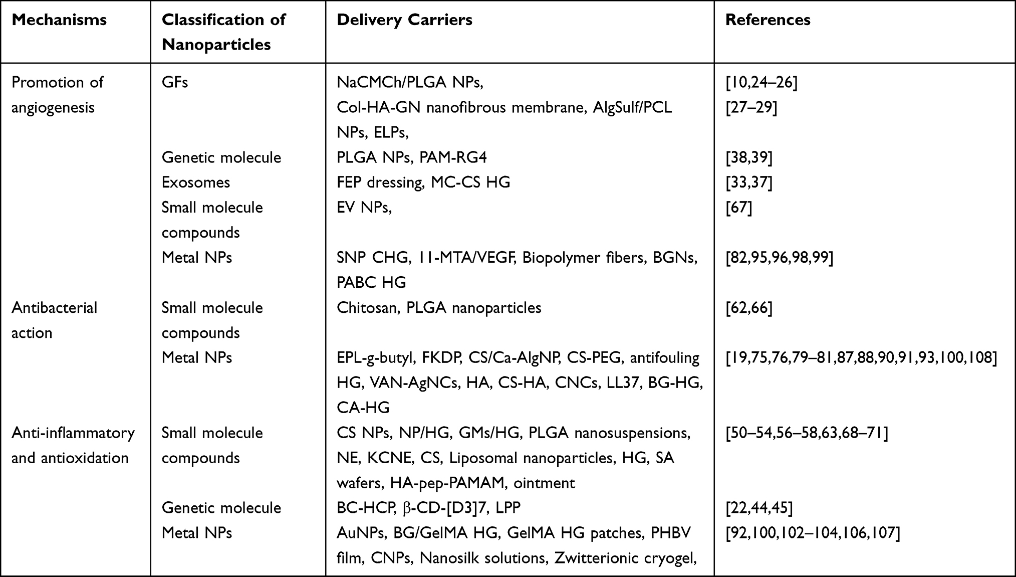 |
Table 3 The Mechanism Underlying the Diabetic Wound Healing of Nanoparticles |
In conclusion, in the future, researchers/scientists who want to research wound healing should aim to explore three mechanisms: the promotion of angiogenesis, anti-inflammatory, and antioxidant effects on the wound site, and antibacterial effects. We can note that the effects of promoting angiogenesis, GFs and some cytokines take the lead as “effective remedies” to promote wound healing. Secondly, most metal ions have a natural antibacterial ability to control wound infection and accelerate healing. Finally, for the inflammatory phase of wound healing, researchers should consider natural or synthetic organic substances that can effectively control the inflammatory response of wounds due to their anti-inflammatory and antioxidant effects. Furthermore, NPs are easily internalized into the body across biological barriers (eg, brain barrier and blood-placental barrier) and their safety depends on their intrinsic properties, prompting the need to develop new nanotechnology-based therapeutic approaches for NPs.9 We look forward to seeing more treatments that will slowly be applied clinically and alleviate patient suffering.
Conclusion
The healing process of chronic wounds arising from diabetes is more difficult compared to normal wound healing. The complex pathophysiological mechanisms of diabetic wounds, impaired cellular function and cytokine secretion, and long-term exposure to the external environment strongly expand the chances of infection, which makes the treatment of diabetic wounds not only complex but also challenging. Based on the characteristics of diabetic wounds and tissue repair mechanisms, we have classified organic molecular nanoparticles (eg, protein-loaded nanoparticles, exosomes, etc.) and inorganic molecular nanoparticles (metals, nonmetals, etc.). Focusing on the mechanisms of the anti-inflammatory, antioxidant and antibacterial properties as well as promoting angiogenesis, the role of a high specific surface area, diffusivity and adsorption with a better controlled and slow-release function brought by nanoparticles in accelerating diabetic wound healing has been discussed based on the good biocompatibility of nanoparticles. Different types of nanoparticles have different strengths, weaknesses, and applicability with regard to the wound healing. In conclusion, many researchers have attempted to use nanocarriers to deliver drugs that not only take full advantage of the biological properties of the nanoparticles to achieve a controlled and slow release of the drug but also help to address the limitations of drug applications.
Recently, academic research and clinical trials on wound healing are emerging, and there is a need to understand more about the biology of diabetic wounds to prepare multifunctional nanosystems using various techniques for targeted therapy to improve healing rates and to try to apply investigated biomaterial nanoparticles in the clinical setting. In the long run, the future direction may be to develop novel nanoparticles with multiple effects (including improving hypoxia, enhancing angiogenesis, reducing oxidative stress, and controlling infection) that not only act in wound healing at all stages of diabetes but also provide a stable physiological environment throughout the wound healing process, thereby reducing potential complications and allowing patients to feel better about their treatment. If possible, multiple nanoparticles or the combination of nanoparticles with other materials to achieve integrated therapeutic effects while ensuring the purity and functionality of nanoparticles. It is essential to further explore how to commercialize the investigated nanomaterials and apply them in the clinical field. In the future, these investigations will not only improve the rate of diabetic wound healing but also lead to further breakthroughs in the field of nanoparticles.
Acknowledgments
This work was supported by the Natural Science Foundation of Heilongjiang Province (LH2019H061), the Natural Science Foundation of China (82072051; 81903233; 81874250), Central Finance support of Local Colleges and Universities Talent Development Funding from Heilongjiang Provincial Department of Finance (2020GSP09), and the Torch Program of Mudanjiang Medical College Science Foundation (2022-MYHJ-006).
Disclosure
The authors report no conflicts of interest in this work.
References
1. Cahill D, Zamboni F, Collins MN. Radiological advances in pancreatic islet transplantation. Acad Radiol. 2019;26(11):1536–1543. doi:10.1016/j.acra.2019.01.006
2. Okonkwo UA, DiPietro LA. Diabetes and wound angiogenesis. Int J Mol Sci. 2017;18:7. doi:10.3390/ijms18071419
3. Kesharwani P, Gorain B, Low SY, et al. Nanotechnology based approaches for anti-diabetic drugs delivery. Diabetes Res Clin Pract. 2018;136:52–77. doi:10.1016/j.diabres.2017.11.018
4. Li SJ, Fan J, Zhou J, Ren YT, Shen C, Che GW. Diabetes mellitus and risk of bronchopleural fistula after pulmonary resections: a meta-analysis. Ann Thorac Surg. 2016;102(1):328–339. doi:10.1016/j.athoracsur.2016.01.013
5. Shen YI, Cho H, Papa AE, et al. Engineered human vascularized constructs accelerate diabetic wound healing. Biomaterials. 2016;102:107–119. doi:10.1016/j.biomaterials.2016.06.009
6. Chen L, Xing Q, Zhai Q, et al. Pre-vascularization enhances therapeutic effects of human mesenchymal stem cell sheets in full thickness skin wound repair. Theranostics. 2017;7(1):117–131. doi:10.7150/thno.17031
7. Dixon D, Edmonds M. Managing diabetic foot ulcers: pharmacotherapy for wound healing. Drugs. 2021;81(1):29–56. doi:10.1007/s40265-020-01415-8
8. Blanco-Fernandez B, Castano O, Mateos-Timoneda MA, Engel E, Perez-Amodio S. Nanotechnology approaches in chronic wound healing. Adv Wound Care. 2021;10(5):234–256. doi:10.1089/wound.2019.1094
9. Haque ST, Saha SK, Haque ME, Biswas N. Nanotechnology-based therapeutic applications: in vitro and in vivo clinical studies for diabetic wound healing. Biomater Sci. 2021;9(23):7705–7747. doi:10.1039/d1bm01211h
10. Qi X, Huan Y, Si H, Zou J, Mu Z. Study of the effect epidermal growth factor nanoparticles in the treatment of diabetic rat ulcer skin and regeneration. J Nanosci Nanotechnol. 2021;21(5):3028–3034. doi:10.1166/jnn.2021.19155
11. Stark WJ, Stoessel PR, Wohlleben W, Hafner A. Industrial applications of nanoparticles. Chem Soc Rev. 2015;44(16):5793–5805. doi:10.1039/C4CS00362D
12. Zhang Y, Luo J, Zhang Q, Deng T. Growth factors, as biological macromolecules in bioactivity enhancing of electrospun wound dressings for diabetic wound healing: a review. Int J Biol Macromol. 2021;193(Pt A):205–218. doi:10.1016/j.ijbiomac.2021.09.210
13. Berger AG, Chou JJ, Hammond PT. Approaches to modulate the chronic wound environment using localized nucleic acid delivery. Adv Wound Care. 2021;10(9):503–528. doi:10.1089/wound.2020.1167
14. Madhukiran D, Jha A, Kumar M, Ajmal G, Bonde GV, Mishra B. Electrospun nanofiber-based drug delivery platform: advances in diabetic foot ulcer management. Expert Opin Drug Deliv. 2021;18(1):25–42. doi:10.1080/17425247.2021.1823966
15. Alavi M, Rai M. Topical delivery of growth factors and metal/metal oxide nanoparticles to infected wounds by polymeric nanoparticles: an overview. Expert Rev Anti Infect Ther. 2020;18(10):1021–1032. doi:10.1080/14787210.2020.1782740
16. Choudhury H, Pandey M, Lim YQ, et al. Silver nanoparticles: advanced and promising technology in diabetic wound therapy. Mater Sci Eng C Mater Biol Appl. 2020;112:110925. doi:10.1016/j.msec.2020.110925
17. Mirza RE, Koh TJ. Contributions of cell subsets to cytokine production during normal and impaired wound healing. Cytokine. 2015;71(2):409–412. doi:10.1016/j.cyto.2014.09.005
18. Han G, Ceilley R. Chronic wound healing: a review of current management and treatments. Adv Ther. 2017;34(3):599–610. doi:10.1007/s12325-017-0478-y
19. Wang Y, Wu Y, Long L, et al. Inflammation-responsive drug-loaded hydrogels with sequential hemostasis, antibacterial, and anti-inflammatory behavior for chronically infected diabetic wound treatment. ACS Appl Mater Interfaces. 2021;13(28):33584–33599. doi:10.1021/acsami.1c09889
20. Bai Q, Han K, Dong K, et al. Potential applications of nanomaterials and technology for diabetic wound healing. Int J Nanomedicine. 2020;15:9717–9743. doi:10.2147/IJN.S276001
21. Desmet CM, Préat V, Gallez B. Nanomedicines and gene therapy for the delivery of growth factors to improve perfusion and oxygenation in wound healing. Adv Drug Deliv Rev. 2018;129:262–284. doi:10.1016/j.addr.2018.02.001
22. Li N, Yang L, Pan C, et al. Naturally-occurring bacterial cellulose-hyperbranched cationic polysaccharide derivative/MMP-9 siRNA composite dressing for wound healing enhancement in diabetic rats. Acta Biomater. 2020;102:298–314. doi:10.1016/j.actbio.2019.11.005
23. Wang F, Zhang W, Li H, Chen X, Feng S, Mei Z. How Effective are nano-based dressings in diabetic wound healing? A comprehensive review of literature. Int J Nanomedicine. 2022;17:2097–2119. doi:10.2147/IJN.S361282
24. Hajimiri M, Shahverdi S, Esfandiari MA, et al. Preparation of hydrogel embedded polymer-growth factor conjugated nanoparticles as a diabetic wound dressing. Drug Dev Ind Pharm. 2016;42(5):707–719. doi:10.3109/03639045.2015.1075030
25. Chu Y, Yu D, Wang P, Xu J, Li D, Ding M. Nanotechnology promotes the full-thickness diabetic wound healing effect of recombinant human epidermal growth factor in diabetic rats. Wound Repair Regen. 2010;18(5):499–505. doi:10.1111/j.1524-475X.2010.00612.x
26. Chereddy KK, Lopes A, Koussoroplis S, et al. Combined effects of PLGA and vascular endothelial growth factor promote the healing of non-diabetic and diabetic wounds. Nanomedicine. 2015;11(8):1975–1984. doi:10.1016/j.nano.2015.07.006
27. Lai HJ, Kuan CH, Wu HC, et al. Tailored design of electrospun composite nanofibers with staged release of multiple angiogenic growth factors for chronic wound healing. Acta Biomater. 2014;10(10):4156–4166. doi:10.1016/j.actbio.2014.05.001
28. Maatouk B, Jaffa MA, Karam M, et al. Sulfated alginate/polycaprolactone double-emulsion nanoparticles for enhanced delivery of heparin-binding growth factors in wound healing applications. Colloids Surf B Biointerfaces. 2021;208:112105. doi:10.1016/j.colsurfb.2021.112105
29. Yeboah A, Cohen RI, Faulknor R, Schloss R, Yarmush ML, Berthiaume F. The development and characterization of SDF1α-elastin-like-peptide nanoparticles for wound healing. J Control Release. 2016;232:238–247. doi:10.1016/j.jconrel.2016.04.020
30. Abdelkader DH, Tambuwala MM, Mitchell CA, et al. Enhanced cutaneous wound healing in rats following topical delivery of insulin-loaded nanoparticles embedded in poly (vinyl alcohol)-borate hydrogels. Drug Deliv Transl Res. 2018;8(5):1053–1065. doi:10.1007/s13346-018-0554-0
31. Azevedo F, Pessoa A, Moreira G, et al. Effect of topical insulin on second-degree burns in diabetic rats. Biol Res Nurs. 2016;18(2):181–192. doi:10.1177/1099800415592175
32. Ribeiro MC, Correa VLR, Silva F, et al. Wound healing treatment using insulin within polymeric nanoparticles in the diabetes animal model. Eur J Pharm Sci. 2020;150:105330. doi:10.1016/j.ejps.2020.105330
33. Wang M, Wang C, Chen M, et al. Efficient angiogenesis-based diabetic wound healing/skin reconstruction through bioactive antibacterial adhesive ultraviolet shielding nanodressing with exosome release. ACS Nano. 2019;13(9):10279–10293. doi:10.1021/acsnano.9b03656
34. Wu Y, Zhang C, Guo R, et al. Mesenchymal stem cells: an overview of their potential in cell-based therapy for diabetic nephropathy. Stem Cells Int. 2021;2021:6620811. doi:10.1155/2021/6620811
35. Hu Y, Rao SS, Wang ZX, et al. Exosomes from human umbilical cord blood accelerate cutaneous wound healing through miR-21-3p-mediated promotion of angiogenesis and fibroblast function. Theranostics. 2018;8(1):169–184. doi:10.7150/thno.21234
36. Liu X, Yang Y, Li Y, et al. Integration of stem cell-derived exosomes with in situ hydrogel glue as a promising tissue patch for articular cartilage regeneration. Nanoscale. 2017;9(13):4430–4438. doi:10.1039/C7NR00352H
37. Wang C, Liang C, Wang R, et al. The fabrication of a highly efficient self-healing hydrogel from natural biopolymers loaded with exosomes for the synergistic promotion of severe wound healing. Biomater Sci. 2019;8(1):313–324. doi:10.1039/C9BM01207A
38. Shi R, Lian W, Han S, et al. Nanosphere-mediated co-delivery of VEGF-A and PDGF-B genes for accelerating diabetic foot ulcers healing in rats. Gene Ther. 2018;25(6):425–438. doi:10.1038/s41434-018-0027-6
39. Kwon MJ, An S, Choi S, et al. Effective healing of diabetic skin wounds by using nonviral gene therapy based on minicircle vascular endothelial growth factor DNA and a cationic dendrimer. J Gene Med. 2012;14(4):272–278. doi:10.1002/jgm.2618
40. Chen G, He L, Zhang P, et al. Encapsulation of green tea polyphenol nanospheres in PVA/alginate hydrogel for promoting wound healing of diabetic rats by regulating PI3K/AKT pathway. Mater Sci Eng C Mater Biol Appl. 2020;110:110686. doi:10.1016/j.msec.2020.110686
41. Li J, Liu Y, Liu B, et al. Mechanisms of aerobic exercise upregulating the expression of hippocampal synaptic plasticity-associated proteins in diabetic rats. Neural Plast. 2019;2019:7920540. doi:10.1155/2019/7920540
42. Zhang P, He L, Zhang J, et al. Preparation of novel berberine nano-colloids for improving wound healing of diabetic rats by acting Sirt1/NF-κB pathway. Colloids Surf B Biointerfaces. 2020;187:110647. doi:10.1016/j.colsurfb.2019.110647
43. Randeria PS, Seeger MA, Wang XQ, et al. siRNA-based spherical nucleic acids reverse impaired wound healing in diabetic mice by ganglioside GM3 synthase knockdown. Proc Natl Acad Sci U S A. 2015;112(18):5573–5578. doi:10.1073/pnas.1505951112
44. Li N, Luo HC, Yang C, et al. Cationic star-shaped polymer as an siRNA carrier for reducing MMP-9 expression in skin fibroblast cells and promoting wound healing in diabetic rats. Int J Nanomedicine. 2014;9:3377–3387. doi:10.2147/IJN.S66368
45. Rabbani PS, Zhou A, Borab ZM, et al. Novel lipoproteoplex delivers Keap1 siRNA based gene therapy to accelerate diabetic wound healing. Biomaterials. 2017;132:1–15. doi:10.1016/j.biomaterials.2017.04.001
46. Tao SC, Rui BY, Wang QY, Zhou D, Zhang Y, Guo SC. Extracellular vesicle-mimetic nanovesicles transport LncRNA-H19 as competing endogenous RNA for the treatment of diabetic wounds. Drug Deliv. 2018;25(1):241–255. doi:10.1080/10717544.2018.1425774
47. Hlaing SP, Kim J, Lee J, et al. S-Nitrosoglutathione loaded poly (lactic-co-glycolic acid) microparticles for prolonged nitric oxide release and enhanced healing of methicillin-resistant Staphylococcus aureus-infected wounds. Eur J Pharm Biopharm. 2018;132:94–102. doi:10.1016/j.ejpb.2018.09.009
48. Hasan N, Cao J, Lee J, et al. PEI/NONOates-doped PLGA nanoparticles for eradicating methicillin-resistant Staphylococcus aureus biofilm in diabetic wounds via binding to the biofilm matrix. Mater Sci Eng C Mater Biol Appl. 2019;103:109741. doi:10.1016/j.msec.2019.109741
49. Meng B, Li J, Cao H. Antioxidant and antiinflammatory activities of curcumin on diabetes mellitus and its complications. Curr Pharm Des. 2013;19(11):2101–2113.
50. Karri VV, Kuppusamy G, Talluri SV, et al. Curcumin loaded chitosan nanoparticles impregnated into collagen-alginate scaffolds for diabetic wound healing. Int J Biol Macromol. 2016;93(Pt B):1519–1529. doi:10.1016/j.ijbiomac.2016.05.038
51. Li F, Shi Y, Liang J, Zhao L. Curcumin-loaded chitosan nanoparticles promote diabetic wound healing via attenuating inflammation in a diabetic rat model. J Biomater Appl. 2019;34(4):476–486. doi:10.1177/0885328219860929
52. Kamar SS, Abdel-Kader DH, Rashed LA. Beneficial effect of curcumin nanoparticles-hydrogel on excisional skin wound healing in type-I diabetic rat: histological and immunohistochemical studies. Ann Anat. 2019;222:94–102. doi:10.1016/j.aanat.2018.11.005
53. Liu J, Chen Z, Wang J, et al. Encapsulation of curcumin nanoparticles with MMP9-responsive and thermos-sensitive hydrogel improves diabetic wound healing. ACS Appl Mater Interfaces. 2018;10(19):16315–16326. doi:10.1021/acsami.8b03868
54. Gourishetti K, Keni R, Nayak PG, et al. Sesamol-loaded PLGA nanosuspension for accelerating wound healing in diabetic foot ulcer in rats. Int J Nanomedicine. 2020;15:9265–9282. doi:10.2147/IJN.S268941
55. Zhang S, Li Y, Qiu X, et al. Incorporating redox-sensitive nanogels into bioabsorbable nanofibrous membrane to acquire ROS-balance capacity for skin regeneration. Bioact Mater. 2021;6(10):3461–3472. doi:10.1016/j.bioactmat.2021.03.009
56. Jee JP, Pangeni R, Jha SK, Byun Y, Park JW. Preparation and in vivo evaluation of a topical hydrogel system incorporating highly skin-permeable growth factors, quercetin, and oxygen carriers for enhanced diabetic wound-healing therapy. Int J Nanomedicine. 2019;14:5449–5475. doi:10.2147/IJN.S213883
57. Shanmugapriya K, Kim H, Kang HW. A new alternative insight of nanoemulsion conjugated with κ-carrageenan for wound healing study in diabetic mice: in vitro and in vivo evaluation. Eur J Pharm Sci. 2019;133:236–250. doi:10.1016/j.ejps.2019.04.006
58. Lopes Rocha Correa V, Assis Martins J, Ribeiro de Souza T, et al. Melatonin loaded lecithin-chitosan nanoparticles improved the wound healing in diabetic rats. Int J Biol Macromol. 2020;162:1465–1475. doi:10.1016/j.ijbiomac.2020.08.027
59. Arantes VT, Faraco AAG, Ferreira FB, et al. Retinoic acid-loaded solid lipid nanoparticles surrounded by chitosan film support diabetic wound healing in in vivo study. Colloids Surf B Biointerfaces. 2020;188:110749. doi:10.1016/j.colsurfb.2019.110749
60. Xia G, Zhai D, Sun Y, et al. Preparation of a novel asymmetric wettable chitosan-based sponge and its role in promoting chronic wound healing. Carbohydr Polym. 2020;227:115296. doi:10.1016/j.carbpol.2019.115296
61. Bairagi U, Mittal P, Singh J, Mishra B. Preparation, characterization, and in vivo evaluation of nano formulations of ferulic acid in diabetic wound healing. Drug Dev Ind Pharm. 2018;44(11):1783–1796. doi:10.1080/03639045.2018.1496448
62. Rozman NAS, Tong WY, Leong CR, et al. Homalomena pineodora essential oil nanoparticle inhibits diabetic wound pathogens. Sci Rep. 2020;10(1):3307. doi:10.1038/s41598-020-60364-0
63. Qayoom A, Aneesha VA, Anagha S, Dar JA, Kumar P, Kumar D. Lecithin-based deferoxamine nanoparticles accelerated cutaneous wound healing in diabetic rats. Eur J Pharmacol. 2019;858:172478. doi:10.1016/j.ejphar.2019.172478
64. Chen H, Xing X, Tan H, et al. Covalently antibacterial alginate-chitosan hydrogel dressing integrated gelatin microspheres containing tetracycline hydrochloride for wound healing. Mater Sci Eng C Mater Biol Appl. 2017;70(Pt 1):287–295. doi:10.1016/j.msec.2016.08.086
65. Chopra P, Nayak D, Nanda A, Ashe S, Rauta PR, Nayak B. Fabrication of poly (vinyl alcohol)-Carrageenan scaffolds for cryopreservation: effect of composition on cell viability. Carbohydr Polym. 2016;147:509–516. doi:10.1016/j.carbpol.2016.04.027
66. Wang T, Zheng Y, Shi Y, Zhao L. pH-responsive calcium alginate hydrogel laden with protamine nanoparticles and hyaluronan oligosaccharide promotes diabetic wound healing by enhancing angiogenesis and antibacterial activity. Drug Deliv Transl Res. 2019;9(1):227–239. doi:10.1007/s13346-018-00609-8
67. Sun M, Xie Q, Cai X, et al. Preparation and characterization of epigallocatechin gallate, ascorbic acid, gelatin, chitosan nanoparticles and their beneficial effect on wound healing of diabetic mice. Int J Biol Macromol. 2020;148:777–784. doi:10.1016/j.ijbiomac.2020.01.198
68. Fan Y, Wu W, Lei Y, et al. Edaravone-loaded alginate-based nanocomposite hydrogel accelerated chronic wound healing in diabetic mice. Mar Drugs. 2019;17:5. doi:10.3390/md17050285
69. Atia NM, Hazzah HA, Gaafar PME, Abdallah OY. Diosmin nanocrystal-loaded wafers for treatment of diabetic ulcer: in vitro and in vivo evaluation. J Pharm Sci. 2019;108(5):1857–1871. doi:10.1016/j.xphs.2018.12.019
70. Zhang D, Huang Q. Encapsulation of Astragaloside with matrix metalloproteinase-2-responsive hyaluronic acid end-conjugated polyamidoamine dendrimers improves wound healing in diabetes. J Biomed Nanotechnol. 2020;16(8):1229–1240. doi:10.1166/jbn.2020.2971
71. Nagai N, Ogata F, Deguchi S, Ueno A, Kawasaki N, Ito Y. Combination ointment containing solid tranilast nanoparticles and dissolved sericin is efficacious for treating skin wound-healing deficits and redness in diabetic rats. Biol Pharm Bull. 2017;40(4):444–450. doi:10.1248/bpb.b16-00812
72. Kaymakcalan OE, Abadeer A, Goldufsky JW, et al. Topical α-gal nanoparticles accelerate diabetic wound healing. Exp Dermatol. 2020;29(4):404–413. doi:10.1111/exd.14084
73. Zheng Z, Liu Y, Huang W, et al. Neurotensin-loaded PLGA/CNC composite nanofiber membranes accelerate diabetic wound healing. Artif Cells Nanomed Biotechnol. 2018;46(sup2):493–501. doi:10.1080/21691401.2018.1460372
74. Xu L, Wang YY, Huang J, Chen CY, Wang ZX, Xie H. Silver nanoparticles: synthesis, medical applications and biosafety. Theranostics. 2020;10(20):8996–9031. doi:10.7150/thno.45413
75. Dai X, Guo Q, Zhao Y, et al. Functional silver nanoparticle as a benign antimicrobial agent that eradicates antibiotic-resistant bacteria and promotes wound healing. ACS Appl Mater Interfaces. 2016;8(39):25798–25807. doi:10.1021/acsami.6b09267
76. Konop M, Czuwara J, Klodzinska E, et al. Evaluation of keratin biomaterial containing silver nanoparticles as a potential wound dressing in full-thickness skin wound model in diabetic mice. J Tissue Eng Regen Med. 2020;14(2):334–346. doi:10.1002/term.2998
77. Kaur P, Sharma AK, Nag D, et al. Novel nano-insulin formulation modulates cytokine secretion and remodeling to accelerate diabetic wound healing. Nanomedicine. 2019;15(1):47–57. doi:10.1016/j.nano.2018.08.013
78. Zheng K, Tong Y, Zhang S, et al. Flexible bicolorimetric polyacrylamide/chitosan hydrogels for smart real‐time monitoring and promotion of wound healing. Adv Funct Mater. 2021;31:34. doi:10.1002/adfm.202102599
79. Choudhary M, Chhabra P, Tyagi A, Singh H. Scar free healing of full thickness diabetic wounds: a unique combination of silver nanoparticles as antimicrobial agent, calcium alginate nanoparticles as hemostatic agent, fresh blood as nutrient/growth factor supplier and chitosan as base matrix. Int J Biol Macromol. 2021;178:41–52. doi:10.1016/j.ijbiomac.2021.02.133
80. Masood N, Ahmed R, Tariq M, et al. Silver nanoparticle impregnated chitosan-PEG hydrogel enhances wound healing in diabetes induced rabbits. Int J Pharm. 2019;559:23–36. doi:10.1016/j.ijpharm.2019.01.019
81. Shi G, Chen W, Zhang Y, Dai X, Zhang X, Wu Z. An antifouling hydrogel containing silver nanoparticles for modulating the therapeutic immune response in chronic wound healing. Langmuir. 2019;35(5):1837–1845. doi:10.1021/acs.langmuir.8b01834
82. Lee YH, Hong YL, Wu TL. Novel silver and nanoparticle-encapsulated growth factor co-loaded chitosan composite hydrogel with sustained antimicrobility and promoted biological properties for diabetic wound healing. Mater Sci Eng C Mater Biol Appl. 2021;118:111385. doi:10.1016/j.msec.2020.111385
83. Tavakoli S, Mokhtari H, Kharaziha M, Kermanpur A, Talebi A, Moshtaghian J. A multifunctional nanocomposite spray dressing of Kappa-carrageenan-polydopamine modified ZnO/L-glutamic acid for diabetic wounds. Mater Sci Eng C Mater Biol Appl. 2020;111:110837. doi:10.1016/j.msec.2020.110837
84. Guthrie KM, Agarwal A, Teixeira LB, et al. Integration of silver nanoparticle-impregnated polyelectrolyte multilayers into murine-splinted cutaneous wound beds. J Burn Care Res. 2013;34(6):e359–367. doi:10.1097/BCR.0b013e31827e7ef9
85. Dhilip Kumar SS, Houreld NN, Abrahamse H. Selective laser efficiency of green-synthesized silver nanoparticles by aloe arborescens and its wound healing activities in normal wounded and diabetic wounded fibroblast cells: in vitro studies. Int J Nanomedicine. 2020;15:6855–6870. doi:10.2147/IJN.S257204
86. Krishnan N, Velramar B, Ramatchandirin B, et al. Effect of biogenic silver nanocubes on matrix metalloproteinases 2 and 9 expressions in hyperglycemic skin injury and its impact in early wound healing in streptozotocin-induced diabetic mice. Mater Sci Eng C Mater Biol Appl. 2018;91:146–152. doi:10.1016/j.msec.2018.05.020
87. Fouda MM, Abdel-Mohsen AM, Ebaid H, et al. Wound healing of different molecular weight of hyaluronan; in-vivo study. Int J Biol Macromol. 2016;89:582–591. doi:10.1016/j.ijbiomac.2016.05.021
88. Anisha BS, Biswas R, Chennazhi KP, Jayakumar R. Chitosan-hyaluronic acid/nano silver composite sponges for drug resistant bacteria infected diabetic wounds. Int J Biol Macromol. 2013;62:310–320. doi:10.1016/j.ijbiomac.2013.09.011
89. Badr G, Hozzein WN, Badr BM, Al Ghamdi A, Saad Eldien HM, Garraud O. Bee venom accelerates wound healing in diabetic mice by suppressing Activating Transcription Factor-3 (ATF-3) and Inducible Nitric Oxide Synthase (iNOS)-mediated oxidative stress and recruiting bone marrow-derived endothelial progenitor cells. J Cell Physiol. 2016;231(10):2159–2171. doi:10.1002/jcp.25328
90. Singla R, Soni S, Patial V, et al. In vivo diabetic wound healing potential of nanobiocomposites containing bamboo cellulose nanocrystals impregnated with silver nanoparticles. Int J Biol Macromol. 2017;105(Pt 1):45–55. doi:10.1016/j.ijbiomac.2017.06.109
91. Singla R, Soni S, Patial V, et al. Cytocompatible anti-microbial dressings of Syzygium cumini cellulose nanocrystals decorated with silver nanoparticles accelerate acute and diabetic wound healing. Sci Rep. 2017;7(1):10457. doi:10.1038/s41598-017-08897-9
92. Ponnanikajamideen M, Rajeshkumar S, Vanaja M, Annadurai G. In vivo type 2 diabetes and wound-healing effects of antioxidant gold nanoparticles synthesized using the insulin plant chamaecostus cuspidatus in albino rats. Can J Diabetes. 2019;43(2):82–89.e86. doi:10.1016/j.jcjd.2018.05.006
93. Wang S, Yan C, Zhang X, et al. Antimicrobial peptide modification enhances the gene delivery and bactericidal efficiency of gold nanoparticles for accelerating diabetic wound healing. Biomater Sci. 2018;6(10):2757–2772. doi:10.1039/c8bm00807h
94. Chen SA, Chen HM, Yao YD, Hung CF, Tu CS, Liang YJ. Topical treatment with anti-oxidants and Au nanoparticles promote healing of diabetic wound through receptor for advance glycation end-products. Eur J Pharm Sci. 2012;47(5):875–883. doi:10.1016/j.ejps.2012.08.018
95. Wei SC, Chang L, Huang CC, Chang HT. Dual-functional gold nanoparticles with antimicrobial and proangiogenic activities improve the healing of multidrug-resistant bacteria-infected wounds in diabetic mice. Biomater Sci. 2019;7(11):4482–4490. doi:10.1039/C9BM00772E
96. Xiao J, Zhu Y, Huddleston S, et al. Copper metal-organic framework nanoparticles stabilized with folic acid improve wound healing in diabetes. ACS Nano. 2018;12(2):1023–1032. doi:10.1021/acsnano.7b01850
97. Wang X, Ding B, Li B. Biomimetic electrospun nanofibrous structures for tissue engineering. Mater Today. 2013;16(6):229–241. doi:10.1016/j.mattod.2013.06.005
98. Wang X, Lv F, Li T, et al. Electrospun micropatterned nanocomposites incorporated with Cu(2)S nanoflowers for skin tumor therapy and wound healing. ACS nano. 2017;11(11):11337–11349. doi:10.1021/acsnano.7b05858
99. Li Y, Xu T, Tu Z, et al. Bioactive antibacterial silica-based nanocomposites hydrogel scaffolds with high angiogenesis for promoting diabetic wound healing and skin repair. Theranostics. 2020;10(11):4929–4943. doi:10.7150/thno.41839
100. Chen YH, Rao ZF, Liu YJ, et al. Multifunctional injectable hydrogel loaded with cerium-containing bioactive glass nanoparticles for diabetic wound healing. Biomolecules. 2021;11:5. doi:10.3390/biom11050702
101. Nourmohammadi E, Khoshdel-Sarkarizi H, Nedaeinia R, et al. Evaluation of anticancer effects of cerium oxide nanoparticles on mouse fibrosarcoma cell line. J Cell Physiol. 2019;234(4):4987–4996. doi:10.1002/jcp.27303
102. Zgheib C, Hilton SA, Dewberry LC, et al. Use of cerium oxide nanoparticles conjugated with MicroRNA-146a to correct the diabetic wound healing impairment. J Am Coll Surg. 2019;228(1):107–115. doi:10.1016/j.jamcollsurg.2018.09.017
103. Augustine R, Zahid AA, Hasan A, Dalvi YB, Jacob J. Cerium oxide nanoparticle-loaded gelatin methacryloyl hydrogel wound-healing patch with free radical scavenging activity. ACS Biomater Sci Eng. 2021;7(1):279–290. doi:10.1021/acsbiomaterials.0c01138
104. Augustine R, Hasan A, Patan NK, et al. Cerium oxide nanoparticle incorporated electrospun poly (3-hydroxybutyrate-co-3-hydroxyvalerate) membranes for diabetic wound healing applications. ACS Biomater Sci Eng. 2020;6(1):58–70. doi:10.1021/acsbiomaterials.8b01352
105. Kobyliak N, Abenavoli L, Kononenko L, Kyriienko D, Spivak M. Neuropathic diabetic foot ulcers treated with cerium dioxide nanoparticles: a case report. Diabetes Metab Syndr. 2019;13(1):228–234. doi:10.1016/j.dsx.2018.08.027
106. Niemiec SM, Louiselle AE, Hilton SA, et al. Nanosilk increases the strength of diabetic skin and delivers CNP-miR146a to improve wound healing. Front Immunol. 2020;11:590285. doi:10.3389/fimmu.2020.590285
107. Sener G, Hilton SA, Osmond MJ, et al. Injectable, self-healable zwitterionic cryogels with sustained microRNA - cerium oxide nanoparticle release promote accelerated wound healing. Acta Biomater. 2020;101:262–272. doi:10.1016/j.actbio.2019.11.014
108. He X, Ding Y, Xie W, et al. Rubidium-containing calcium alginate hydrogel for antibacterial and diabetic skin wound healing applications. ACS Biomater Sci Eng. 2019;5(9):4726–4738. doi:10.1021/acsbiomaterials.9b00547
109. Turner CT, McInnes SJ, Melville E, Cowin AJ, Voelcker NH. Delivery of flightless I neutralizing antibody from porous silicon nanoparticles improves wound healing in diabetic mice. Adv Healthc Mater. 2017;6:2.
110. Ren X, Han Y, Wang J, et al. An aligned porous electrospun fibrous membrane with controlled drug delivery - An efficient strategy to accelerate diabetic wound healing with improved angiogenesis. Acta Biomater. 2018;70:140–153. doi:10.1016/j.actbio.2018.02.010
111. Gan J, Liu C, Li H, et al. Accelerated wound healing in diabetes by reprogramming the macrophages with particle-induced clustering of the mannose receptors. Biomaterials. 2019;219:119340. doi:10.1016/j.biomaterials.2019.119340
112. Zhang Q, Oh JH, Park CH, Baek JH, Ryoo HM, Woo KM. Effects of dimethyloxalylglycine-embedded poly (ε-caprolactone) fiber meshes on wound healing in diabetic rats. ACS Appl Mater Interfaces. 2017;9(9):7950–7963. doi:10.1021/acsami.6b15815
113. López-Goerne T, Ramírez P, Alvarez D, et al. Physicochemical properties and in vivo evaluation of Pt/TiO(2)-SiO(2) nanopowders. Nanomedicine. 2018;13(17):2171–2185. doi:10.2217/nnm-2018-0078
114. Tang L, Li X, Bai Y, Wang P, Zhao Y. MicroRNA-146a negatively regulates the inflammatory response to Porphyromonas gingivalis in human periodontal ligament fibroblasts via TRAF6/p38 pathway. J Periodontol. 2019;90(4):391–399. doi:10.1002/JPER.18-0190
115. Feng Y, Chen L, Luo Q, Wu M, Chen Y, Shi X. Involvement of microRNA-146a in diabetic peripheral neuropathy through the regulation of inflammation. Drug Des Devel Ther. 2018;12:171–177. doi:10.2147/DDDT.S157109
116. Pivodová V, Franková J, Galandáková A, Ulrichová J. In vitro AuNPs’ cytotoxicity and their effect on wound healing. Nanobiomedicine. 2015;2:7. doi:10.5772/61132
117. Franková J, Pivodová V, Vágnerová H, Juráňová J, Ulrichová J. Effects of silver nanoparticles on primary cell cultures of fibroblasts and keratinocytes in a wound-healing model. J Appl Biomater Funct Mater. 2016;14(2):e137–142. doi:10.5301/jabfm.5000268
 © 2022 The Author(s). This work is published and licensed by Dove Medical Press Limited. The full terms of this license are available at https://www.dovepress.com/terms.php and incorporate the Creative Commons Attribution - Non Commercial (unported, v3.0) License.
By accessing the work you hereby accept the Terms. Non-commercial uses of the work are permitted without any further permission from Dove Medical Press Limited, provided the work is properly attributed. For permission for commercial use of this work, please see paragraphs 4.2 and 5 of our Terms.
© 2022 The Author(s). This work is published and licensed by Dove Medical Press Limited. The full terms of this license are available at https://www.dovepress.com/terms.php and incorporate the Creative Commons Attribution - Non Commercial (unported, v3.0) License.
By accessing the work you hereby accept the Terms. Non-commercial uses of the work are permitted without any further permission from Dove Medical Press Limited, provided the work is properly attributed. For permission for commercial use of this work, please see paragraphs 4.2 and 5 of our Terms.