")
Back to Journals » Clinical, Cosmetic and Investigational Dermatology » Volume 14
A Case of Surgical Removal of a Giant and Multiple Acquired Plate-Like Osteoma Cutis After Implantation of a Dilator
Authors Wu F , Qiao W, Sun X, Li C, Pan H, He R
Received 24 June 2021
Accepted for publication 3 September 2021
Published 22 September 2021 Volume 2021:14 Pages 1331—1335
DOI https://doi.org/10.2147/CCID.S325501
Checked for plagiarism Yes
Review by Single anonymous peer review
Peer reviewer comments 3
Editor who approved publication: Dr Jeffrey Weinberg
Fangfang Wu,1,* Weilong Qiao,2,* Xiangdong Sun,2 Chengzhi Li,2 Huiqing Pan,3 Renliang He4
1Department of Dermatology, Dermatology Hospital, Southern Medical University, Guangzhou, Guangdong, People’s Republic of China; 2Department of Burn and Plastic Surgery,The First People’s Hospital of Kashgar Prefecture, Qeshqer Shehiri, Kashgar, Xinjiang, People’s Republic of China; 3Department of Pathology, Dermatology Hospital, Southern Medical University, Guangzhou, Guangdong, People’s Republic of China; 4Department of Dermatologic Surgery and Skin Tumors, Dermatology Hospital, Southern Medical University, Guangzhou, Guangdong, People’s Republic of China
*These authors contributed equally to this work
Correspondence: Renliang He
Department of Dermatologic Surgery and Skin Tumors, Dermatology Hospital, Southern Medical University, Guangzhou, Guangdong, People’s Republic of China
Tel +8613660089583
Email [email protected]
Abstract: Osteoma cutis (OC) is a group of rare skin ossification diseases, most of which are secondary to inflammation, scarring, trauma, or tumors, but a small portion are primary. Plate-like osteoma cutis is rare, especially after puberty. This report documents a case of a 30-year-old female, who presented with multiple stone-hard plates on the forehead and bilateral temples, with no relevant family history, or abnormalities in metabolism. These lesions showed slow progression over the last 11 years. The pathological diagnosis confirmed osteoma cutis. The forehead lesions were treated surgically due to aesthetic problems. In addition, long-term follow-up and observations are still needed to determine progression to deeper levels of tissue.
Keywords: plate-like osteoma cutis, giant, dilator implantation, surgery
Background
Osteoma cutis (OC) is the aberrant formation of mature lamellar bone within the dermis and subcutaneous tissue. Cutaneous osteoma is different from cutaneous calcification. Calcification is the deposition of amorphous, insoluble calcium salts, while ossification is the deposition of calcium phosphate skin on a proteinaceous matrix to form hydroxyapatite crystals, which sometimes extend into deeper tissues. Classification of osteoma cutis separates primary and secondary osteoma cutis. Primary osteoma cutis accounts for 15% of cases, including fibrodysplasia ossificans progressiva (FOP), progressive osseous heteroplasia (POH), plate-like osteoma cutis (PLOC), and Albright hereditary osteodystrophy (AHO). Secondary osteoma cutis accounts for 85% of cases and a variety of diseases such as basal cell carcinoma, pigmented nevus, acne, dermatomyositis, such as scarring.1 Most plate-like osteoma cutis occurs in childhood and is related to GNAS gene mutations.2 Herein, we report the case of a female patient who presented with scalp with multiple plate-like indurations for more than 11 years, in which induration slowly increased. We removed the lamellar induration through surgery, and the patient recovered well.
Case Report
A 30-year-old Chinese woman was evaluated for gradually enlarging localized plates over the scalp hairline, which later coalesced into large plaques that were present over the past 11 years. New lesions had progressively appeared on the bilateral temporalis over the last 5 years, and these lesions increased and merged into plate-like plaques. She is 160 cm tall and weighs 70 kg. No history of previous traumatic or inflammatory processes was determined. Family history denied a history of skin and subcutaneous ossification.
On dermatological examination, three hard, painless, and free movable irregular pinked plaques were located in the forehead region, in the left temple region and in the right temple region. These lesions were approximately 9.5 cm x 9 cm, 7.5 cm x 5 cm and 7 cm x 7 cm in size (Figure 1). There was a surrounding pink-red nodule. She had no features of acromegaly or myxedema, and her toes and long bones are of normal thickness. Serum calcium and phosphate, renal function, parathyroid hormone, and vitamin D levels were in the normal range. Skin biopsy showed lamellar bone tissue distributed in the dermis. Osteocytes were in the bone tissue, and osteoblasts were seen around the bone tissue (Figure 2). The pathological diagnosis was osteoma cutis. Owing to the paucity of associated findings, a diagnosis of acquired plate-like osteoma cutis was made.
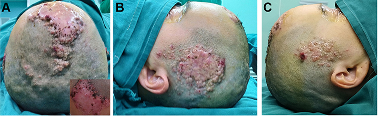 |
Figure 1 Three well-delimited, infiltrated and hard plaques involving the scalp (A), left temple (B) and right temple (C). |
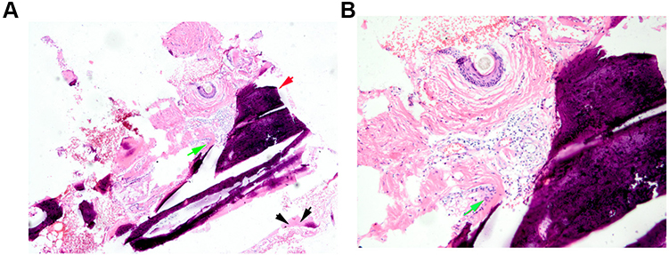 |
Figure 2 Biopsy, H&E staining, and lamellar bone tissue distributed in the dermis (A). Osteocytes were in the bone tissue, and osteoblasts were seen around bone tissue (B). Black arrows: osteoblast; Green arrows: osteoclast; Red arrow: Lamellar bone tissue. |
Discussion
PLOC is a special type of primary skin osteoma that occurs without underlying skin diseases and abnormal calcium and phosphorus metabolism.3 Plaque-like osteoma was first identified in 1978 by Worret and Burgdorf and has the following characteristics: ① skin lesions appear at birth or in the first year after birth; ② at one bone plate appears with or without other skin osteomas; ③ no abnormal calcium or phosphorus metabolism; and ④ no infection or trauma or other triggering events.4 Our patient had no underlying endocrine or genetic disorders, and the lesions met these diagnostic criteria. This disease needs to be differentiated from multiple miliary osteoma cutis on the face. The latter is often secondary to female patients with severe acne. The clinical manifestations are multiple small changes in skin tones or light blue firm papules on the face.5
PLOC mostly occurs in childhood, and there have been a few reports of onset after puberty in recent years.6 Skin osteoma is most commonly found on the head,1,6 followed by the face.5,7 For the first time, we report a case of a giant and multiple PLOC in an adult patient. Since the onset of the condition in the patient, the skin lesions have gradually increased and merged into plate-like plaques. Long-term follow-up observations are still needed to determine the progression to deeper levels of tissue.
The current treatment methods for osteoma cutis include topical retinoids (0.025–0.1%), CO2 laser, and surgical excision, etc.7–9 Some researchers have reported using microneedle trephine to treat miliary osteoma cutis of the face, with relatively small skin trauma and less pigmentation.10
If ossification has formed, surgical excision of new bone is still the main treatment method. The patient’s skin lesions were large and scattered, and dilator implantation and staged surgery were considered. In view of aesthetic issues, the dilator was implanted first (Figure 3); then, the large skin lesions on the forehead and top of the head were surgically removed and stitched with palm-flap transfer repair and adjacent flap transfer (Figure 4). Six months after surgery, there was no recurrence (Figure 5). Postoperative patients need to regularly monitor their serum calcium and phosphate, renal function, parathyroid hormone and vitamin D levels and other related indicators using skin CT or skin ultrasound a.1
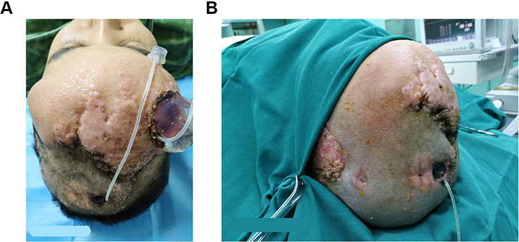 |
Figure 3 One month after implantation of the dilator. (A) The total amount was 300mL when the water was injected for one month, and the dilator was partially exposed, and the local aseptic bandage was performed. (B) The condition of the skin flap when the expander is removed three months later. |
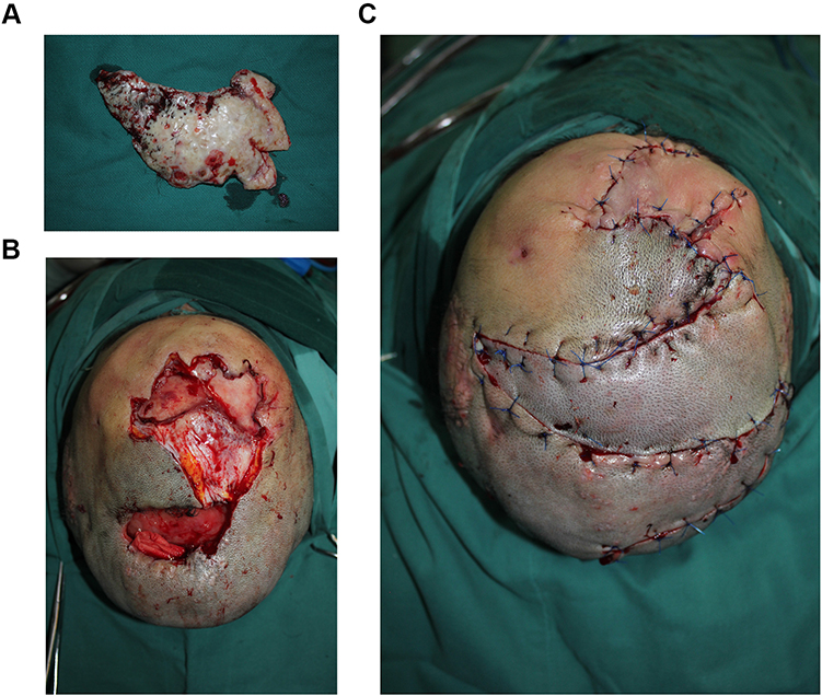 |
Figure 4 Surgical removal of large skin lesions (A), surgical incisions (B), postoperative sutures (C). |
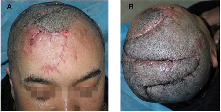 |
Figure 5 A light red striped scar can be seen on the forehead (A) and top of the head (B) after surgery. |
In summary, we report a patient with three lesions of plaque-like osteoma cutis, which were acquired in adulthood. The first treatment option for giant plate-like cutaneous osteoma is dilator implantation and fractional surgery, which have good results and a low recurrence rate.
Data Sharing Statement
The data that support the findings of this study are available from the corresponding author upon reasonable request.
Ethics Approval and Consent to Participate
This study was approved by the Research Ethics Committee of Dermatology Hospital, Southern Medical University and Kashgar Prefecture First Peoples Hospital. The study was written informed consent was obtained from patient. The patient gave informed consent for the publication of their case details and any accompanying images. The study protocol was complied with the 1975 Declaration of Helsinki.
Funding
No funding sources.
Disclosure
The authors declare no conflicts of interest.
References
1. Gloria O, Roberto S, Stefano P. A case of secondary osteoma cutis associated with lichen planopilaris. Acta Derm Venereol. 2019;99(12):1190–1191.
2. Huh JY, Kwon MJ, Seo KY, Kim MK, Kim DH. Novel nonsense GNAS mutation in a 14‐month‐old boy with plate‐like osteoma cutis and medulloblastoma. J Dermatol. 2014;41(4):319–321. doi:10.1111/1346-8138.12284
3. Hernandez-Martin A, Perez-Mies B, Torrelo A. Congenital plate-like osteoma cutis in an infant. Pediatr Dermatol. 2009;26(4):479–481. doi:10.1111/j.1525-1470.2009.00962.x
4. Worret WI, Burgdorf W. [Congenital, plaque-like osteoma of the skin in an infant]. Hautarzt. 1978;29(11):590–596. German.
5. Painsi C, Tarmann R, Georg WF, et al. Multiple miliary osteoma cutis of the scalp. J Dtsch Dermatol Ges. 2015;13(11):1185–1187.
6. Orme CM, Hale CS, Meehan SA, Long W. Plate-like osteoma cutis. Dermatol Online J. 2014;20(12).
7. Duarte BM, Pinheiro RR, Cabete J. Multiple miliary osteoma cutis: a comprehensive review and update of the literature. Eur J Dermatol. 2018;28(4):434–439.
8. Kim D, Franco GA, Shigehara H, Asaumi J, Hildenbrand P. Benign miliary osteoma cutis of the face: a common incidental CT finding. Am J Neuroradiol. 2017;38(4):789. doi:10.3174/ajnr.A5096
9. Berbert ALCV, Mantese SADO, Hiraki KRN, Loyola AM, Queiroz NP. Multiple cutaneous miliary osteomas of the face: a case report. Surg Cosmet Dermatology. 2012;4(4):360–363.
10. Baskan E, Turan H, Tunali S, Toker SC, Adim SB, Bolca N. Miliary osteoma cutis of the face: treatment with the needle microincision-extirpation method. J Dermatolog Treat. 2007;18(4):252–254. doi:10.1080/09546630701287878
 © 2021 The Author(s). This work is published and licensed by Dove Medical Press Limited. The full terms of this license are available at https://www.dovepress.com/terms.php and incorporate the Creative Commons Attribution - Non Commercial (unported, v3.0) License.
By accessing the work you hereby accept the Terms. Non-commercial uses of the work are permitted without any further permission from Dove Medical Press Limited, provided the work is properly attributed. For permission for commercial use of this work, please see paragraphs 4.2 and 5 of our Terms.
© 2021 The Author(s). This work is published and licensed by Dove Medical Press Limited. The full terms of this license are available at https://www.dovepress.com/terms.php and incorporate the Creative Commons Attribution - Non Commercial (unported, v3.0) License.
By accessing the work you hereby accept the Terms. Non-commercial uses of the work are permitted without any further permission from Dove Medical Press Limited, provided the work is properly attributed. For permission for commercial use of this work, please see paragraphs 4.2 and 5 of our Terms.