")
Back to Journals » OncoTargets and Therapy » Volume 8
Primary malignant lymphoma of the uterus and broad ligament: a case report and review of literature
Authors Chen R, Yu Z, Zhang H, Ding J, Chen B
Received 26 November 2014
Accepted for publication 20 December 2014
Published 29 January 2015 Volume 2015:8 Pages 265—268
DOI https://doi.org/10.2147/OTT.S78171
Checked for plagiarism Yes
Review by Single anonymous peer review
Peer reviewer comments 3
Editor who approved publication: Professor Daniele Santini
Runzhe Chen, Zhengping Yu, Hongming Zhang, Jiahua Ding, Baoan Chen
Department of Hematology and Oncology (Key Department of Jiangsu Medicine), Zhongda Hospital, Medical School, Southeast University, Nanjing, Jiangsu Province, People’s Republic of China
Abstract: Primary malignant lymphoma of the uterus and broad ligament is rare. Here, we present a rare case of primary diffuse large B-cell lymphoma (DLBCL) of uterus and broad ligament in a 63-year-old female. The patient presenting with lower abdominal distention was referred to our hospital. Subsequent abdominal and pelvic ultrasound revealed the presence of a large mass, which was highly suspected as subserosal uterine leiomyoma. A large tumor was found with unclear boundary with right posterior wall, broad ligament and bilateral adnexa during surgery. Her uterus and the tumor of a broad ligament and bilateral adnexa were all excised as a result. Postoperative pathological examination showed DLBCL in uterus and broad ligament. Further examinations excluded metastatic diseases, which supported the diagnosis of primary DLBCL of the uterus and broad ligament. The patient received six cycles of R-CHOP (21 days) regimen. During the 8 months follow-up, no evidence of disease recurrence was identified. As the prevalence of primary extranodal lymphoma is increasing, the details of this rare case may highlight the importance and facilitate treatment of similar diseases. A summary focusing on the presentation and prognosis as well as a review of current management is also discussed.
Keywords: diffuse large B-cell lymphoma, primary uterine and broad ligament lymphoma, extranodal lymphoma, diagnosis, therapy
Introduction
Primary malignant lymphoma of the genital tract is a rare disease accounting for only 1% of extranodal lymphomas, and the frequent sites occurring includes in the ovary, uterus, cervix, vagina, and the vulva.1 Over the 30 years, less than 150 cases of primary uterine lymphoma have been reported.2 The clinical presentations of primary cervical lymphoma are very similar to that of leiomyoma, which often include abnormal bleeding and have a large, bulky cervix on pelvic examination.1 As a result, it is very important to make the correct diagnosis of this uncommon disease, as the treatment of cervical lymphomas differs from more common primary cervical cancers.3 In order to help clinicians be aware of this diagnosis when encountering a cervical mass, aid them in treating similar patients, and highlight the importance of this disease, we report a rare case of primary uterine and broad ligament malignant lymphoma misdiagnosed as leiomyoma, and the associated literature is also discussed.
Case report
A 63-year-old multiparous female, post-menopausal for 15 years, was presented to the Department of Obstetrics and Gynecology of the Affiliated Zhongda Hospital of Southeast University complaining of a 2-month history of abdominal bloating, increased abdominal girth, and difficulty with urination. Vaginal examination showed the same size as the 10-month uterus of the mass with extension and fixation to the posterior cervix. The other clinical examination was unremarkable. The patient’s review of systems was negative for fever, chills, weight loss, night sweats, nausea, vomiting, or bowel or bladder symptoms, and her past medical and surgical history was unremarkable. Subsequent abdominal and pelvic ultrasound revealed the presence of a 15.9 cm ×12.0 cm ×8.6 cm mass, with no definite evidence of local infiltration and lymphadenopathy, which was highly suspected as subserosal uterine leiomyoma (Figure 1). Considering the large size of the leiomyoma, hysteromyomectomy was performed. During the surgery, a large, solid mass and dumbbell-shaped mass were detected, with diameters of 14 cm and 8 cm, respectively. The boundary of the mass was not clear with right posterior wall, broad ligament, and bilateral adnexa. Since malignant tumor was considered, her uterus, tumor of broad ligament, and bilateral adnexa were all excised. Unexpectedly, a diagnosis of diffuse large B-cell lymphoma (DLBCL) was determined as a result of the postoperative immunohistochemical profile analysis of the mass and broad ligament. Immunohistological examination (Figure 2) revealed CD3 (−), CD5 (+/−), CD7 (−), CD10 (+), CD20 (+), CD30 (−), CD79a (+), Bcl-6 (+), EBER (−), Ki67 approximately 80% (+), LCA (+), Mum-1 (−), PAX5 (+), and Pan CK (−), and suggested putative germinal center B-cell origin. Thus, finally, the diagnosis of DLBCL of uterus and broad ligament was established, and the patient was referred to the Department of Hematology and Oncology. Bone marrow biopsy was negative for the evidence of lymphoma. Detection results of lactate dehydrogenase, serum electrolytes, liver function, serum electrophoresis, immunoglobulins, cancer marker screening, and complete blood counts were all found unremarkable. A staging chest computed tomography (CT) scan only showed very small (<1 cm) mediastinal lymph nodes. Positron emission tomography (PET)/CT scan showed flourodeoxyglucose-18 (FDG)-avid disease affecting the cervix and uterus with partial obstruction of the right ureter. She was considered to have Ann Arbor stage IEA (stage 1 extranodal without B symptoms) DLBCL with high intermediate of the international prognostic index (risk factors: age, stage, and more than one extranodal involvement). Since the tumor was more than 10 cm large, she was treated with six cycles of R-CHOP (21 days interval) chemotherapy (combination rituximab – a monoclonal antibody directed against the CD20 antigen with cyclophosphamide, doxorubicin, vincristine, and prednisolone). A restaging PET/CT scan showed complete response with disappearance of the previously FDG avid pelvic mass, and her CT scan of the abdomen and pelvis became normal after four cycles of R-CHOP. No major side effects were experienced during the course of treatment. During the 8 months of follow-up, no evidence of disease recurrence was identified. At present, the patient is scheduled for regular follow-up appointments.
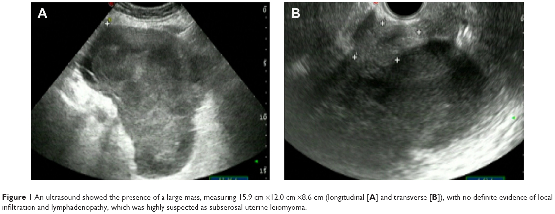 | Figure 1 An ultrasound showed the presence of a large mass, measuring 15.9 cm ×12.0 cm ×8.6 cm (longitudinal [A] and transverse [B]), with no definite evidence of local infiltration and lymphadenopathy, which was highly suspected as subserosal uterine leiomyoma. |
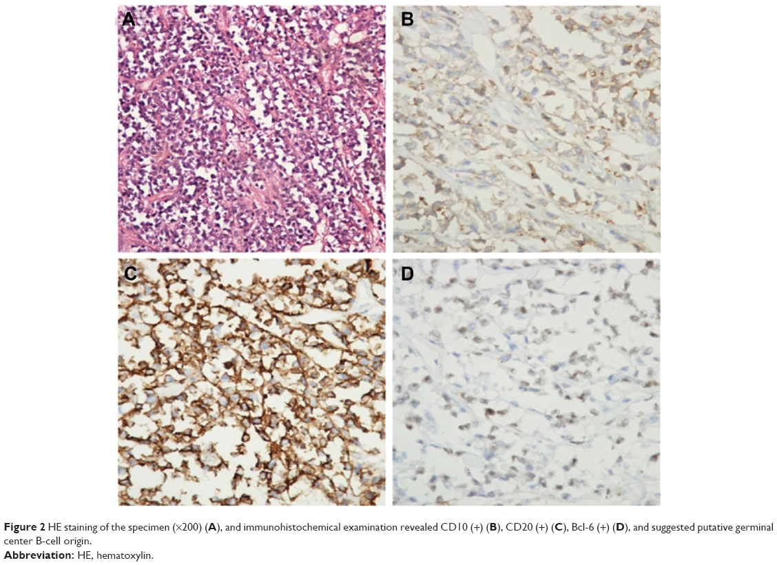 | Figure 2 HE staining of the specimen (×200) (A), and immunohistochemical examination revealed CD10 (+) (B), CD20 (+) (C), Bcl-6 (+) (D), and suggested putative germinal center B-cell origin. |
Discussion
Primary malignant lymphoma of the gynecologic organs is a very rare occurrence. The clinical workup leading to the appropriate diagnosis is also very challenging. Patients tend to be asymptomatic or have nonspecific abdominal symptoms, including abdominal bloating, pressure, and discomfort. According to reports, only 17% of non-Hodgkin’s lymphoma patients have constitutional symptoms such as fever, night sweats, and weight loss.1
The most common presenting feature of primary uterine lymphoma is bleeding from the vagina, which is similar to that of cervical carcinoma.2 As a result, this type of lymphoma is always neglected by physicians, leading to incorrect diagnosis such as sarcoma, chronic inflammation, or poorly differentiated carcinoma.4 The diagnosis of primary uterine lymphoma could be made by various procedures, including abnormal Pap smear, cervical or endometrial curettage, biopsy, conization, and hysterectomy.5 Hysterectomy is not recommended, and it is important for gynecological oncologists to be aware of this neoplastic disease and to include cervical or vaginal lymphoma in the differential diagnosis of patients presenting with examinations consistent with cervical or vaginal cancer. Therefore, a correct diagnosis could lead to the appropriate therapy, and radical gynecological surgery can be avoided for primary cervical and vaginal lymphoma.6,7 Immunohistochemical examination is necessary to determine the type and subtype of lymphoma,8 and most primary lymphomas are in Ann Arbor stage IE or IIE.9 In stages III and IV, it is sometimes difficult to determine whether uterine involvement is primary or secondary to disseminated lymphoma.10 The lymphoma stage of our patient is IV EA, because of focal infiltration to uterine adnexa of DLBCL and the patient having no B symptoms. CT could facilitate accurate measurement of both tumor size and extent, and provide useful information to plan an appropriate therapeutic regimen and follow response to treatment. In CT, the lesion usually appears as an enhancing mass in the uterine cervix, and no calcification could be detected. Since these lesions are often bulky, involvement of the vagina, bladder, rectum, and adjacent lymph nodes should also be considered. In cases of primary uterine lymphoma including ours, however, bulky lymphadenopathy is rarely present.11 The latest technique for assessing lymphoma is FDG-PET, which allows the detection of viable tumor cells independent of morphology and differential diagnosis of the disease, and areas in which the utility of FDG-PET imaging is being evaluated include staging of disease, treatment monitoring, and detection of recurrence.11,12
Optimal treatment for primary DLBCL of uterine and broad ligament in the elderly has not been so clearly defined, as a consequence of its low incidence.8 R-CHOP with or without radiotherapy is recommended as the first-line therapy. Though rituximab may cause an additional cost compared with CHOP alone as the first-line treatment of DLBCL, it is also associated with an increased life expectancy.13–15 Besides, according to a recent research,8 a full course of R-CHOP treatment and an abbreviated R-CHOP with radiation resulted in similar survival, but the latter may be better tolerated than the former. Combination chemotherapy with tailored radiotherapy is the preferred treatment option for patients for tumors more than 10 cm in diameter or in advance stage. In our case, after only receiving six cycles of R-CHOP regimen, a restaging PET/CT scan of the patient showed complete response and her CT scan of the abdomen and pelvis was normal, so the patient was not given radiotherapy. R-CHOP was proven effective, as significant improvement was observed in our patient with no evidence of metastasis up to date. However, specialists still recommend regular follow-up of these women by the hematologists and gynecological oncologists to monitor systemic and local relapse, respectively.5
Conclusion
Primary malignant lymphoma of uterus and broad ligament is a rare disease, and the diagnosis is typically made with an immunohistochemical analysis. The first-line therapy of DLBCL is R-CHOP with or without radiotherapy. This presentation shows that primary uterine lymphoma presents with symptoms similar to that of more common gynecologic diseases, including leiomyoma; highlights how important it is when diagnosing a patient presenting with common symptoms in gynecology; and reviews the diagnostic therapy of primary DLBCL in uterus and broad ligament.
Acknowledgments
This work was supported by the National Natural Science Foundation of People’s Republic of China (grant no 81170492 and 81370673), National High Technology Research and Development Program 863 of People’s Republic of China (grant no 2012AA022703), National Key Basic Research Program 973 of People’s Republic of China (grant no 2010CB732404), Key Medical Projects of Jiangsu Province (grant no BL2014078), and Key Discipline of Jiangsu Province (2011–2015). Sincere gratitude is given to Dr Pei Zhang for his technical assistance.
Disclosure
The authors have no conflict of interest.
References
Ahmad AK, Hui P, Litkouhi B, et al. Institutional review of primary non-Hodgkin lymphoma of the female genital tract: a 33-year experience. Int J Gynecol Cancer. 2014;24(7):1250–1255. | ||
Hariprasad R, Kumar L, Bhatla DM, Kukreja M, Papaiah S. Primary uterine lymphoma. Report of 2 cases and review of literature. Am J Obstet Gynecol. 2006;195(1):308–313. | ||
Chan JK, Loizzi V, Magistris A, et al. Clinicopathologic features of six cases of primary cervical lymphoma. Am J Obstet Gynecol. 2005;193 (3 pt 1):866–872. | ||
Kendrick JE 4th, Straughn JM Jr. Two cases of non-Hodgkin’s lymphoma presenting as primary gynecologic malignancies. Gynecol Oncol. 2005;98(3):490–492. | ||
Upanal N, Enjeti A. Primary lymphoma of the uterus and cervix: two case reports and review of the literature. Aust N Z J Obstet Gynaecol. 2011;51(6):559–562. | ||
Ustaalioglu BB, Bilici A, Seker M, et al. Primary non-Hodgkin lymphoma of cervix successfully treated with rituximab: positron emission tomography images before and after therapy: a case report. Leuk Res. 2010;34(4):e108–e110. | ||
Cohn DE, Resnick KE, Eaton LA, deHart J, Zanagnolo V. Non-Hodgkin’s lymphoma mimicking gynecological malignancies of the vagina and cervix: a report of four cases. Int J Gynecol Cancer. 2007;17(1):274–279. | ||
Odejide OO, Cronin AM, Davidoff AJ, LaCasce AS, Abel GA. Limited stage diffuse large B-cell lymphoma: comparative effectiveness of treatment strategies in a large cohort of elderly patients. Leuk Lymphoma. 2014:1–9. | ||
Mandato VD, Palermo R, Falbo A, et al. Primary diffuse large B-cell lymphoma of the uterus: case report and review. Anticancer Res. 2014;34(8):4377–4390. | ||
Novotny S, Ellis T, Stephens J. Primary B-cell lymphoma of the cervix presenting with bilateral hydronephrosis. Obstet Gynecol. 2011;117 (2 pt 2):444–446. | ||
Mylam KJ, Kostakoglu L, Hutchings M, et al. FDG-PET/CT after one cycle of chemotherapy in patients with diffuse large B-cell lymphoma: results of a Nordic/US intergroup study. Leuk Lymphoma. 2014:1–28. | ||
Ab Hamid S, Wastie ML. Primary non-Hodgkin’s lymphoma presenting as a uterine cervical mass. Singapore Med J. 2008;49(3):e73–e75. | ||
Gisselbrecht C. Futility of relapsed diffuse large B cell lymphoma transplantation? Biol Blood Marrow Transplant. 2014;20(11):1667–1669. | ||
Khor S, Beca J, Krahn M, et al. Real world costs and cost-effectiveness of Rituximab for diffuse large B-cell lymphoma patients: a population-based analysis. BMC Cancer. 2014;14:586. | ||
Cunningham D, Hawkes EA, Jack A, et al. Rituximab plus cyclophosphamide, doxorubicin, vincristine, and prednisolone in patients with newly diagnosed diffuse large B-cell non-Hodgkin lymphoma: a phase 3 comparison of dose intensification with 14-day versus 21-day cycles. Lancet. 2013;381(9880):1817–1826. |
 © 2015 The Author(s). This work is published and licensed by Dove Medical Press Limited. The full terms of this license are available at https://www.dovepress.com/terms.php and incorporate the Creative Commons Attribution - Non Commercial (unported, v3.0) License.
By accessing the work you hereby accept the Terms. Non-commercial uses of the work are permitted without any further permission from Dove Medical Press Limited, provided the work is properly attributed. For permission for commercial use of this work, please see paragraphs 4.2 and 5 of our Terms.
© 2015 The Author(s). This work is published and licensed by Dove Medical Press Limited. The full terms of this license are available at https://www.dovepress.com/terms.php and incorporate the Creative Commons Attribution - Non Commercial (unported, v3.0) License.
By accessing the work you hereby accept the Terms. Non-commercial uses of the work are permitted without any further permission from Dove Medical Press Limited, provided the work is properly attributed. For permission for commercial use of this work, please see paragraphs 4.2 and 5 of our Terms.