")
Back to Journals » Neuropsychiatric Disease and Treatment » Volume 11
Catatonia in Down syndrome; a treatable cause of regression
Authors Ghaziuddin N, Nassiri A, Miles J
Received 10 November 2014
Accepted for publication 27 January 2015
Published 2 April 2015 Volume 2015:11 Pages 941—949
DOI https://doi.org/10.2147/NDT.S77307
Checked for plagiarism Yes
Review by Single anonymous peer review
Peer reviewer comments 4
Editor who approved publication: Dr Roger Pinder
Neera Ghaziuddin,1 Armin Nassiri,2 Judith H Miles3
1Department of Psychiatry, University of Michigan, Ann Arbor, Michigan, 2Community Psychiatry, San Jose, California, 3Thompson Center for Autism and Neurodevelopmental Disorders and Department of Child Health, University of Missouri, Columbia, Missouri, USA
Objective: The main aim of this case series report is to alert physicians to the occurrence of catatonia in Down syndrome (DS). A second aim is to stimulate the study of regression in DS and of catatonia. A subset of individuals with DS is noted to experience unexplained regression in behavior, mood, activities of daily living, motor activities, and intellectual functioning during adolescence or young adulthood. Depression, early onset Alzheimer’s, or just “the Down syndrome” are often blamed after general medical causes have been ruled out. Clinicians are generally unaware that catatonia, which can cause these symptoms, may occur in DS.
Study design: Four DS adolescents who experienced regression are reported. Laboratory tests intended to rule out causes of motor and cognitive regression were within normal limits. Based on the presence of multiple motor disturbances (slowing and/or increased motor activity, grimacing, posturing), the individuals were diagnosed with unspecified catatonia and treated with anti-catatonic treatments (benzodiazepines and electroconvulsive therapy [ECT]).
Results: All four cases were treated with a benzodiazepine combined with ECT and recovered their baseline functioning.
Conclusion: We suspect catatonia is a common cause of unexplained deterioration in adolescents and young adults with DS. Moreover, pediatricians and others who care for individuals with DS are generally unfamiliar with the catatonia diagnosis outside schizophrenia, resulting in misdiagnosis and years of morbidity. Alerting physicians to catatonia in DS is essential to prompt diagnosis, appropriate treatment, and identification of the frequency and course of this disorder.
Keywords: electroconvulsive therapy, benzodiazepines, adolescence, young adulthood
Introduction
Down syndrome (DS), the most common genetic disorder in the USA, occurs in around one in 800 live births, affording a prevalence of 83,000 children.1 DS is caused by trisomy 21 due either to chromosome nondisjunction (94%) or, less often, translocations. Though not well documented in the literature, adolescents and young adults with DS have been reported to suffer from functional deterioration.2–8 When medical illnesses, including seizures, sleep apnea, anemias, and thyroid and cardiac disease have been ruled out, regressive symptoms are typically attributed to depression, early onset Alzheimer’s, or just the progression of DS.2–4,9,10
Here, we present evidence that catatonia not only occurs in the adolescents reported, but also very likely explains much of the functional deterioration previously reported. Unlike dementia, which shows progressive, irreversible decline in memory, intellectual functioning, and personality, DS patients with catatonia respond to treatment and may even regain baseline functioning when treated appropriately. We suggest that DS patients as a group may be at heightened risk for developing catatonia and that early recognition by the broad medical community is key to successful treatment.
“Catatonia” is a neuropsychiatric syndrome that typically presents with motor signs and has a characteristic response to benzodiazepines and electroconvulsive therapy (ECT).11 The main symptoms of catatonia are a change in motor activity (reduced or less often increased motor activity), unusual movements (stereotypies, grimacing, freezing, ambitendency, infrequent blinking, motor or vocal tics, posturing, automatic obedience), changes in speech (reduced meaningful speech; mutism; echolalia; “verbigeration”, or senseless repetition of words or phrases; increased latency), changes in oral intake (reduced appetite and/or slowing down of food intake), decline in activities of daily living (ADL), bladder or bowel incontinence, negativism, and disruptions in cognition. Symptoms may present with dysregulation of mood; sleep; and the appearance of psychotic, quasi-psychotic, or obsessional preoccupations.12 The condition may be diagnosed based on eliciting relevant history; examination of motor and other symptoms; and completion of a standardized rating scale, such as the Bush-Francis Catatonia Rating Scale (BFCRS), which measures the severity of catatonia symptoms.13
Historically, catatonia was regarded as a complication of schizophrenia. However, recent evidence indicates catatonia’s association with schizophrenia is merely historical and that most cases diagnosed in a psychiatric setting are associated with mood disorders.14 Though catatonia was described as linked with neurological and medical conditions since its description,15 only in the last decade has catatonia been associated with autoimmune, infective, metabolic, and genetic disorders. There is preliminary evidence, using positron emission tomography scan of the brain, that the underlying pathophysiological process may include hypometabolism of the occipitotemporal and thalamic regions.16 The recently published Diagnostic and Statistical Manual of Mental Disorders: DSM-5 (DSM-5) includes “catatonia NOS [not otherwise specified]” (298.89), which was proposed by a group of experts as a unique diagnostic category,17 in the absence of other psychopathology.18 Some practitioners in the field also regard this as a temporary diagnosis, to allow for treatment to proceed until another underlying disorder can be identified.19 These changes in the understanding of catatonia have not reached most physicians, due to catatonia’s entrenched historical association with schizophrenia and because almost all reports are in the psychiatric literature.20,21
We previously reported DS and catatonia in two adolescent females.22 We now report four additional cases and their treatment outcomes. All four individuals are diagnosed with trisomy 21 DS and comorbid unspecified catatonia. and have been successfully treated with ECT due to the failure to achieve baseline function with a benzodiazepine alone. All four cases were treated with ECT due to failure to achieve baseline function with a benzodiazepine alone. ECT was used based on the American Academy of Child and Adolescent Psychiatry guidelines.23 Bilateral electrode placement was used in all cases using the MECTA device (MECTA Corporation, Tualatin, OR, USA) or the Thymatron®. Induction medications used were methohexital, succinylcholine, and glycopyrrolate.
Methods
Institutional review board approval was obtained at both institutions, data collected are part of standard clinical care, and fictitious identifiers are used in describing the cases.
Cases
Table 1 summarizes the demographics; Table 2, the presenting symptoms; Table 3, the medical findings; and Table 4, the treatment and clinical outcomes following benzodiazepine treatment and ECT of the four patients described in this report.
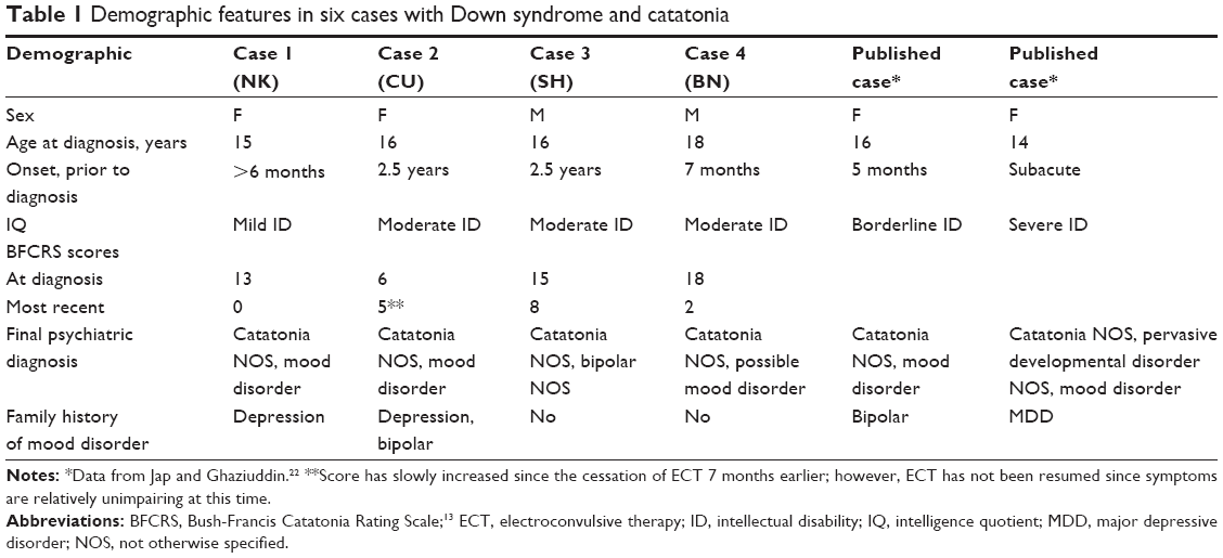 | Table 1 Demographic features in six cases with Down syndrome and catatonia |
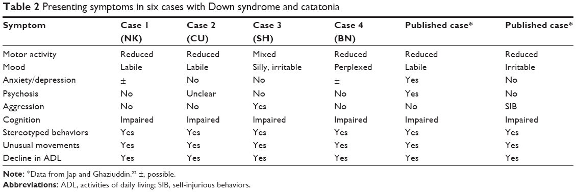 | Table 2 Presenting symptoms in six cases with Down syndrome and catatonia |
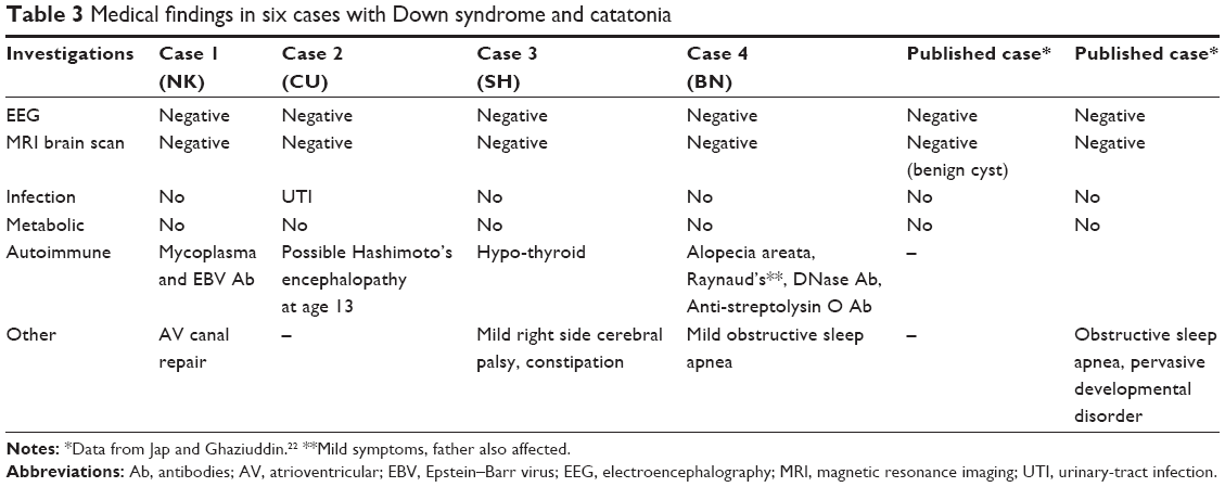 | Table 3 Medical findings in six cases with down syndrome and catatonia |
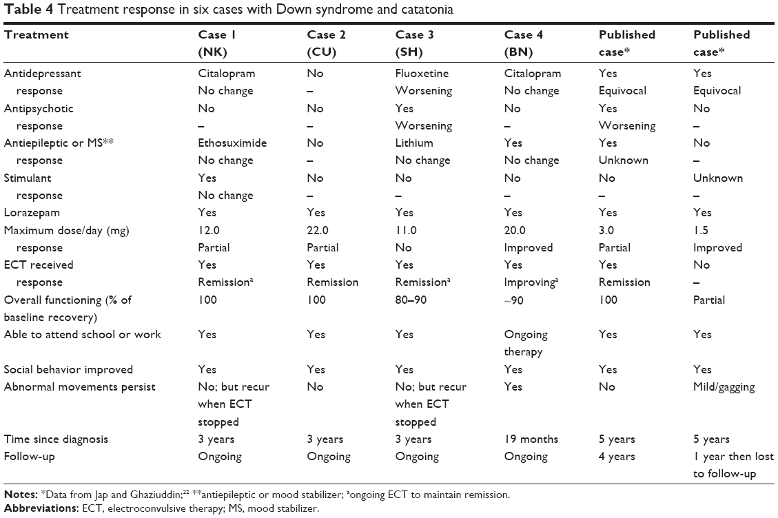 | Table 4 Treatment response in six cases with Down syndrome and catatonia |
Case 1: NK
NK is a Caucasian female, 15 years old, in ninth grade at presentation, with mild intellectual disability (ID), (intellectual quotient [IQ] =55–69), atrioventricular canal repair at age 6 months, and no previous psychiatric history. She presented with a 6-month history of gradual development of slow motor activity, reduced speech with increased latency, unusual movements such as repetitive movements and posturing, blunted and inappropriate affect, mood lability, sleep disturbance, slow eating, and a general decline in previously attained skills, all of which supported the catatonia diagnosis. Family history was significant for maternal anxiety. Previous medical work-up included brain magnetic resonance imaging (MRI), electroencephalograms (EEGs), computed tomography scan of sinuses, thyroid function tests, and polysomnogram to rule out sleep apnea, all with normal findings. The only exceptions were evidence of previous Mycoplasma and Epstein–Barr virus infections. Recent stressors included transition to high school and the death of a grandmother a year earlier.
At presentation, she had received low-dose clonazepam for 1 month with some improvement in symptoms. Previous medication trials included ethosuximide for 2 weeks (no effect); a trial of lisdexamfetamine (resulted in agitation, mood and sleep dysregulation, and possible hallucinations); and citalopram 20 mg, which may have led to further decline in ADL. Based on this presentation, NK was diagnosed with unspecified catatonia.24 Treatment with lorazepam 1.5 mg three times daily was initiated, and gradually titrated to a total dosage of 12 mg daily with some improvement in functioning. However, psychomotor slowing and mood lability persisted and was impairing. She was therefore hospitalized and started on ECT; lorazepam was continued and citalopram discontinued. She responded well to ECT and continued a tapering course of ECT, ending having had a total of 26 treatments. During this time, she was also started on lithium carbonate and amiloride 5 mg for renal protection. When her symptoms recurred 3 months later, she received a second course of 21 bilateral ECT over 7 months with 100% recovery.
She continues to receive maintenance ECT at 2-week intervals, primarily to control motor symptoms, and has received a total of 73 treatments over the past 31 months. Lithium was discontinued due to lack of response and side effects (polyuria and weight gain).
Case 2: CU
CU is a Caucasian female, aged 16 years old at presentation, with moderate ID (IQ =40–54), no previous psychiatric history, and a family history of anxiety in a first-degree relative. At baseline, CU attended school and functioned at preschool level. Recent stressors included parental marital stress and family relocation. Symptoms were first noted 2.5 years prior to presentation, when she was diagnosed with a presumed Hashimoto’s encephalopathy at an outside institution. At that time, her symptoms included grimacing, eye blinking, and decline in functioning. She was noted to have elevated thyroglobulin antibodies (711 IU/mL) and marginally increased thyroid peroxidase antibodies (45 units/mL) but normal thyroid stimulating hormone, and free T4 and T3 levels. Routine spinal fluid analysis, EEGs, and brain MRI were normal. Oral prednisone was initiated with some improvement, although it is unclear how long she remained well. Two years later, she presented to our institution, with reoccurrence of symptoms of 7 months’ duration. Symptoms included periods of prolonged immobility, agitation, repetitive and darting eye movements, involuntary movements of the mouth and flicking movements of her fingers, depressed mood, decline in ADL, apparent loss of hedonic capacity, staring, and decreased speech output.
In addition to routine medical tests, thyroid functions, thyroglobulin antibody, and thyroid microsomal antibody; single-nucleotide polymorphism chromosomal microarray; creatinine phosphokinase; hepatitis A, B and C antibodies; paraneoplastic antibody (immunoglobulin G N-methyl-D-aspartate receptor); 25 hydroxy vitamin D; brain MRI; and, EEG disclosed no significant abnormalities.
CU was diagnosed with unspecified catatonia.24 Symptoms supporting the catatonia diagnosis included reduced motor activity, episodes of increased activity, grimacing, stereotyped finger movements, staring, reduced speech, and decline in skill level. Mood symptoms, not specific to the catatonia diagnosis, were also noted. An oral challenge dose of lorazepam 1 mg resulted in an immediate but short-lived improvement. Lorazepam was gradually increased to a maximum dose of 5.5 mg four times daily with limited improvement in symptoms. Lamotrigine was later added and titrated up to 150 mg daily for mood stabilization with no significant impact. Due to progressive decline, ECT was initiated. At the time of writing this report, CU has received 86 bilateral ECT treatments over a 6-month period with notable improvement. Her mother believes CU has regained 100% of her baseline functioning. There are no reported symptoms 5 months after discontinuing ECT. She continues to receive lorazepam 10 mg/day and memantine 20 mg/day and is closely monitored for recurrence of symptoms.
Case 3: SH
SH is a biracial male, aged 16 years old at presentation, with moderate ID (IQ =40–54) whose only medical problems were hypothyroidism diagnosed at age 5 years, constipation, and mild congenital right-sided cerebral palsy. At 13 years of age, SH developed uncharacteristic aggression and an inability to perform previously mastered tasks. Numerous antipsychotic agents were tried, all of which failed to stop his progressive decline. His parents and referring clinician attributed his decline to a trial of fluoxetine that they associated with periods of immobility lasting several minutes, chewing but not swallowing, inability to complete simple tasks, and high-frequency self-injurious behaviors. These symptoms improved but did not reverse upon discontinuation of fluoxetine. At 16 years of age, SH presented for a second opinion due to acute exacerbation of symptoms in the previous 8 to 10 weeks. Presenting symptoms included increased motor activity interspersed with episodic cessation of all activity for a few seconds, vocal stereotypies, staring spells, jerky head movements (tics), unprovoked aggression, reduced sleep, slowed and reduced food intake, and weight loss.
SH was diagnosed with unspecified catatonia.24 Symptoms supporting the diagnosis included episodes of both reduced and increased activity, periodic cessation of all motor activity, repetitive and meaningless utterances (also known as “verbal stereotypies” or “verbigeration”), staring, decline in skills, and a markedly slow food intake rate that resulted in progressive weight loss. Laboratory tests (complete blood count, comprehensive metabolic panel, thyroid and liver function tests, antinuclear antibodies, thyroid antibodies, 24-hour EEGs with video recording, and MRI) were within normal limits.
Lorazepam treatment was initiated but a maximum dose of 11 mg/day did not result in consistent improvement. Subsequently, he was treated with bilateral ECT with excellent response. The parents believe that he has regained 100% of his baseline function. At the time of writing this report, he has received a total 76 bilateral electrode placement (BL ECT) over the past 18 months and continues to receive weekly maintenance treatment. So far, attempts to taper the frequency of ECT have resulted in return of symptoms. A trial of lithium, administered at a therapeutic dose and over a 1-year period, was discontinued due to the lack of additional benefit. A future trial with memantine may be considered, although this agent was not helpful in the past when administered concurrently with an antipsychotic agent.
Case 4: BN
BN is a Caucasian male with no previous psychiatric problems, who was aged 18 years old at presentation. His only medical problems were alopecia areata successfully treated at age 12 years and mild Raynaud’s phenomenon. Despite moderate-range ID (based on IQ testing), he demonstrated higher-than-expected adaptive functioning, including playing the piano, reading, and working part-time. Family history was positive for adult-onset hypothyroidism and Raynaud’s in the father. There was no history of stressors.
Symptoms emerged at age 17 years, 5 months, with progressive psychomotor slowing, episodic immobility, freezing, reduced speech and eye-blink rate, loss of hedonic capacity, grimacing and holding his tongue out, repeated shrugging and turning of the shoulders, slow eating, and stereotyped movements of the fingers. There were no sleep problems and minimal snoring. Neurological examination was normal except for overall motor slowing. Neurologic testing was unremarkable, including brain MRI, routine and 24-hour EEGs. Laboratory testing included comprehensive metabolic profile (CMP), complete blood count, B12, B6, and folate levels; iron, total iron binding capacity, and iron saturation; thyroid panel including anti-thyroglobulin and thyroid peroxidase antibodies; rapid plasma reagin (RPR); creatinine phosphate; ceruloplasmin; sedimentation rate; and a comprehensive immune function evaluation. Immune studies were positive for mildly elevated antinuclear, anti-streptococcal, and anti-Mycoplasma antibodies; elevated serum immunoglobulin G levels against dopamine D1 receptor, dopamine D2 receptor, and tubulin; and elevated serum induction of Ca2+/calmodulin-dependent kinase II in human neuronal cell lines.
A lorazepam (1.5 mg, IV) challenge resulted in marked improvement in motor symptoms lasting approximately 24 hours. Based on these findings and due to the lack of evidence to suggest another underlying medical or neuropsychiatric disorder, BN was diagnosed with unspecified catatonia.24 Symptoms supporting the catatonia diagnosis were progressive motor slowing, periodic cessation of all motor activity (freezing episodes), grimacing, posturing (a posture or movement maintained for a prolonged period), staring, loss of previously gained skills, markedly slowed food intake, negativism, and a loss of hedonic capacity. Lorazepam was titrated to a maximum dose of 20 mg/day resulting in 80% of baseline function. ECT was initiated due to failure to achieve baseline. At the time of writing, the patient has had an immediate and robust response to 12 bilateral treatments, with more spontaneous speech and fluidity of movement. The plan is to continue ECT until return to baseline function is attained.
Discussion
Here, we report four new cases of adolescents with DS who presented with unexplained regression and impairing symptoms across multiple domains (motor, speech, behavior, mood, and daily living skills) consistent with catatonia. Although mood symptoms were variably present, catatonia diagnosis was based on the presence of prominent motor and other regressive symptoms (ADL, speech, social). Furthermore, antidepressants failed to elicit a meaningful response in three of the new cases who underwent a selective serotonin re-uptake inhibitor trial (Table 4), and Case 3 experienced a marked deterioration. We report these cases in conjunction with data from our two published cases to illustrate similarities in presentation, treatment response, and outcomes.22 We suspect that catatonia may be a relatively common but unrecognized cause of reversible decline in adolescents with DS.
It would be valid for a reader to ask why these cases were not diagnosed with a primary affective disorder instead of catatonia. Affective symptoms were indeed present, which led us to include this in our final diagnoses (Table 1). These included sadness (Cases 2, 4); a labile mood (cases 1, 3); loss of hedonic capacity (Cases 1, 2, 4); impaired sleep (Cases 1–3); and reduced food intake, albeit related to reduced speed, chewing, or swallowing (Cases 1, 3). Ultimately, the catatonia diagnosis was made based on the predominance of motor symptoms in addition to the lack of response to antidepressant or mood stabilizers. Motor disturbances were prominent and impairing in each case and could not be explained on the basis of a primary affective illness. They included severe motor retardation (Cases 1–4); motor freezing (Cases 1, 2, 4); persistent or periodic agitation or increased motor activity (Cases 1–3); abnormal eye movements such as staring, reduced blink rate or eye flickering (cases 2, 3, 4); and a variety of other movements, such as holding out one’s tongue for no apparent reason (Case 3), finger stereotypies (Case 4), facial grimacing, or posturing (Cases 1, 2, 4). These motor and behavioral symptoms, which are considered pathognomonic for catatonia,25 indicated catatonia as the appropriate primary diagnosis. Periods of virtual immobility, extreme withdrawal alternating with periodic agitation, seen in Cases 1, 2, and 3, are also considered virtually pathognomonic of catatonia. Furthermore, the three cases treated with antidepressants prior to ECT either failed to respond or deteriorated, arguing against an atypical affective-disorder presentation. During ongoing ECT, we attempted trials of lithium in Cases 1 and 3, but this did not reduce their reliance on ECT and rather caused unacceptable side effects necessitating discontinuation. Ultimately, the diagnosis was supported by the fact that each individual only responded to the established anti-catatonic treatments benzodiazepine and ECT. These individuals experienced no significant medical complications prior to or during their anti-catatonia treatments. All were cared for at home by the involved families and despite their reduced speed, reduced food intake, and decline in functioning, remained well fed and were kept relatively active.
The diagnosis of unspecified catatonia in all cases was reached following comprehensive psychiatric and neurological examinations and laboratory investigations, which were nondiagnostic of other precipitating causes. One young woman (CU) who had been diagnosed with Hashimoto’s encephalitis 2.5 years prior to her regression and had responded to steroid treatment, showed no evidence of re-emergence of Hashimoto’s encephalitis on presentation to us. Of note, Hashimoto’s encephalitis is one of several autoimmune disorders that has been associated with development of catatonia.26 Subjects SH and BN had also been diagnosed previously with disorders commonly attributed to autoimmune activation (hypothyroidism, alopecia areata, Raynaud’s). Both SH and BN had positive immune responses to previous infections (Mycoplasma, Epstein–Barr, Streptococcus); BN had significant immune responses to neurotransmitter receptors. Though immune disorders are significantly more common in DS (hypothyroidism in 35% and alopecia areata in 6%) than in the general population, the significance of these co-occurring immune disorders and reactions is unclear. No other catatonia risk factors were identified.
Each of our patients responded as expected to established catatonia treatment protocols, including an affirmative response to benzodiazepines. All cases required ECT to achieve full recovery to baseline or close-to-baseline functioning, despite an initial response to a benzodiazepine. The need for ECT in patients with catatonia and DS to achieve and maintain full return to baseline is similar to that experienced by individuals with autism and other cognitive disabilities, including a recently reported woman with DS and catatonia in Australia.27–29 Unlike our standard protocol, in which we routinely compare pre-ECT objective memory with post-ECT functions, pre-ECT cognitive assessment was not possible due to the severity of symptoms prior to the start of treatment. However, in each case, the parents and teachers have resoundingly agreed that the patients have not only returned to their baseline functioning but also are continuing to acquire new information. Another noteworthy feature of response to ECT in this patient group is the patients’ need for prolonged maintenance ECT. Prolonged maintenance ECT has not been reported in the literature in typically developing individuals. Although, the cases we report achieved remission (defined as no or minimal symptoms), we also note a recurrence of symptoms when ECT is discontinued (with the exception of Case 2 and one of two previously published cases; Table 4). Need for such prolonged maintenance ECT raises questions about the possibility of difference in pathophysiological mechanisms, which may underlie catatonia in DS and other developmentally delayed individuals versus those with typical development. Experience is insufficient to speculate on whether this is due to patients’ underlying developmental disorders or to delays in recognition of catatonia and treatment initiation. The three cases who are undergoing maintenance ECT continue to live in their homes, are maintaining their baseline function with ongoing ECT and do not show any evidence of side effects. One previously published case, who was treated with ECT approximately 4 years earlier, is also functioning close to her baseline but without needing maintenance ECT.
We suggest that neither regression nor catatonia in DS is a rare or new phenomenon. Evidence is accumulating that a significant number of adolescents and young adults with DS experience life-changing regression.3 Prasher, in England, reported that “a significant minority” of young adults with DS between 15 and 30 years of age develop a “specific regressive/disintegrative disorder”.4 He observed gradual-but-severe deterioration in function, including loss of previously acquired skills in the areas of cognition; expressive and receptive language; mobility; adaptive and social skills; plus changes in personality, behavior, and mood. The individuals may become mute; withdrawn, with reduced social interaction; show decreased interests and activities; and mild-to-moderately severe low mood. Based on a lack of diagnostic clues and normal laboratory testing, imaging and EEGs, Prasher called the disorder “Young Adults with a Disintegrative Syndrome” (YADS). The similarities between catatonia and YADS are remarkable. Recently, Akahoshi et al in Japan, reported 13 DS individuals with good premorbid function who developed acute neuropsychiatric symptoms as adolescents or young adults.5 Their symptoms, including psychomotor slowness in 100% and stupor or catatonia in 15%, strongly suggest catatonia syndrome. Their official diagnoses were major depressive (54%), obsessive, psychotic NOS, anxiety, and adjustment disorders. Devenny et al examined the clinical records of children and adolescents with DS attending a medical clinic for individuals with ID. Of individuals who met the study criteria, 44% (14/34) showed evidence of severe regression, described as relatively rapid onset of decline in functioning following typical development for children with DS.30 Symptoms included loss of adaptive skills, cessation of learning, increased difficulty with communication and mutism, depression, anxiety, mood swings, noncompliance, withdrawal, hyperactivity, aggression, sleep disturbance, ritualistic behaviors, self-absorption, difficulties with transitions, incontinence, change in food habits, and obesity. Review of the early DS literature, primarily from physicians working in institutions for the mentally retarded, reveals the DS catatonia association was appreciated. In 1934, Earl reported “cataplexy” in six DS cases and suggested a tendency for DS individuals to display catalepsy.7 Rollin described catatonia in 28/73 institutionalized individuals with DS in 1946.8 These studies and personal communications from other clinicians indicate that, though not well studied, relatively abrupt and severe regression in previously acquired skills occurs in DS with a significant frequency. Nevertheless, the true risk for catatonia in adolescents and young adults with DS is unknown and awaits a systemic follow-up of all children followed in a group of DS clinics.
We suggest a number of factors conspired to thwart recognition of the DS/catatonia association, with the historical –albeit erroneous – classification of catatonia as a complication of schizophrenia the most liable.31 Only recently has a concerted effort by recognized experts in the study of catatonia32 revitalized the catatonia diagnosis and spurred the recent changes in the DSM-5.24 The DSM-5 now includes the diagnosis of “unspecified catatonia”, which is a useful category when there is no underlying neuropsychiatric or medical diagnosis. Unfortunately, the DSM-5 has failed to emphasize that the condition may occur across the age span, including in children and adolescents.
A number of entrenched misconceptions in the DS literature may also have contributed to the delayed recognition of catatonia. Foremost is the almost universal awareness that anatomical evidence of Alzheimer brain changes may occur in DS by age 40 years.33 Unfortunately, few are aware of recent studies indicating dementia in DS seldom occurs below the age of 50 and that Alzheimer symptoms in DS are qualitatively similar to Alzheimer symptoms of the general population.30,34–37 Lack of communication between specialists is also undoubtedly a factor. Almost all recent reports of catatonia have been published in psychiatry journals and the well-respected DS adult treatment guides neither discuss the risk of significant regression in adolescents and young adults nor mention catatonia.38–45 In addition, clinicians with little experience of DS commonly assume the symptoms reported by families are just part of DS, a phenomenon psychiatrists term “therapeutic blindness”.46
Recommendations and future perspectives
We offer persuasive preliminary evidence that clinicians should consider catatonia when presented with regression in adolescents and young adults with DS. Steps that assist with the diagnosis include familiarity with the symptoms, especially the psychomotor slowing and mutism. A lorazepam challenge test (1–2 mg orally or by intravenous infusion) done in the clinic may confirm the diagnosis. Since regression and catatonia both evoke numerous etiologies, an inclusive medical work-up is warranted for every patient presenting with regression. Thanks to medical progress, the life expectancy of persons with DS has increased from an average of 35 years in 1965 to about 60 years today.47 Recognition of catatonia, leading to prompt and appropriate treatment, has the potential of preventing prolonged morbidity and will allow us to prevent life-threatening complications including malignant catatonia.20,48
It must be kept in mind that catatonia is a syndrome defined on the basis of clinical symptoms and response to therapy. Though the underlying etiology is considered to be a disruption of the gamma-aminobutyric acid (GABA)/glutamate neurotransmission functions, especially within the basal ganglia and frontal lobe circuitry, the causes are unclear.21 Current evidence suggests a number of etiologic models including immune dysfunction, endocrine disruption, and underlying seizures.49 We expect that linking catatonia to DS, which is a well-studied genetic disorder, with data on autoimmune dysfunction, plus well-characterized mouse models, will speed research leading to understanding the catatonia syndrome, its etiologies, treatments, and prevention.50 Large-scale prospective studies are needed to identify the extent of both regression and catatonia in DS and identify optimum treatment protocols.
Disclosure
The authors declare no conflicts of interest in this work.
References
Shin M, Besser LM, Kucik JE, Lu C, Siffel C, Correa A; Congenital Anomaly Multistate Prevalence and Survival Collaborative. Prevalence of Down syndrome among children and adolescents in 10 regions of the United States. Pediatrics. 2009;124(6):1565–1571. | ||
Stein D, Munir K, Karweck A, Davidson E, Stein M. Developmental regression, depression, and psychosocial stress in an adolescent with Down syndrome. J Dev Behav Pediatr. 2013;34(3):216–218. | ||
Devenny DA, Matthews A. Regression: atypical loss of attained functioning in children and adolescents with Down syndrome. In: Hodapp RM, editor. International Review of Research in Developmental Disabilities. Vol 41. San Diego, CA; Waltham, MA; and London: Academic Press; 2011:233–264. | ||
Prasher V. Disintegrative syndrome in young adults. Ir J Psychol Med. 2002;19:101–102. | ||
Akahoshi K, Matsuda H, Funahashi M, Hanaoka T, Suzuki Y. Acute neuropsychiatric disorders in adolescents and young adults with Down syndrome: Japanese case reports. Neuropsychiatr Dis Treat. 2012;8:339–345. | ||
Krinsky-McHale S, Silverman W. Dementia and mild cognitive impairment in adults with intellectual disability: issues of diagnosis. Dev Disabil Res Rev. 2013;18(1):31–42. | ||
Earl CJ. The primitive catatonic psychosis of idiocy. Br J Med Psychol. 1934;14(3):230–253. | ||
Rollin HR. Personality in mongolism with special reference to the incidence of catatonic psychosis. Am J Ment Defic. 1946;51(2):219–237. | ||
Dykens EM. Psychiatric and behavioral disorders in persons with Down syndrome. Ment Retard Dev Diabil Res Rev. 2007;13(3):272–278. | ||
Capone G, Goyal P, Ares W, Lannigan E. Neurobehavioral disorders in children, adolescents, and young adults with Down syndrome. Am J Med Genet C Semin Med Genet. 2006;142C(3):158–172. | ||
Dhossche DM, Wachtel LE. Catatonia is hidden in plain sight among different pediatric disorders: a review article. Pediatr Neurol. 2010;43(5):307–315. | ||
Ghaziuddin N, Dhossche D, Marcotte K. Retrospective chart review of catatonia in child and adolescent psychiatric patients. Acta Psychiatr Scand. 2012;125(1):33–38. | ||
Bush G, Fink M, Petrides G, Dowling A. Catatonia. I. Rating scale and standardized examination. Acta Psychiatr Scand. 1996;93(2):129–136. | ||
Fink M, Shorter E, Taylor MA. Catatonia is not schizophrenia: Kraepelin’s error and the need to recognize catatonia as an independent syndrome in medical nomenclature. Schizophr Bull. 2010;36:314–320. | ||
Kahlbaum K. Die Katatonie oder das Spannungsirresein [Catatonia or tension insanity]. Berlin: Verlag August Hirschwald; 1874. German. | ||
Breker DN, Bohnen NI. Single case study of brain FDG PET imaging in a patient with catatonia. Clin Nucl Med. 2013;38(7):297–298. | ||
Francis A, Fink M, Appiani F, et al. Catatonia in Diagnostic and Statistical Manual of Mental Disorders, Fifth Edition. J ECT. 2010;26(4):246–247. | ||
Fink M. Catatonia from its creation to DSM-V: Considerations for ICD. Indian J Psychiatry. 2011;53(3):214–217. | ||
Tandon RH, Heckers S, Bustillo J, et al. Catatonia in DSM 5. Schizophr Res. 2013;150(1):26–30. | ||
Fink M. Rediscovering catatonia: the biography of a treatable syndrome. Acta Psychiatr Scand. 2013;(441):1–47. | ||
Daniels J. Catatonia: clinical aspects and neurobiological correlates. J Neuropsychiatry Clin Neurosci. 2009;21(4):371–380. | ||
Jap SN, Ghaziuddin N. Catatonia among adolescents with Down syndrome: a review and 2 case reports. J ECT. 2011;27(4):334–337. | ||
Ghaziuddin N, Kutcher SP, Knapp P; American Academy of Child and Adolescent Psychiatry Work Group on Quality Issues. Summary of the practice parameter for the use of electroconvulsive therapy with adolescents. J Am Acad Child Adolesc Psychiatry. 2004;43(1):119–122. | ||
American Psychiatric Association (APA). Diagnostic and Statistical Manual of Mental Disorders: DSM-5. 5th ed. Washington DC: APA; 2013. | ||
Pommepuy N, Januel, D. [Catatonia: resurgence of a concept. A review of the international literature.] Encephale. 2002;28(6 Pt 1):481–492. French. | ||
Armangue T, Petit-Pedrol M, Dalmau J. Autoimmune encephalitis in children. J Child Neurol. 2012;27(11):1460–1469. | ||
Wachtel LE, Hermida A, Dhossche DM. Maintenance electroconvulsive therapy in autistic catatonia: a case series review. Prog Neuropsychopharmacol Biol Psychiatry. 2010;34(4):581–587. | ||
Collins J, Halder N, Chaudhry N. Use of ECT in patients with an intellectual disability: review. Psychiatrist. 2012;36:55–60. | ||
Torr J, D’Abrera JC. Maintenance electroconvulsive therapy for depression with catatonia in a young woman with Down syndrome. J ECT. 2014;30(4):332–336. | ||
Devenny DA, Krinsky-McHale SJ, Sersen G, Silvermann WP. Sequence of cognitive decline in dementia in adults with Down’s syndrome. J Intellect Disabli Res. 2000;44(Pt 6):654–665. | ||
Fink M. Hidden in plain sight: catatonia in pediatrics: “An editorial comment to Shorter E. “Making childhood catatonia visible (Separate from competing diagnoses”, (1) Dhossche D, Ross CA, Stoppelbein L. ‘The role of deprivation, abuse, and trauma in pediatric catatonia without a clear medical cause’, (2) Ghaziuddin N, Dhossche D, Marcotte K. ‘Retrospective chart review of catatonia in child and adolescent psychiatric patients’ (3)”. Acta Psychiatr Scand. 2012;125(1):11–12. | ||
Francis A, Fink M, Appiani F, et al. Catatonia in Diagnostic and Statistical Manual of Mental Disorders, Fifth Edition. J ECT. 2010;26(6):246–247. | ||
Wisniewski K, Wisniewski H, Wen G. Occurrence of neuropathological changes and dementia of Alzheimer’s disease in Down’s syndrome. Ann Neurol. 1985;17(3):278–282. | ||
Zigman WB, Schupf N, Urv T, Zigman A, Siverman W. Incidence and temporal patterns of adaptive behavior change in adults with mental retardation. Am J Ment Retard. 2002;107(3):161–174. | ||
Urv TK, Zigman WB, Silverman W. Psychiatric symptoms in adults with Down syndrome and Alzheimer’s disease. Am J Intellect Dev Disabil. 2010;115(4):265–276. | ||
Mann DM. Association between Alzheimer’s disease and Down syndrome: Neuropathological observations. In: Berg JM, Karlinsky H, Holland AJ, editors. Alzheimer Disease, Down Syndrome and their Relationship. Oxford: Oxford University Press; 1993:69–71 | ||
Ball SL, Holland AJ, Hon J, Huppert FA, Treppner P, Watson PC. Personality and behaviour changes mark the early stages of Alzheimer’s disease in adults with Down’s syndrome: findings from a prospective population-based study. Int J Geriatr Psychiatry. 2006;21(7):661–673. | ||
Bull MJ; Committee on Genetics. Health supervision for children with Down syndrome. Pediatrics. 2011;128(2):393–406. | ||
Van Cleve S, Cannon S, Cohen WI. Part II: Clinical Practice Guidelines for adolescents and young adults with Down Syndrome: 12 to 21 Years. J Pediatr Health Care. 2006;20(3):198–205. | ||
Davidson M. Primary care for children and adolescents with Down syndrome. Pediatr Clin North Am. 2008;55(5):1099–1111. | ||
Baum RA, Nash PL, Foster JE, Spader M, Ratliff-Schaub K, Coury DL. Primary care of children and adolescents with down syndrome: an update. Curr Probl Pediatr Adolesc Health Care. 2008;38(8):241–261. | ||
Malt E, Dahl R, Haugsand T, et al. Health and disease in adults with zDown syndrome. Tidsskr Nor Laegeforen. 2013;133(3):290–294. | ||
Määttä T, Määttä J, Tervo-Määttä T, Taanila A, Kaski M, Iivanainen M. Healthcare and guidelines: a population-based survey of recorded medical problems and health surveillance for people with Down syndrome. J Intellect Dev Disabil. 2011;36(2):118–126. | ||
Weijerman ME, de Winter JP. Clinical practice. The care of children with Down syndrome. Eur J Pediatr. 2010;169(12):1445–1452. | ||
Roizen NJ, Patterson D. Down’s syndrome. Lancet. 2003;361(9365):1281–1289. | ||
Kocmur M, Vodopivec Z. Reappearance of catatonia. A case report. Neuro Endocrinol Lett. 2006;27(1–2):260–262. | ||
Zigman WB, Lott IT. Alzheimer’s disease in Down syndrome: neurobiology and risk. Ment Retard Dev Disabil Res Rev. 2007;13(3):237–246. | ||
Clinebell KA, Azzam PN, Gopalan P, Haskett R. Guidelines for preventing common medical complications of catatonia: case report and literature review. J Clin Psychiatry. 214;75(6):644–651. | ||
Dhossche DM, Stoppelbein L, Rout UK. Etiopathogenesis of catatonia: generalizations and working hypotheses. J ECT. 2010;26(4):253–258. | ||
Cramer N, Galdzicki Z. From abnormal hippocampal synaptic plasticity in down syndrome mouse models to cognitive disability in down syndrome. Neural Plast. 2012;2012:101542. |
 © 2015 The Author(s). This work is published and licensed by Dove Medical Press Limited. The full terms of this license are available at https://www.dovepress.com/terms.php and incorporate the Creative Commons Attribution - Non Commercial (unported, v3.0) License.
By accessing the work you hereby accept the Terms. Non-commercial uses of the work are permitted without any further permission from Dove Medical Press Limited, provided the work is properly attributed. For permission for commercial use of this work, please see paragraphs 4.2 and 5 of our Terms.
© 2015 The Author(s). This work is published and licensed by Dove Medical Press Limited. The full terms of this license are available at https://www.dovepress.com/terms.php and incorporate the Creative Commons Attribution - Non Commercial (unported, v3.0) License.
By accessing the work you hereby accept the Terms. Non-commercial uses of the work are permitted without any further permission from Dove Medical Press Limited, provided the work is properly attributed. For permission for commercial use of this work, please see paragraphs 4.2 and 5 of our Terms.